![]()
![]()
![]()
Use LEFT and RIGHT arrow keys to navigate between flashcards;
Use UP and DOWN arrow keys to flip the card;
H to show hint;
A reads text to speech;
228 Cards in this Set
- Front
- Back
|
What is the angle of Louis? |
It is the manubriosternal notch (or angle of Louis) and it joins the manubrium of the sternum to the body of the sternum. |
|
|
What is the area below the Xiphoid process in between the ribs called? |
Costal angle. |
|
|
Which vertebra do you find the vertebra prominens? |
C7 |
|
|
Where on the skeleton can you find the apex of the lungs? |
At the vertebra prominens or C7. |
|
|
Where does the inferior angle of the scapula end? |
At the 8th rib |
|
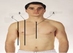
What is A? |
A is the midsternal line |
|

What is B? |
B is the midclavicular line |
|

What is C? |
C is the anterior axillary line |
|
|
What is the percussion sound heard over most of the lungs? |
Resonance |
|
|
What are the percussion sounds? |
- tympanic - hyperresonant - normal - dull - flat |
|
|
What does a tympanic percussion sound sound like? |
Drum like sounds heard over air filled structures during the abdominal examination |
|
|
What does a hyperresonant percussion sound sound like? |
Is said to sound similar to percussion of puffed up cheeks. May hear it over a pneumothorax. |
|
|
What does a normal percussion sound sound like? |
They are the resonant sounds produced by percussing a normal chest. |
|
|
What does a dull percussion sound sound like? |
Sounds similar to percussion of a mass such as a liver |
|
|
What does a flat percussion sound sound like? |
It is heard when percussion over bone |
|
|
What is the lining of the lungs called? |
Visceral pleura |
|
|
What is a barrel chest? |
Is typical of a patient with COPD. Is a loss of lung elasticity and flattening of the diaphragm. |
|
|
What kind of breath sounds are auscultated over the lower lobes of a healthy patient's lungs? |
Vesicular breath sounds |
|
|
Which of these situations would indicate decreased or absent breath sounds? A. When the bronchial tree is obstructed B. When the lungs are already hyperinflated from emphysema C. When there is air or fluid in the pleural space which obstructs the sound. |
All of them. |
|
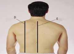
What is D? |
D is the scapular line |
|

What is E? |
E is the vertebral line |
|

What is F? |
F. is the anterior axillary line |
|
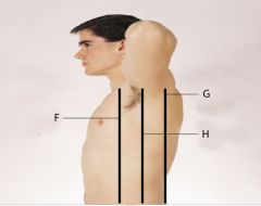
What is G? |
G is the posterior axillary line |
|

What is H? |
H is the midaxillary line |
|
|
Where is the mediastinum and what does it contain? |
It is the middle section of the thoracic cavity and it contains the esophagus, trachea, heart, great vessels and the right and left pleural cavities. |
|
|
Describe the lobes of the lungs. |
- paired but not quite symmetrical - right is shorter than the left due to the liver - left is narrower than the right due to the heart - right has 3 lobes - left has 2 lobes - lobes are separated by fissures |
|
|
Describe the right upper lobe of the lung from the anterior view. |
Apex is above the scapula, the bottom, medial edge ends at the 4th rib and the bottom lateral edge ends at the 5th rib along the right midaxillary line. |
|
|
Describe the right middle lobe of the lung from the anterior view. |
Top medial edge starts at the 4th rib, the top lateral edge starts at the 5th rib along the right midaxillary line and the bottom ends at the 6th rib along the right midclavicular line. |
|
|
Describe the right bottom lobe of the lung from the anterior view. |
The top edge starts at the 5th rib along the right midaxillary line and the bottom medial edge ends at the 6th rib along the right midclavicular line and the lateral edge ends at the 8th rib. |
|
|
What is the name of the fissure that separates the two lower lobes of the right lung? |
The oblique fissure. |
|
|
Describe the upper lobe of the left lung from the anterior view. |
The apex is above the clavicle and ends at the 6th rib along the left midclavicular line. |
|
|
Describe the lower lobe of the left lung from the anterior view. |
The lower lobe of the left lung can start to be seen at the 5th rib on the lateral side and the bottom edge on the medial side at the 6th rib along the left midclavicular line and the bottom edge on the lateral side is at the 8th rib. |
|
|
Describe the upper lobe of the left lung from the posterior view. |
The upper edge begins at the vertebral prominens and the oblique fissure starts at T3. The oblique fissure ends at T4 on the lateral side. |
|
|
When inflated from the posterior view, how far down does the diaphragm go? |
T12 |
|
|
Describe the trachea and bronchial tree |
- the trachea is anterior to the esophagus and is 10-11cm long in an adult - the trachea begins at the level of the cricoid cartilage in the neck - the trachea bifurcates just below the sternal angle into the left and right bronchi (posteriorly bifurcates at T4) - the right main bronchus is shorter, wider and more vertical than the left main bronchus |
|
|
What is the bronchial tree lined with? |
- Goblet cells that secrete mucus - cilia that filter and protect |
|
|
What is dead space? |
Space that is filled with air but not available for gaseous exchange. |
|
|
What is an acinus? |
A functional respiratory unit. The alveolar sac that contains multiple alveoli. Resembles a blackberry in form. |
|
|
Where does gaseous exchange occur in the human body? |
Occurs across the respiratory membrane in the alveolar ducts and alveoli. |
|
|
What is hypoventilation and what are the results of hypoventilation? |
Hypoventilation is shallow breathing (below 12 breaths per minute). This can cause the blood to have too much CO2 and not enough O2. If this happens, then the pH of the blood will drop. |
|
|
What is hyperventilation and what are the results of hyperventilation? |
Hyperventilation is rapid, deep breathing (faster than 20 breaths per minute). The can cause the blood to have too little CO2 and too much O2. If this happens, then the pH of the blood will increase. |
|
|
What is the respiratory center in the brain stem? |
The pons and the medulla |
|
|
What is hypercapnia? |
Too much CO2 in the blood. |
|
|
What is hypoxemia? |
Not enough O2 in the blood. |
|
|
What is respiration? |
The physical act of breathing. |
|
|
What is inspiration? |
Air rushing into the lungs (chest size increases) |
|
|
What is expiration? |
Air is expelled from the lungs (chest recoils - like letting go of a rubber band). It is passive. |
|
|
What muscles are used in inspiration? |
- external intercostals elevate the ribs - diaphragm descends as it contracts - sternocleidomastoid muscle elevates the sternum when extra breath is needed - the scalenus muscles can also elevate the ribs when extra breath is needed |
|
|
What muscles are used in forced expiration? |
- internal intercostals - external oblique and abdominal rectus both depress the lower ribs and compress the viscera |
|
|
What happens in the aging adult regarding respiration? |
- less mobile thorax (doesn't move as well) - respiratory muscle strength declines - there is a decrease in elastic properties within the lungs - less distensible (less stretch) and less ability to recoil - decreased vital capacity - increased residual volume - loss of intraalveolar septa (membranes) - decreased number of alveoli - lung bases become less ventilated - greater risk for post op atelectasis and infection |
|
|
What is vital capacity? |
The maximum amount of air that a person can expel from the lungs after filling the lungs to maximum. A decrease in this means less distensible. |
|
|
What is residual volume? |
The amount of air remaining in the lungs even after the most forceful expiration. An increase in this means there is less ability for recoil. |
|
|
What is atelectasis? |
Lung collapse |
|
|
Tb tends to run higher for what ethnic group? |
Asian-Americans |
|
|
What is FEV1/ FVC and what is it normally for a healthy adult? |
It is the ratio of forced expiratory volume for 1 second to forced vital capacity. It is normally 75% to 80%. |
|
|
If a patient has a cough, what questions would be asked regarding that cough? |
- how long have you had it? - is it dry or productive? - what color is the sputum? - how much sputum is there? - is there shortness of breath? Is it affected by position? (3 pillow orthopnea) - is there chest pain? - is there a past history of respiratory infection? - smoking history? - is the work or living environment dangerous? - when was the last Tb test, flu vaccine or chest X-ray?
|
|
|
What is normal shape for posterior chest? |
- spinous processes are in a straight line - the thorax is symmetrical - cross section would be an elliptical shape - the ribs slope downward - the scapula are symmetrical in each hemithorax |
|
|
What is hemoptysis? |
Bloody mucus |
|
|
What is scoliosis? |
A curvature of the spine to one side or the other. |
|
|
What is kyphosis? |
A curvature of the spine forward. Common in the elderly. |
|
|
What is "barrel chest"? |
Rounded, bulging chest that resembles a barrel. The ribs become horizontal (instead of sloping downward). Chest appears to be in continuous inspiration. Caused by breathing issues such as emphysema or COPD. |
|
|
What would be abnormal regarding a chest/ breathing assessment? |
- Overly developed (hypertrophied) neck muscles - tripod position (leaning forward with arms braced against the knees - helps with expiration) |
|
|
What is normal for the skin of the posterior chest? |
- skin color consistent with ethnicity - no lesions - no change in any nevus (moles) |
|
|
What would be abnormal for the skin of the chest? |
Cyanosis or pallor |
|
|
What is symmetric expansion? |
As the patient inhales deeply, your thumbs should move apart symmetrically. Normal for assessment of the posterior chest. |
|
|
What would be abnormal regarding chest expansion and what could it mean? |
Unequal chest expansion could mean - atelectasis - pneumonia - thoracic trauma such as fractured ribs - pneumothorax - pain if pleura are inflamed |
|
|
What is the angle of Louis? |
The manubriosternal notch. |
|
|
What is bradypnea? |
Abnormally slow respiration |
|
|
Describe bronchial breath sounds. |
- harsh - high pitched - loud |
|
|
Describe bronchiovesicular sounds. |
- medium in loudness - medium in pitch |
|
|
Describe vesicular sounds. |
Lowest in pitch of all the lung sounds. |
|
|
What are fine crackles? |
Adventitious lung sounds that sound like short popping sounds, high pitched and are usually in the bases of the lower lobes. This is often associated with an inflammation or infection of the small bronchi, bronchioles or alveoli. |
|
|
What are coarse crackles? |
Adventitious lung sounds that sound like short crackling sounds, low pitched and are usually located in the trachea or large bronchi. Can often be heard with pneumonia, atelectasis, pulmonary fibrosis and acute bronchitis as well as congestive heart failure and pulmonary edema. |
|
|
What is rhonchi? |
Adventitious lung sounds that sound like long snoring sounds, low pitched and usually located in the bronchi. Can be heard with asthma, bronchitis, pneumonia, emphysema, etc. |
|
|
What are wheezes? |
Adventitious lung sounds that sound like long musical notes, high pitched and located throughout the lungs. This is often heard with asthma. |
|
|
What is stridor? |
Adventitious lung sounds that sound like long crowing sounds, high pitched and located in the trachea. Can often be heard without a stethoscope. Caused by a blockage in the trachea or larynx. Needs immediate attention. |
|
|
What would fine crackles in a vesicular region indicate? |
It would likely indicate pneumonia. |
|
|
What is bronchitis? |
inflammation of the membrane lining of the bronchial tubes |
|
|
What's the difference between acute and chronic bronchitis? |
Acute is one incidence that happened and has cleared up. Chronic is for two months or longer for 2 years in a row. |
|
|
What is bronchophony? |
Abnormal voice sounds. When the patient repeats the word "99" and it can be heard clearly through the stethoscope. Indicates consolidation or compression of lung tissue. |
|
|
What is egophony? |
Abnormal voice sounds. When the patient says "eee" and it sounds like a long "A" sound. Indicates something is wrong. |
|
|
What is COPD? |
Chronic Obstructive Pulmonary Disease. Usually involves emphysema and chronic bronchitis. |
|
|
What is the costal angle? |
The angle created by the ribs as they slope away from the xiphoid process. |
|
|
What is dyspnea? |
Difficulty, or labored, breathing |
|
|
What is emphysema? |
Due to loss of elasticity, lungs are already hyperinflated. Lung sounds are absent. |
|
|
What is eupnea? |
Normal relaxed breathing. |
|
|
What are fissures? |
The borders that separate the lobes of the lungs. |
|
|
How many fissures are there in the lungs? |
2 in the right (3 lobes) 1 in the left (2 lobes) |
|
|
What is tactile, or vocal, fremitus? |
Using the balls of the fingers, touching the person's chest, there is a palpable vibration when the patient says the word "99" |
|
|
What is hypercapnia? |
Too much CO2 in the blood. |
|
|
What is hyperresonance? |
A lower pitched booming sound (lower than resonance). |
|
|
What is resonance? |
Low pitch, clear hollow sound heard over normal lung tissue. |
|
|
What is hyperventilation? |
Excessively deep and rapid breathing (faster than 24 breaths per minute). Often caused by stress. |
|
|
What is hypoxemia? |
Not enough oxygen in the blood. |
|
|
What is kyphosis? |
A forward curvature of the spine. Common in the elderly. |
|
|
What is orthopnea? |
A position taken to aid in breathing. A tripod stance with arms braced against the legs and leaning forward. Often used by people with COPD. |
|
|
What is paroxysmal nocturnal dyspnea. |
Waking up for a shortness of breath. |
|
|
What is Pleural friction rub? |
An adventitious lung sound caused by an inflammation of the pleura of the lung. It common in pneumonia, pulmonary embolism and pleurisy. |
|
|
What is pneumonia? |
Inflammation of the lungs caused by an infection |
|
|
What is a pneumothorax? |
The presence of air or gas in the pleural spaces of the lungs. High risk of lung collapse. Caused by a hole in the lung or the chest wall (trauma) or a ruptured cyst on the surface of the lung. |
|
|
What is retraction? |
The pulling in of the muscles in an effort to breathe (using the accessory muscles). |
|
|
What is scoliosis? |
A lateral curvature of the spine. |
|
|
What is the suprasternal notch? |
Also known as the jugular notch, it is found at the superior border of the manubrium of the sternum. |
|
|
What is tachypnea? |
Excessively rapid, shallow, respiration, faster than 24 breaths per minute. |
|
|
Name the anterior thoracic landmarks. |
- suprasternal notch - sternum - sternal angle (angle of Louis) - costal angle |
|
|
Name the posterior thoracic landmarks. |
- vertebra prominens - spinous processes - inferior border of the scapula - twelfth rib - reference lines (scapular & vertebral) |
|
|
Name the components of the thoracic cage. |
- the sternum - 12 pairs of ribs - 12 thoracic vertebrae - diaphragm is the floor of the thoracic cavity - first 7 ribs attach directly to the sternum - ribs 8, 9, 10 attach to the costal cartilage - ribs 11 & 12 are "floating" |
|
|
Describe the borders of the lung from the anterior view. |
The apex is 3 to 4 cm above the inner third of the clavicles. The base rests on the diaphragm at the the 6th rib in the midclavicular line. |
|
|
Describe the borders of the lung from the lateral view. |
The lung extends from the apex of the axilla down to the 7th or 8th rib. |
|
|
Describe the borders of the lung from the posterior view. |
The location of C7 marks the apex of lung tissue and around T10 marks the base. Deep inspiration can make the lungs expand down to T12. |
|
|
Describe the pleurae. |
They are the serous membranes that form an envelope between the lungs and chest wall. The visceral pleura lines the outside of the lungs. The parietal pleura lines the inside of the chest wall and diaphragm. In between is called the pleural cavity which is filled with a small amount of lubricating fluid. |
|
|
How dangerous is second-hand cigarette smoke? |
Very. It increases an adults risk for cancer and heart disease. It also increases the risk for sudden infant death syndrome, ear infections and asthma attacks in children. There is no safe level of exposure to second hand smoke. |
|
|
What are the four major functions of respirations? |
1. supplying O2 to the body to produce energy 2. removing CO2 as a waste product 3. maintaining homeostasis (acid/base balance) 4. maintaining heat exchange |
|
|
What are the functions of the trachea and bronchial tree? |
They transport gases between the environment and the lung tissue. They constitute the dead space of the person. The bronchial tree is lined with mucus and cilia to protect the body from small particles. |
|
|
What is the normal respiratory rate for an adult? |
10 to 20 breaths per minute. |
|
|
What is Cheyne-Stokes respiration? |
A cycle of respiration where the rate increases, decreases then stops and starts over again. |
|
|
What causes Cheyne-Stokes respiration? |
The most common causes are; - severe heart failure - renal failure - meningitis - drug overdose - increased cranial pressure (ICP). |
|
|
What is Biot's respiration? |
A respiration pattern similar to Cheyne-Stokes, but irregular in rate and depth. |
|
|
What often happens with chronic emphysema? |
Hypertrophy of the abdominal muscles. |
|
|
When would there be decreased or absent breath sounds? |
- when the bronchial tree is obstructed by secretions or a foreign body - in emphysema, the loss of elasticity and hyperinflated lungs cause the inhaled air to not make much noise. |
|
|
When would there be increased breath sounds? |
They occur with pneumonia or fluid in the intrapleural space. The sound is conducted better when the tissue is dense. |
|
|
What is colostrum? |
A yellowish liquid, rich in immunities secreted by the mammary gland of female mammals a few days before and after giving birth. |
|
|
What are Cooper's ligaments? |
The connective tissue in the breast that help maintain structural integrity. |
|
|
What is galactorrhea? |
An abnormally abundant flow of milk from a lactating woman or secretion of milk from a non-lactating person. |
|
|
What is gynecomastia? |
An abnormal enlargement of breast tissue in a male. |
|
|
What is the inframammary ridge? |
Strands of fibrous tissue that surround the breast and make a ridge along the bottom. |
|
|
What does Lactiferous mean? |
Producing or secreting milk. |
|
|
What is lymphadenopathy? |
Swollen lymph nodes. |
|
|
What is mastalgia? |
Breast pain. |
|
|
What is mastitis? |
Inflammation of the breast. |
|
|
What are montgomery's glands? |
Sebaceous glands in the areola of the nipple that help protect the nipple. |
|
|
What is Paget's disease? |
Breast cancer that affects the skin of the breast looking similar to eczema. |
|
|
What is peau d'orange? |
The condition describing the look of Paget's disease on a breast. |
|
|
What is supernumerary nipple? |
A third nipple (or triple nipple). |
|
|
What is the tail of Spence? |
An extension of breast tissue that extends into the axilla. |
|
|
What is tanner staging? |
Staging to determine sexual maturity. |
|
|
Name the internal structures of the human breast. |
- Glandular tissue (15-20 lobes) - Cooper's ligaments that contract in breast cancer that cause pits and dimples in overlying skin. - adipose tissue - 4 quadrants (upper outer quadrant that has the axillary tail of Spence - site of most breast cancers) - 4 groups of axillary nodes |
|
|
Name the 4 groups of axillary nodes. |
- central axillary nodes - pectoral (anterior) nodes - subscapular (Posterior) nodes - lateral (along the humerus) |
|
|
What age related changes happen in the female breast? |
- decrease in estrogen and progesterone - breast tissue atrophies - decreased breast size & elasticity (lumps become more prominent) - breasts droop and sag - decreased axillary hair |
|
|
What questions should be asked during a breast assessment? |
- Ever get breast pain? (cyclic? when? where?) - lumps (redness, dimpling, swelling, warmth) - discharge (clear? bloody? what meds taking?) - rash (Paget's disease -eczema starts on areola) - swelling? (one spot or all over?) r/t period, pregnancy, breastfeeding - trauma (hematoma, edema) - history of breast disease (surgeries, mammoplasty? - surgery - Self care behaviors (BSE monthly) |
|
|
Describe Breast self exams (BSE). |
- should start at age 20 - should be done monthly after menses - arm up of the side being checked - vertical strip pattern is best |
|
|
When should women start getting annual mammograms? |
At age 40. |
|
|
What are the different techniques for palpation of the breast? |
- vertical strip (which is best) - concentric circles - spokes on a wheel - all should include tail of Spence (into axilla) |
|
|
When inspecting the breast, what does a nurse look for? |
- symmetry (slight asymmetry is normal) - size - shape - skin (smooth, even color, "orange skin") - edema |
|
|
When palpating the nipple, what is done and why? |
Squeeze the nipple between thumb and forefinger for discharge, inversion, skin changes and compare to other side. |
|
|
When a breast lump is documented, what information needs to be on the documentation - 11 items? (test question may ask what's missing from documentation) |
- location (using hands on a clock) - distance (in cm. from nipple) - size - shape - consistency (firm, hard, soft, mushy) - movable or fixed - distinctiveness (solitary or mulitple) - nipple - skin over the nipple (red?dimpled?) - tenderness - lymphadenopathy? |
|
|
What is afterload? |
The opposing pressure the ventricle must generate to open the aortic valve against the higher aortic pressure. At the end of diastole, the ventricle pressure is 5-10mmHg and the aortic pressure is 70-80mmHg. The ventricle must tense up to overcome this pressure. |
|
|
What is apical impulse? |
Also known as the Point of Maximum impulse. It is the furthermost point laterally and inferiorly from the sternum that the cardiac impulse can be felt. |
|
|
What is an arrhythmia? |
Also known as a dysrhythmia, it is an irregular heartbeat. |
|
|
What is automaticity? |
The ability of the cardiac muscle cells to general pacemaking stimuli. |
|
|
What is a bruit? |
An unusual sound that blood makes when it rushes past an obstruction in an artery when the sound is auscultated with the bell of a stethoscope. |
|
|
Define Cardiac output. |
The amount of blood pumped by the heart ventricles in 1 minute. Normal CO is about 4-6L/min. The amount is calculated by the equation CO=SV x HR. |
|
|
What is stroke volume? |
The amount of blood ejected from the ventricle with one cardiac cycle (heartbeat). |
|
|
What is diastole? |
The heart at rest - in between contractions (about 2/3 of it's time). |
|
|
What is systole? |
The heart contraction - about 1/3 of it's time. |
|
|
What is DOE? |
Dyspnea on exertion. Shortness of breath which occurs with effort. Usually a sign of heart failure or ischemia. |
|
|
What is a functional murmur? |
A murmur without any cardiac disease. |
|
|
Define hepatojugular reflux |
Distention of the jugular vein induced by applying pressure over the liver. It suggests insufficiency of the right side of the heart. |
|
|
What is an innocent murmur? |
A functional murmur. |
|
|
What is orthostatic hypotension? |
An abnormal decrease in blood pressure as the patient stands up from a sitting or lying position. |
|
|
Define pericardial friction rub. |
A grating or scraping noise heard with a stethoscope with the heart beat caused by an inflammation of the pericardium. |
|
|
What is precordium? |
The region of the anterior surface of the body covering the heart and lower thorax. |
|
|
What is preload? |
The volume of blood in the ventricles at the end of diastole ('stretch'). |
|
|
What is pulse deficit? |
A peripheral pulse less than the actual heart rate indicating a lack of peripheral perfusion. |
|
|
What is pulse pressure? |
The difference between systolic and diastolic pressures. (Widens in the elderly). |
|
|
What is sinus arrhythmia? |
An irregular cardiac rhythm in which the heart rate usually increases with inspiration and decreases with expiration. Is common in children and young adults and has no clinical significance except in the elderly. |
|
|
Anteriorly, where is the apex of the heart? |
5th intercostal space along the midclavicular line |
|
|
Anteriorly, where is the right atrium? |
To the right and above the right ventricle on the anterior cardiac surface and the right cardiac border. |
|
|
Anteriorly, where is the left ventricle? |
Behind the right ventricle on the left cardiac border. |
|
|
Anteriorly, where is the left atrium? |
Behind the right atrium. |
|
|
Anteriorly, where are the great vessels? |
At the base of the heart. |
|
|
Where is the superior and inferior vena cavae? |
Behind the right side of the heart - superior, above, and inferior, below. |
|
|
Describe the pulmonary arteries. |
They receive deoxygenated blood from the right ventricle of the heart and carries blood to the lungs. The only arteries to carry deoxygenated blood besides umbilical arteries in the fetus. |
|
|
Describe pulmonary veins. |
They carry oxygenated blood from the lungs to the left atrium of the heart. Of the very few veins that carry oxygenated blood. There are four - two from either lung. |
|
|
Describe the superior vena cava. |
The vena cava are the largest veins bringing blood back from the body. The superior drains blood back to the heart from the head, neck and arms. |
|
|
Describe the inferior vena cava. |
The vena cava are the largest veins bringing blood back from the body. The inferior brings blood back to the heart from the lower part of the body. |
|
|
What is S1? |
Closure of the tricuspid and mitral valves. "Lub" |
|
|
What is S2? |
Closure of the aortic and pulmonic valves. "Dup" |
|
|
What is S3? |
A ventricular "gallop". Vibrations during diastole. Think of the heart sounds as though saying the word "Ken-TUCK-y" and the y represents S3 and can be heard best towards the apex of the heart (T & M). |
|
|
What is S4? |
An atrial "gallop". Vibrations at the end of diastole. A low frequency sound that occurs in late diastolic filling due to atrial contraction. If loud, it indicates pathology. Think of the heart sounds as though saying "TEN-nes-see" and "ten" respresents S4 and can be heard best at the apex of the heart (M). |
|
|
Name three conditions that can cause murmurs. |
1. Velocity of blood increases (flow murmur) such as in exercise or thyrotoxicosis -hyperthyroidism which increases the metabolic rate and the heart rate. 2. Viscosity of blood decreases such as anemia 3. Structural defects in the valves. |
|
|
What are the characteristics of heart sounds? |
1. Frequency - the relative pitch 2. Intensity - loud or soft 3. Duration - heart sounds are shorter than silent periods 4. Timing - systole or diastole |
|
|
How does the electrical current spread through the heart? |
The electrical impulse starts with the SA node (the pacemaker of the heart) and travels through the atria to the AV node where it then travels down through the bundle of HIS, down the right and left bundle branches and the Purkinje fibers in the ventricles. |
|
|
Describe the carotid artery pulse. |
A pressure wave generated by each systole pumping blood into the aorta. |
|
|
Why is it important to assess the jugular veins? |
The jugular veins empty direction into the superior vena cava which empties into the right atrium of the heart. If there is problems with the right side of the heart, the blood will back up causing the jugular veins to become distended. Very important information! |
|
|
What is the function of the foramen ovale? |
To bypass the lungs and send the blood directly into the left side of the heart and to the body of the fetus. The blood is oxygenated from mother's body and does not need to go through the lungs of the fetus. |
|
|
What is the function of the ductus arteriosus? |
To bypass the lungs from the pulmonary artery through the ductus arteriosus to the aorta. The fetus' blood is already oxygenated and does not need to go through the fetus' lungs. |
|
|
What are the risk factors associated with heart disease and stroke? |
- age - stress - smoking - chronic alcoholic use - lack of exercise - poor diet |
|
|
What are the hemodynamic changes with aging? |
- widening of pressure pulse, due to the stiffening of the large arteries - ventricular wall thickness increases - decreased ability of the heart to respond to increased need for oxygen. (exercise intolerance). |
|
|
Where can the valves be heard during auscultation? |
Aortic Valve - second right interspace Pulmonic valve - second left interspace Erb's point - third left interspace Tricuspid valve - left lower sternal border Mitral valve area - fifth interspace at around left midclavicular line |
|
|
What is a heart murmur? |
A blowing, swooshing sound that occurs with turbulent blood flow in the heart or great vessels. Is typically abnormal and is graded on it's severity. |
|
|
What could jugular distention be a symptom of? |
Right sided heart failure. |
|
|
Regarding the heart, what could SOB be a symptom of? |
Left sided heart failure. |
|
|
How is jugular venous distention estimated? |
With the patient lying at a 45 degree angle a ruler is placed at the sternal angle. A straight edge is placed at the top of where the jugular vein can be seen and leveled across to the ruler. The measurement on the ruler estimates the jugular venous distention. Normal is less than 3cm. |
|
|
What is the Allen test? |
A test of adequate circulation by holding down the radial and ulnar arteries at the wrist, allowing the hand to lose color. Pressure is released over the ulnar artery only and the hand should return to normal color in 2 to 5 seconds. |
|
|
What is arteriosclerosis? |
When peripheral blood vessels grow more rigid and lose elasticity with age. |
|
|
What is an artery? |
A blood vessel carrying oxygenated (in most instances) blood from the heart to the body. |
|
|
What is atherosclerosis? |
The deposit of fatty plaques on the intima (innermost lining) of the arteries. |
|
|
What is capillary refill? |
Blanching the fingernail beds and waiting for them to return to the normal pink color. Usually it returns in a fraction of a second, but up to 2 seconds is considered normal. |
|
|
What is claudication distance? |
The distance a person can walk before claudication takes place (pain caused by peripheral arterial disease). This typically takes place in the calf, but can also happen in the feet, thigh or buttocks. |
|
|
What is deep vein thrombophlebitis? |
A deep vein blocked by a thrombus (a clot), causing inflammation, blocked venous return, cyanosis and edema |
|
|
What is a doppler? |
An ultrasonic stethoscope. |
|
|
What is edema? |
Fluid buildup in the interstitial spaces. |
|
|
What is the Homan's sign? |
When a patient dorsi-flexes their toes and bends their knee, causing pain in the calf muscle. It is called a positive Homan sign that they have DVT. |
|
|
What is ischemia? |
A deficient supply of oxygenated blood to tissues caused by obstruction of a blood vessel. |
|
|
What is a Lymph node? |
Any of the small bodies located along the lymph vessels that filter bacteria and foreign bodies from lymphatic fluid. |
|
|
What is lymphedema? |
Tender, swollen lymph nodes. |
|
|
What is pitting edema? |
When edema is pressed and the pit remains after pressure is removed. |
|
|
What is the pulse? |
With every heart beat, the heart pumps blood into the aorta. The force flares the arterial walls and generates a pressure wave that is palpable called the pulse. |
|
|
What is pulsus paradoxus? |
Beats having a weaker amplitude with inspiration and stronger with expiration. Commonly found with cardiac tamponade, also found in severe bronchospasm of acute asthma. |
|
|
Describe the spleen. |
Located in the upper left quadrant of the abdomen and has four functions; 1. destroys old red blood cells 2. produces antibodies 3. stores red blood cells 4. filters microorganisms from the blood |
|
|
What is the thymus? |
A flat, pink-gray gland located in the superior mediastinum behind the sternum and in front of the aorta (on top of the heart). Is important in developing T-lymphocytes of the immune system. |
|
|
Describe the tonsils. |
The tonsils are part of the lymphatic system by responding to local infection. The are located at the entrance to the respiratory and GI tracts. |
|
|
What are varicose veins? |
A condition in which the superficial veins in the legs become tortuous, knotted and swollen caused by a defect in the venous valves or the venous pump that normally moves blood out of the legs while standing for long periods of time. |
|
|
What is a vein? |
A blood vessel carrying deoxygenated from the body toward the heart, with the exception of the pulmonary vein, which carries oxygenated blood to the left atrium of the heart. |
|
|
What is a venous, or stasis, ulcer? |
Wounds that occur due to improper functioning of the venous valves, usually of the legs. |
|
|
Name the pulses palpable for examination. |
1. brachial pulse in the arm 2. radial pulse in the forearm 3. the abdominal aorta in thinner people 4. the femoral artery in the upper thigh 5. the popliteal artery behind the knee 6. the posterior tibial artery near the ankle 7. dorsalis pedis artery on the top of the foot |
|
|
What are the mechanisms that keep blood moving toward the heart in the venous system? |
1. The pumping heart 2. Adequate blood pressure in the arteries 3. Semilunar valves in the veins to keep blood from flowing backwards. 4. Contraction of skeletal muscles which squeezes veins, producing a sort of pumping action. 5. Changing pressures in the chest cavity during breathing that produce a kind of pumping action in the veins in the thorax. |
|
|
What are capacitance vessels? |
Another name for veins due to their capacity to stretch and hold more blood when blood volume increases. |
|
|
What are the risk factors for venous stasis? |
- Long periods of immobility like driving, flying, bed rest or an orthopedic cast. - hypercoagulable states and vein wall trauma |
|
|
What is the structure of the lymphatic system? |
They start out as open ended capillaries that siphon interstitial fluid. The capillaries converge to form vessels that drain into larger ones. They have valves like veins so flow is one way from the tissues to the blood stream and is very slow. The flow is propelled by contracting skeletal muscles like the venous system. |
|
|
What are the three functions of the lymphatic system? |
1. to conserve fluid and plasma proteins that leak from capillaries 2. to form a major part of the immune system that defends the body against disease 3. to absorb lipids from the intestinal tract |
|
|
What are the locations of the superficial groups of lymph nodes that are palpable during examination? |
1. cervical nodes 2. axillary nodes 3. epitrochlear node in the antecubital fossa (inside of the elbow) 4. inguinal nodes in the groin |
|
|
What are the related organs and functions of the lymphatic system? |
The spleen - store or destroy RBCs, make antibodies, filter blood The tonsils - respond to local inflammation The thymus - making T lymphocytes |
|
|
What differences affect the skin with arterial and venous insufficiency? |
Arterial - skin is shiny and free of hair and temp is cool Venous - skin is flaky and dry and temp is normal |
|
|
What differences are there regarding ulcers with arterial and venous insufficiency? |
Arterial - the ulcers are extremely painful, appear on the lower third of the leg and toes, lack of granulation tissue, edges are smooth and well defined and they are deep Venous - the ulcers have mild to moderate pain, are irregularly shaped and are shallow. |
|
|
If a patient was to get an ulcer on the ankle, where would it be if it were from arterial insufficiency? Venous insufficiency? |
Arterial insufficiency would cause an ulcer on the lateral malleolus whereas a venous insufficiency would cause an ulcer on the medial malleolus. |
|
|
What difference is there regarding pulses with arterial and venous insufficiency? |
With arterial insufficiency, the pulses are not palpable (or reduced) With venous insufficiency, the pulses are normal |