![]()
![]()
![]()
Use LEFT and RIGHT arrow keys to navigate between flashcards;
Use UP and DOWN arrow keys to flip the card;
H to show hint;
A reads text to speech;
164 Cards in this Set
- Front
- Back
|
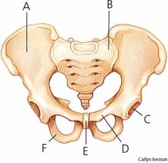
What is an innominate bone? |
A bone also known as hip bone which is irregularly shaped and fuses the illium, pubis and ischium in the pelvis. |
|
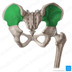
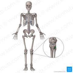
What is the bone highlighted? |
The Illium |
|
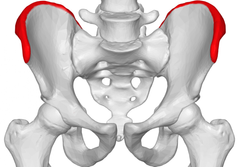
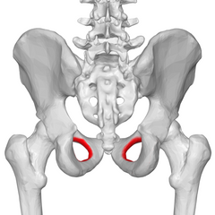
What bone is highlighted? |
The Illiac Crest |
|
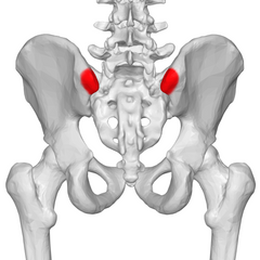
What bone is highlighted? |
ASIS |
|
|
What does ASIS stand for? |
Anterior Superior Illiac Spine |
|
What bone is highlighted? |
PSIS |
|
|
What does PSIS stand for? |
Posterior Superior Illiac Spine |
|
What bone is highlighted? |
AIIS |
|
|
What does AIIS stand for |
Anterior Inferior Illiac Spine |
|
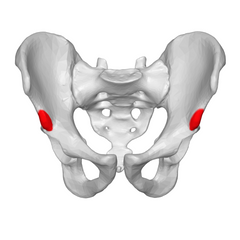
What bone is highlighted? |
Ischial Tuberosity |
|
What bone is highlighted? |
Obturator Foramen |
|
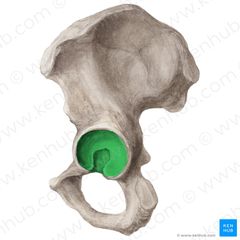
what is shown here? |
The aecetabulum |
|
|
What is the acetabulum? |
The socket part, of the ball and socket joint where the head of the femur sits. |
|
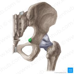
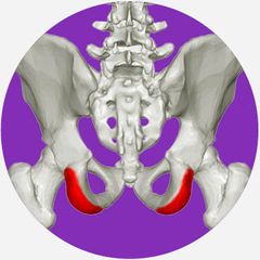
What is shown here? |
The ischial spine |
|
|
How many bones make up the pelvic girdle? |
4 |
|
|
What are the bones that make up the pelvic girdle? |
the sacrum, the coxyx, the illim, the ischium and the pubis |
|
|
How many joints are in the hip? |
3 |
|
|
Which are the joints in the hip? |
Lumbosacral, symphysis pubis and sacroilliac |
|
|
What bones meet at the lumbosacral joint? |
The last vertebrae of the lumbar region of the spine and the first vertebrae of the Sacrum region of the spine. |
|
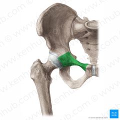
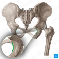
What joint is being shown here? |
The lumbosacral Joint |
|
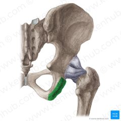
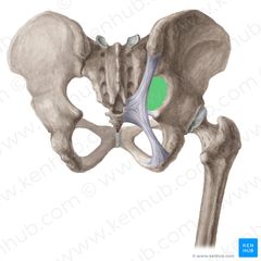
What joint is being show here? |
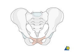
Symphysis pubis |
|
|
What two bones meet at the symphysis pubis? |
The left and right pubic |
|
|
what type of joint is the symphysis pubis? |
cartilaginous |
|
|
What two types of cartilage is the symphysis pubis? |
hylaine and fibrocartilage |
|
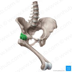
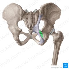
What joint is shown here? |
Sacroilliac |
|
|
what bones meet at the sacroilliac joint? |
The illium and sacrum |
|
|
What type of joint is the sacroilliac |
Synovial joint |
|
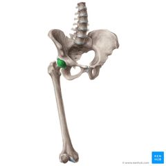
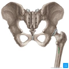
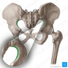
What joint is shown here? |
The acetabulfemoral joint |
|
|
What articulations are at the acetabulofemoral joint? |
The head of the femur and the acetabulum |
|
|
What is the acetabulum made up of? |
the ischium, illium and pubis |
|
|
What type of joint is the acetabulofemoral joint? |
synovial |
|
|
what is the classification of the acetabulofemoral joint |
multi-axial |
|
|
what is a multi-axial joint? |
a joint which can provide movement along three or more axis |
|
|
what is a mono-axial joint? |
Where movement occurs across one plane of movement. |
|
|
What is a bi-axial joint? |
where movement occurs across two planes of movement. |
|
|
What movements are available at the acetabulofemoral joint? |
Flexion, Extension, Abduction, Adduction, Medial Rotation and Lateral Rotation |
|
|
What plane of movement does the acetabulofemoral joint allow? |
Sagittal, Frontal and Transverse |
|
|
What axis of movement does the acetabulofemoral joint allow? |
Medial-Lateral, Anterior-Posterior and Superior-Inferior |
|
|
What is a 'fibrous joint'? |
Where the adjacent bones are connected by fibrous connective tissue. |
|
|
What is a 'cartilaginous joint'? |
The articulating surfaces are joined by fibrocartilage or hyaline cartilage. |
|
|
What is a 'synovial joint' |
Where the articulating surfaces are indirectly connected by a joint cavity filled with lubricating fluid called 'synovial' fluid. |
|
|
What is a snyarthrosis joint? |
A nearly immobile joint- doesn't provide movement. |
|
|
What is the nature of a snyarthrosis joint? |
To provide a strong union between the two articulations- important for offering protection for vital organs |
|
|
What is an example of a snyarthrosis joint? |
The sutures in the skull |
|
|
What is an 'ampiathrosis joint'? |
A joint which has very limited mobility. |
|
|
What is an example of an ampiathrosis joint? |
The cartilaginous joint between vertebrae or the pubic symphasis. |
|
|
What is the role of an ampiathrosis joint? |
Provide a strong connection between the adjacent bones and provides protection of vital organs. |
|
|
What is a 'diathrosis joint?' |
A freely movable joint also known as a synovial joint |
|
|
What is the nature of a diathrosis joint? |
Allow joints a wide range of movement |
|
|
What is an example of a diathrosis joint? |
The ball and socket joint at the hip (acetabulofemoral joint) |
|
|
What is the classification of a diathrosis/synovial joint? |
multiaxial |
|
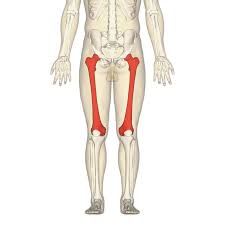
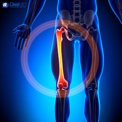
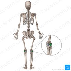
What bone is highlighted here? |
The femur |
|
what is highlighted here? |
head of the femur |
|
what is highlighted here? |
The neck of the femur |
|
What part of a bone is highlighted yellow? |
Body of the femur |
|
What part of a bone is shown here? |
The greater trochanter |
|
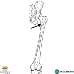
What is the arrow pointing to? |
The lesser trochanter |
|
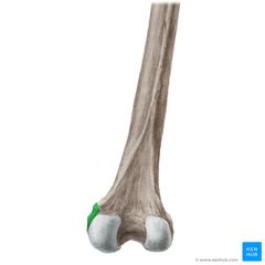
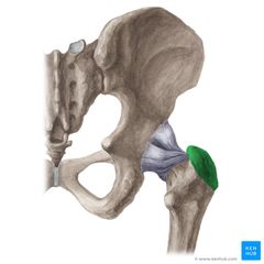
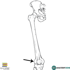
What is highlighted green here? |
The medial epicondyle |
|
What is the arrow pointing to? |
The lateral epicondyle |
|
what highlighted in green here? |
Lateral condyle |
|
What is shown here? |
Medial condyle |
|
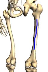
what is the green colour highlighting? |
The pectineal line |
|
|
Where is the pectineal line located? |
Runs below the lesser trochanter, diagonally towards the linea aspera |
|
|
What is the role of the pectineal line? |
Provides attatchment for the adductor brevis |
|
|
Where is the head of the femur located? |
At the proximal end of the femur |
|
|
Where is the neck of the femur located? |
Between the head and both trochanters |
|
|
where is the body of the femur located? |
between each end of the bond ends (head |
|
|
Where is the greater trochanter located? |
Laterally between the neck and body of the femur |
|
|
What is the role of the greater trochanter? |
Provides attachment for the gluteus maximus and minimus and most deep rotator muscles |
|
|
Where is the lesser trochanter located? |
Medially and posteriorly, distal to the greatertrochanter. |
|
|
What is the role of the lesser trochanter? |
Providing attachment for the iliopsoas muscle |
|
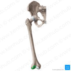
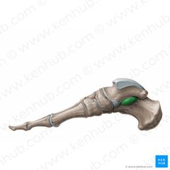
what is shown here? |
The adductor tubercle |
|
|
Where is the adductior tubercle located? |
Proximal to the medial epicondyle. |
|
|
What is the role adductor tubercle? |
Provides attachment for a portion of the adductor magnus. |
|
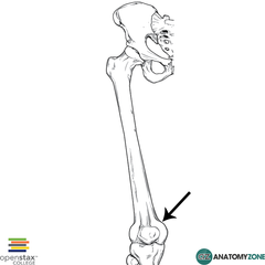
What is highlighted in blue? |
The linea aspera of the femur |
|
|
Where is the linea aspera located? |
Runs the length of the body of the femur |
|
|
What is the role of the linea aspera? |
Provides muscle attachment |
|
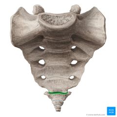
What joint is highlighted green? |
The sacrococcygeal joint |
|
|
What are the two articulations in the sacrococcygeal joint? |
The last vertebrae of the sacrum region of the spine and the coccygeal |
|
|
What type of joint it the sacrococcygeal joint? |
An ampiathrosis joint. |
|
|
What is the function of the pelvic girdle? |
Transfers the weight from the top half of the body, to the lower limbs while keeping the body balanced. |
|
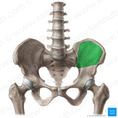
What is shown here? |
The illiac fossa |
|
|
What is a fossa? |
An area, usually in bone, where there a depression or hollowing which is usually smooth. |
|
|
Give an example of a fossa? |
the fossa of the scapular or the cranial fossa. |
|
What is shown here? |
The superior ramus of pubic bone |
|

What is shown here? |
Inferior pubic ramus |
|
What is shown here? |
The tubersoity of the ischium |
|
What is the letter F showing? |
Ramus of the ischium |
|
What is the image showing here? |
The greater sciatic notch |
|
What is being shown here? |
The lesser sciatic notch |
|
What is being shown here? |
The greater sciatic foramen |
|
Whats is being shown here? |
The lesser sciatic foramen |
|
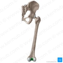
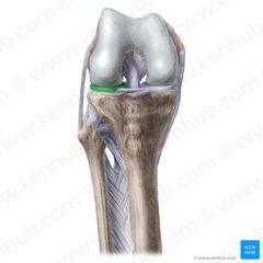
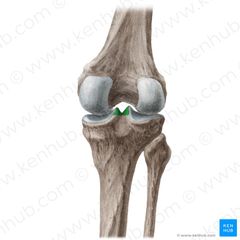
What is shown in green? |
The intercondylar fossa of the femur |
|
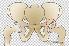
What is circled here? |
The acetabular labrum |
|
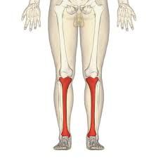
What bone is shown here? |
The tibia |
|
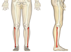
What bone is shown here? |
The fibular |
|
What is shown here? |
Medial meniscus |
|
What is shown here? |
Lateral meniscus |
|
What is shown here? |
Medial condyle of the tibia |
|
What is shown here? |
Lateral condyle |
|
What is shown here? |
Tibial plateau |
|
What is shown here? |
The intercondylar eminence of the tibia |
|
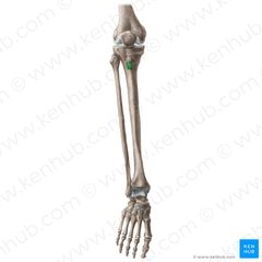
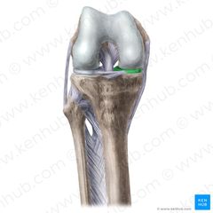
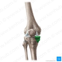
What is highlighted green? |
Tibial tuberosity |
|
What is shown here? |
Medial malleolus |
|
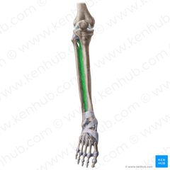
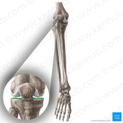
What is shown here? |
The interossous membrane |
|
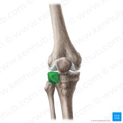
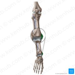
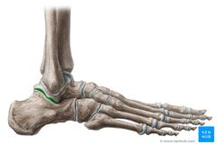
What joint is shown here? |
the proximal tibiofibular joint |
|
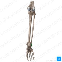
What is shown here? |
Lateral malleolus |
|
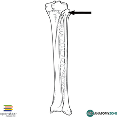
What is the arrow pointing to? |
Head of the fibular |
|
|
How many tibiofibular joints are there? |
3 |
|
|
What are the tibiofibular joints? |
Proximal tibiofibular, intermediate (interossous membrane) tibiofibular and the distal tibiofibular |
|
|
how many tarsals is the foot made up of? |
7 |
|
|
What are the names of the tarsals? |
lateral, intermediate and medial cuneiforms, cuboid, talus, calaneous and navicular |
|
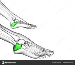
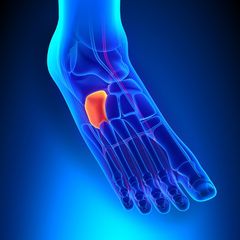
What bone is highlighted green? |
The calcaneous (heel bone) |
|
What bone is highlighted here? |
The talus |
|
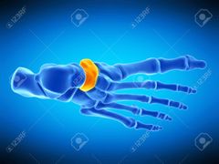
What bone is highlighted here? |
The cuboid |
|
What bone is shown here? |
The medial cuneiform |
|
What bone is highlighted red? |
Lateral cuneiform |
|
What bone is shown in red? |
The intermediate cuneiform |
|
What bone is shown in yellow? |
The navicular |
|
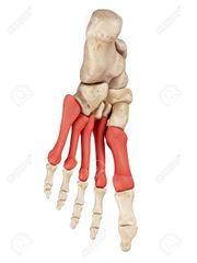
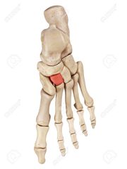
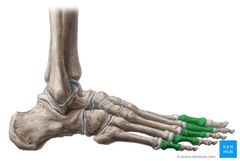
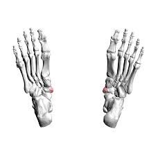
What bones are highlighted red? |
Metatarsals 1- 5 |
|
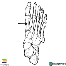
What metatarsal is shown here? |
the first metetarsal |
|
what metatarsal is shown here? |
The fifth metatarsal |
|
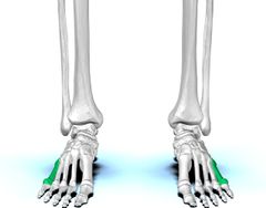
What is highlighted green? |
Proximal Phalanhges |
|
What is shown in green? |
the distal phalanges |
|
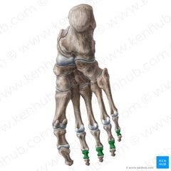
What phalanges are shown here? |
The intermediate phalanges |
|
What is highlighted green? |
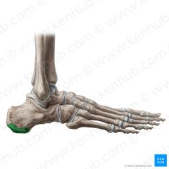
Calcaneal tuberosity |
|
What is shown here? |
The navicular tuberosity |
|
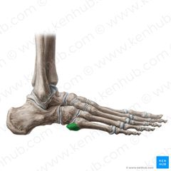
What is highlighted green? |
the sustentalculum tali |
|
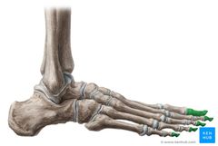
What is shown here? |
Tuberosity of the 5th metatarsal |
|
|
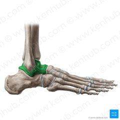
what is the true ankle joint? |
the talocrural joint |
|
|
what are the articulating bones in the talocrural joint? |
tibia, fibular and talus |
|
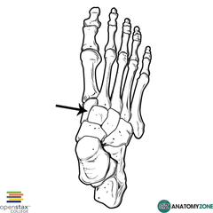
What joint is shown here? |
The subtalar joint |
|
|
what two bones meet at the subtalor joint? |
the talus and the calcaneous |
|
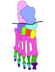
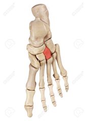
What is the line showing? |
the midtarsal joint line also known as the transverse |
|
|
What are the articulations at the midtarsal joint line? |
The calcanealcuboid (calcaneous + Cuboid) and the Talonavicular (Talus + Navicular) |
|
|
What is the classification of the talocrural joint? |
synovial hinge joint |
|
|
What movements are available at the talocrural joint? |
plantar and dorsi flexion |
|
What plane of movement is this? |
Sagittal plane |
|
|
What axis does a sagittal plane of movement have? |
Frontal axis |
|
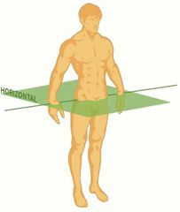
What plane of movement is this? |
Transverse plane |
|
|
What axis does a transverse plane have? |
longitudinal axis |
|
What plane of movement is shown here? |
Frontal plane |
|
|
What axis does the frontal plane have? |
sagittal axis |
|
|
meaning of anterior |
at the front or in front |
|
|
meaning of posterior |
at the back or behind |
|
|
meaning of superior |
above |
|
|
meaning of inferior |
below |
|
|
meaning of lateral |
towards the outer part of the body, away from the midline. |
|
|
meaning of medial |
towards the inner side of the body, towards the midline of the body. |
|
|
meaning of distal |
further away from the trunk or root of the limb |
|
|
meaning of proximal |
closer to the trunk or root of the limb |
|
|
meaning of superficial |
closer to the skin |
|
|
meaning of deep |
further from the skin. |
|
|
What is compact bone? |
Bone tissue which is more dense |
|
|
what is spongy bone? |
a softer bone tissue found at the epiphysis (end) of bones |
|
|
Whats the role of a ligament? |
Connects bone to bone |
|
|
Whats the function of the skeleton? |
- provides shape and support - enables you to move - protects your organs - produces blood cells - stores vitamins and minerals |
|
|
What is the diaphysis of the bone? |
The shaft (middle of the bone) |
|
|
What is the epiphysis of the bone? |
The end of the bones |
|
|
Whats the name of the cartilage found at the end of the sternum? |
xiphoid process |
|
|
What cartilage is the acetabulum covered in? |
hayline |
|
|
What is the role of the acetabular labrum? |
To deepen the socket |
|
|
What cartilage is the acetabular labrum? |
Fibrocartilage |
|
|
What are the fixed joints of the pelvis? |
Sacroilliac, lumbosacral and symphasis pubis |