![]()
![]()
![]()
Use LEFT and RIGHT arrow keys to navigate between flashcards;
Use UP and DOWN arrow keys to flip the card;
H to show hint;
A reads text to speech;
53 Cards in this Set
- Front
- Back
|
What is blood made up of |
Plasma (water like) 55% White blood cells (WBCs) // platelet cells (clotting factors) 1% Red blood cells (RBCs) 44% |
|
|
Plasma |
92% water, 8% dissolved molecules Proteins Macronutrients: glucose, amino acids, fats Micronutrients: vitamins, minerals (ions) Gases: Oxygen and carbon dioxide Waster products Hormones |
|
|
Albumin |
Plasma protein Osmotic pressure gradient for nutrient exchange in capillaries |
|
|
Globulins |
Plasma protein Protect against invading microbes |
|
|
Erythrocytes (RBCs) structure |
Biconcave disks No nucleus (enucleated) - blood can carry more hemoglobin = more oxygen Short lifespan -> 120 days Contain hemoglobin - pigment (red) - contains iron -> absorbs oxygen |
|
|
Erythrocytes function |
RBC’s carry oxygen to tissue - exchange for CO2 to be repaired Formed from stem cells in the bone marrow |
|
|
Anemia |
RBC deficiency Not enough iron = decreases oxygen to cells Low energy levels |
|
|
Where do blood cells come from |
Stem cells located in the bone marrow |
|
|
RBC production |
RBC formation is stimulated by erythropoietin (hormone) secreted by kidneys Lifespan of RBC is 120 days - broken down in liver and spleen |
|
|
Hemoglobin |
Defective Causes RBC’s to bend into sickle shape and causes blood to be blocked at capillaries |
|
|
Leukocytes (WBC) |
Ratio of red to white = 700:1 Has a nucleus, largest blood cell Engulfs invading cells through phagocytosis - digest microbe - forms pus Makes antibodies When you’re delicious you body makes more leukocytes Also originate from stem cells |
|
|
Types of WBC’s |
B cells (lymphocyte) - memory B cells T cells (lymphocyte) 4 types - Helper T, Killer T, Suppressor T, Memory T Macrophages - big eaters, largest of leukocytes - engulfs foreign invaders |
|
|
Leukaemia |
Cancer of the WBCs: the number of leukocytes in the blood increases, but the cells don’t function normally |
|
|
Platelets |
Small, fragile, contain specialized proteins (thromboplastin) Starts clotting - join with calcium in plasma - 1st step in clotting Also originate from stem cells |
|
|
Platelets blood clotting |
Fibrinogen (blood protein) + Thromboplastin (from platelet) + Ca 2+ (in blood) -> Fibrin (blood clot) 1. Platelets hit a rough edge of an injured blood vessel, rupture and release thromboplastin 2. Thromboplastin and Ca 2+ convert fibrinogen into fibrin 3. Fibrin is a stretchy net that traps RBC’s and seals wound |
|
|
After clotting |
Fibrin threads contract, pulling edges of the clot closer together until the injured vessel is closed Plasmin (plasma protein) will dissolve the clot once the blood vessel wall is repaired |
|
|
Thrombus |
Problems with clotting molecules may cause a thrombus to form inside a closed vessel |
|
|
Embolus |
If a thrombus dislodges and travels in the circulatory system |
|
|
Hemophilia |
Genetic disorder where clotting factors are missing from plasma: no clotting |
|
|
Antigen |
Foreign protein maker found on a cell
Stimulate the formation of antibodies |
|
|
Blood types |
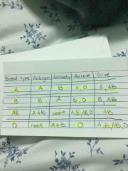
Back (Definition) |
|
|
Wrong blood types during a transfusion |
Blood will clump (clot) - agglutinate Blockages will occur |
|
|
Antibodies |
Proteins produced by WBCs in response to an invader Attach to antigens: causes clumping |
|
|
Universal acceptor vs donor |
Universal donor : O -no antigens present, won’t bond to antibodies, only accept O and donates blood to A,B,AB,O Universal acceptor: AB -both antigens present, only donate to AB, can accept blood from A,B,AB,O |
|
|
Rhesus factor (Rh) |
Another antigen in RBCs Present: RH+ -> 85% of people Absent: RH- -> 15% of people Humans have no natural antibodies for Rh - but they can be produced later on in life |
|
|
Rh and pregnancy |
If Rh- mother and Rh+ father baby can be Rh+ 1st child Rh+ (no problems) - No mixing of blood until birth. During birth blood will mix. Mothers immune system creates Rh+ antibodies. No harm to baby 2nd child Rh+ (problems) - Mother has Rh+ antibodies. If they enter baby, blood will clump. Reduced oxygen delivery. “Blue baby” - solution: transfuse baby with Rh- blood |
|
|
Pathogens |
A bacteria, fungi, virus or protozoan that can cause disease |
|
|
1st line of defence against infection |
Skin: layers of dead cells -> oil and sweat Digestive system: stomach acid, enzyme Gas exchange: hair, cilia, mucus, coughing and sneezing Urinary system: mucus, periodic flow of urine |
|
|
Defence: chemical responses |
Some fatty acids in the skin are toxic to bacteria Acids in stomach kill bacteria Lysozyme’s in tears break down the cell wall of bacteria Other bacteria in intestines and reproductive system use up the nutrient so new invaders starve (competitive inhibition) |
|
|
2nd line of defence: inflammation |
Microbe penetrate body’s first line of defence Triggers series of changes leading to inflammation at point of entry Phagocytosis ingest bacteria |
|
|
2nd line of defence: inflammation |
Microbe penetrate body’s first line of defence Triggers series of changes leading to inflammation at point of entry Phagocytosis ingest bacteria |
|
|
Immune response |
Triggered when both defence systems fail: pathogen gains access to the body 1. When foreign antigens enter the body, it causes lymphocytes to make antigens, antibodies are antigen specific. When an antibody attaches to a foreign antigen it destroys the cell attaches to it 2. Antibodies cause several aboriginal to clump together, making it easier for macrophages to rupture them |
|
|
Immune response |
Triggered when both defence systems fail: pathogen gains access to the body 1. When foreign antigens enter the body, it causes lymphocytes to make antigens, antibodies are antigen specific. When an antibody attaches to a foreign antigen it destroys the cell attaches to it 2. Antibodies cause several antigens to clump together, making it easier for macrophages to rupture them |
|
|
Step 1 |
Bacteria enters the body with antigen on surface |
|
|
Step 1 |
Bacteria enters the body with antigen on surface |
|
|
Killer T cells |
Puncture membrane of virus |
|
|
B cells |
Makes antibodies |
|
|
Memory T cells |
Remember the antigen so that antibodies are produced faster next time |
|
|
Suppressor T cells |
Stop the response |
|
|
Allergies |
Occurs when your immune system mistakes harmless cells for harmful invaders Increased tissue swelling, mucus secretions and constructed airways are common responses |
|
|
Mast cells |
Protects us from harmful particles in the air we breath |
|
|
Auto immune diseases |
Lymphocytes attack the body’s own cells and the suppressor T cells don’t stop |
|
|
Vaccine |
Dead or weakened virus is injected into the body Body produces antibodies to prevent future infections Memory cells are in place if this microbe ever enters the body again |
|
|
Antibiotics |
Drug made from bacteria or fungi - used only to kill diseases caused by bacteria Overuse danger - overtime bacteria becomes antibiotics resistant - antibodies can’t cure viruses - can cause serious side effects like allergic reactions |
|
|
Step 2 |
Macrophage engulfs antigen through phagocytosis and pushes antigen marker to the outer surface |
|
|
Step 3 |
Helper T cells copy the antigen shape |
|
|
Step 4 |
Helper T cells tell B cells to make antibodies |
|
|
Step 5 |
Antibodies attach to antigens |
|
|
Step 6 |
Macrophages engulfs antigen Or Killer T cells puncture membrane of virus |
|
|
Step 7 |
Suppressor T cells stop the response |
|
|
Step 8 |
Memory T cells remember the antigen If the antigen ever enters the bloodstream again the memory T cells immediately triggers an immune response Should prevent you from getting the same infection twice |
|
|
Macrophage |
Engulfs antigen and pushes antigen marker to the outer surface |
|
|
Helper T cells |
Copy the antigen shape |