![]()
![]()
![]()
Use LEFT and RIGHT arrow keys to navigate between flashcards;
Use UP and DOWN arrow keys to flip the card;
H to show hint;
A reads text to speech;
69 Cards in this Set
- Front
- Back
|
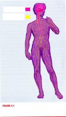
The nervous system can be divided into two divisions, what are they?
|
CNS (Central Nervous System)
PNS (Peripheral Nervous System) |
|
|
The location of the CNS is located in _____
|
The skull and spine/vertebrae column
|
|
|
The two parts of the CNS are _____
|
The brain and the spinal cord
|
|
|
What are the two parts of the PNS and what are their purpose?
|
Somatic interacts with EXTERNAL ENVIRONMENTS
Autonomic interacts with INTERNAL ENVIRONMENTS |
|
|
The CNS is composed of what nerves, and what are their functions?
|
Afferent, which brings SENSORY INFORMATION into the CNS
Efferent, which carries MOTOR COMMANDS away from the CNS |
|
|
The autonomic division of the PNS is composed of what branches, and what are their functions?
|
Sympathetic, fight or flight response
Parasympathetic, sooth-relax-comfort |
|
|
Where is the location of the Sympathetic nerves?
|
Projects from CNS to LUMBAR (small of back) and THORACIC REGIONS (chest)
|
|
|
Where is the location of the Parasympathetic nerves?
|
Projects from BRAIN and SACRAL (very lower back) region of the spinal cord
|
|
|
What special group of PNS nerves leaves the CNS from the brain through the skull, rather than from the spinal cord?
|
Cranial nerves, 12 of them, mostly for sensory
|
|
|
The brain and spinal cord are well-protected by the _____ and _____, and by membranes called _____
|
Skull and Vertebrae
Meninges: Dura Mater (tough mother: outside) Arachnoid (spidery mother: middle) Pia Mater (gentle mother: inside) |
|
|
The subarachnoid meninges is also called _____ and contains _____ and _____, located directly on top of the _____
|
Pia Mater
large blood vessels and CSF CNS/cortex |
|
|
Cerebrospinal fluid (CSF) is manufactured by the _____ - capillary networks that protrude into the ventricles.
|
Choroid Plexuses
|
|
|
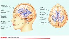
CSF circulates through the _____ of the brain, the _____ of the spinal cord, and the _____, and is absorbed into large channels called _____ in the dura mater and then into the blood stream.
|
Ventricular system
Central Canal Subarachnoid space Sinuses |
|
|
What is hydrocephalus? Explain reaction.
|
When the flow CSF is blocked, CSF fills the subarachnoid space and the central canal of the spinal cord, as well as the cerebral ventricles (4 large inner chambers of the brain - 2 lateral ventricles, and 3rd and 4th ventricle)
|
|
|
Explain Blood-brain barrier.
|
Most blood vessels of the brain do not readily allow compounds to pass from the general circulation into the brain, include many toxins or molecules, due to the tightly-packed nature of the cells of these blood vessels.
|
|
|
The gross structures of the nervous system are made up of hundreds of billions of different cells that are either _____ or _____ or _____
|
Neurons or Glia or Satellite cells
|
|
|
What are the fundamental functional units of the nervous system?
|
Cells that are specialized for the RECEPTION, CONDUCTION, and TRANSMISSION of electrochemical signals.
|
|
|
What are the parts of a multipolar motor neuron?
|
1) Semipermeable Cell Membrane
2) Cell Body (aka SOMA) 3) Dendrites 4) Axon 5) Axon Hillock 6) Myelin Sheaths 7) Nodes of Ranvier 8) Buttons 9) Synapses |
|
|
What is the function of Semipermeable Cell Membrane?
|
The cell membrane is semipermeable because of special proteins that allow chemical to cross the membrane. This semipermeable is critical to the normal activity of the neuron. The inside of the cell is filled with CYTOPLASM.
|
|
|
What is the function of Cell Body (aka SOMA)?
|
The metabolic center of the cell. The soma also contains the NUCLEUS of the neuron, which contains the cell's DNA.
|
|
|
What is the function of Dendrites?
|
The shorter processes that emanate from the cell body and receive input from synaptic contacts with other neurons.
|
|
|
What is the function of Axon?
|
A projection from the cell body, may be as long as a meter.
|
|
|
What is the function of Axon Hillock?
|
The junction between cell body and axon, is a critical structure in the conveyance of electrical signals by the neuron.
|
|
|
What is the function of Myelin Sheaths?
|
Formed by the OLIGODENDROGLIA in the CNS and SCHWANN cells in the PNS; they insulate the axon and assists in the conduction of electrical energy.
|
|
|
What is the function of Nodes of Ranvier?
|
The small spaces between adjacent myelin sheaths.
|
|
|
What is the function of Buttons?
|
The branched endings of the axon that releases chemicals that allow the neuron to communicate with other cells.
|
|
|
What is the function of Synapses?
|
The points of communication between the neuron and other cells (neurons, muscle fibers).
|
|
|
Define the physiological makeup of a multipolar neuron.
|
Most commonly drawn in textbooks, has multiple dendrites and an axon extending from soma.
|
|
|
Define the physiological makeup of a unipolar neuron.
|
One process that combines both axon and dendrites.
|
|
|
Define the physiological makeup of a bipolar neuron.
|
Two processes: a single axon and a single dendrite.
|
|
|
Define the physiological makeup of a interneuron.
|
Has no axons.
|
|
|
The most common type of cells in the nervous system are _____ and _____; they outnumber neurons _____ to _____.
|
GLIA and SATELLITE CELLS
10 to 1 |
|
|
Glial cells are found in the _____ and satellite cells are found in the _____; they provide physical and functional support to neurons.
|
CNS
PNS |
|
|
The glial cells and satellite cells that form the myelin sheaths of axons in the CNS and PNS are _____ and ______ respectively.
|
Glia cells = Oligodendrocytes cells
Satellite cells = Schwann cells |
|
|
_____ are the largest of the glial cells; they support and provide nourishment for neurons.
|
Astroglia
|
|
|
Glial and satellite cells play a key role in the function of the nervous system. Why?
|
They help SEND CHEMICAL SIGNAL between neurons, and ESTABLISH and MAINTAIN connections between neurons.
|
|
|
The spinal cord cross section, the _____ (cell bodies) forms a butterfly inside of the _____ (myelinated axons).
|
Grey Matter
White Matter |
|
|
The spinal cord cross section, the upper (dorsal; posterior) wings of the butterfly are called the _____; the lower (ventral; anterior) wings are called the _____.
|
Dorsal horns
Ventral horns |
|
|
How many pairs of nerves are attached to the spinal cord?
|
31
|
|
|
31 pairs of nerves are attached to the spinal cord. As they near the cord, they split into _____ or _____.
|
Dorsal Roots
Ventral Roots |
|
|
Describe the Dorsal Roots.
|
Sensory axons; cell bodies lie just outside the spinal cord in the Dorsal Root Ganglia
|
|
|
Describe the Ventral Roots.
|
Motor axons; cell bodies lie inside the Ventral Horns
|
|
|
How many divisions of the mammalian brain, and what are they?
|
5
Diencepahlon, Telencephalon, Mesencephalon, Myelencephalon, and Mentencephalon |
|
|
The nervous system is first recognizable in the developing embryo as the _____. The brain develops from these swellings at one end of the neural tube; the _____, the _____, and the _____.
|
Neural tube
Forebrain, Midbrain, Hind Brain |
|
|
The hind brain develops into the _____.
|
Myelencephalon and the Mentencephalon
|
|
|
The forebrain develops into the _____ also called _____.
|
Diencepahlon and the Telencephalon
Cerebral Hemisphere |
|
|
The term _____ refers to the stem on which the cerebral hemisphere rest.
|
Brain stem
|
|
|
What are the components of the brain stem.
|
Mesencephalon + Metencephalon + Myelencephalon + Diencephalon
|
|
|
The myelencephalon is commonly called the _____; it is composed of major _____ and ______ tracts ---- sleep, attention, muscle tone, cardiac function, and respiration.
|
Ascending and descending
|
|
|
The core network of nuclei is the _____; arousal system.
|
Reticular formation
|
|
|
The metencephalon has two parts: the _____ and _____.
|
Cerebellum (little brain)
Pons (bridge) |
|
|
The cerebellum has both _____ and _____ functions.
|
Sensorimotor and Cognitive
|
|
|
The pons is visible as a swelling on the inferior surface and also contains the _____.
|
Reticular formation
|
|
|
The mesencephalon is composed of the _____ and _____.
|
Tectum and Tegmentum
|
|
|
In mammals, the tectum consists of the _____ and the _____; in lower vertebrates, there is simply a single _____.
|
Superior colliculi (visual relay) and the Inferior colliculi (auditory relay)
Optic tectum |
|
|
The tegmentum contains the reticular formation, the _____ , the _____; and the _____.
|
Red nucleus (sensorimotor)
Substantia nigra (sensorimotor cell bodies here die in patients with Parkinson's disease) Periaqueductal gray (mediates analgesia) |
|
|
Define analgesia.
|
The ability to feel pain
|
|
|
What sensorimotor cell bodies here die in patients with Parkinson's disease.
|
Substantia nigra
|
|
|
What are the two main structures of the diencephalons?
|
The thalamus and hypothalamus
|
|
|
Some thalamic nuclei are _____; describe the types and functions.
|
Sensory relay nuclei
- Lateral geniculate nuclei (vision) - Medial geniculate nuclei (audition) - Ventral posterior nuclei (touch) |
|
|
The _____ is just below the thalamus ("hypo" means below) and the _____ (snot gland) is suspended from the _____. The _____ are two small bumps visible on the inferior surface, just behind the hypothalamus.
|
Hypothalamus
Pituitary gland Mammalian bodies |
|
|
The _____ is the X-shaped part of the optic nerves that lies just in front of the pituitary gland; axons that originate from the nasal half of each retina cross over (_____).
|
Optic chiasm
Decussate |
|
|
Telencephalon mediates most _____. The telencephalon is the largest division of human brain. Large tracts called _____ connect the two hemispheres and the _____ is the largest _____.
|
Complex cognitive functions
Commissures Corpus Callosum Commissures |
|
|
About 90% of human cortex is _____, comprised of ______ cell layers of _____ and ______.
|
Neocortex
6 cell layers Pyramidal cells and Stellate cells |
|
|
The hippocampus is not neocortex; instead, it is a three-layer cortical area that lies in the _____.
|
Medial temporal lobe
|
|
|
How many lobes of the cerebral hemisphere are defined by the fissure of the cerebral cortex? Describe their locations.
|
4 lobes
- Frontal lobe: superior to lateral fissure and anterior to central fissure - Temporal lobe: inferior to lateral fissure - Parietal lobe: posterior to central fissure - Occipital lobe: posterior to temporal lobe and parietal lobe |
|

|

|
|

|

|
|

|

|

