![]()
![]()
![]()
Use LEFT and RIGHT arrow keys to navigate between flashcards;
Use UP and DOWN arrow keys to flip the card;
H to show hint;
A reads text to speech;
210 Cards in this Set
- Front
- Back
|
What is immunity based on? |
The recognition of self and destruction of foreign material |
|
|
What is an antigen? |
Any foreign molecule that can trigger an immune response |
|
|
What are the most common antigens? |
Proteins and very large polysaccharides |
|
|
Where can antigens be found? |
On the surface of cancer cells, parasites and bacteria, on pollen grains, and on envelopes of viruses |
|
|
What are blood groups based on? |
The presence of certain types of antigens on the surface of red blood cells |
|
|
Why is the knowledge of antigens on blood cells important? |
A blood transfusion given to an individual with the wrong blood type can stimulate an immune response called agglutination (clumping of blood cells) followed by hemolysis where red blood cells are destroyed and blood may coagulate in vessels |
|
|
What does blood typing involve? |
Mixing samples of blood with antibodies |
|
|
What does it mean if somebody is blood type A? |
They have antigen A on the surface and contain anti-B antibodies in their plasma. Their cells will destroy blood cells with the B-antigen on the surface |
|
|
What happens when a pathogen enters the blood? |
The specific antigen on the surface of the membrane is identified. Then phagocytes called macrophages recognise a pathogen as foreign, engulfs it and partially destroys it. |
|
|
What doe macrophages do with the antigens from pathogens? |
They display them in the plasma membrane of the macrophages |
|
|
What kind of cells recognise antigens on macrophages? |
Lymphocytes called helper T cells - they each have an antibody-like receptor protein in their plasma membrane, which can bind to antigens displayed on the surface of macrophages (specific helper T cells have receptor proteins for that antigen) |
|
|
When the helper-T cells bind to antigen, what is said to have happened? |
They have been activated by the macrophage
|
|
|
What happens to activated helper T cells? |
They bind to lymphocytes called B cells (the B cell receptors are specific to antigens). The helper T cell activates the B cell by binding and also by releasing a signalling protein |
|
|
What happens when B cells are activated? |
They begin to clone themselves, producing cloned plasma B cells that produce the same antibodies |
|
|
What do plasma cells (mature B cells) contain? |
Extensive amount of rER, ribosomes and mitochondria |
|
|
What name is given to the process by which B cells generate large numbers of plasma cells that produce the same antibody? |
Clonal selection |
|
|
What happens to the antibodies produced by B cells? |
They are secreted and help destroy the pathogen - they are only in the body for a few weeks or months - the plasma cells that produced them are also gradually lost once the infection has been overcome |
|
|
If B cells do not become plasma cells, what do they become? |
Memory cells, which remain long after the infection |
|
|
Memory cells remain inactive until when? |
Until the same pathogen infects the body again, in which case they become active and respond rapidly |
|
|
What are the 5 ways that antibodies aid the destruction of pathogens? |
- opsonisation
- neutralisation of viruses + bacteria - neutralisation of toxins - activation of complement - agglutination |
|
|
What is opsonisation? |
Antibodies make the pathogen more recognisable to phagocytes so they are more readily engulfed - once bound, they can link the pathogen to the phagocyte |
|
|
What is neutralisation of viruses and bacteria? |
Antibodies can prevent viruses from docking to host cells so that they cannot enter the cells |
|
|
What is neutralisation of toxins? |
Some antibodies can bind to the toxins produced by pathogens, preventing them from affecting susceptible cells |
|
|
What is activation of complement? |
The complement system = a collection of proteins which lead to perforation of the membranes of pathogens. - Antibodies bound to the surface of a pathogen activate a complement cascade, leading to the formation of a "membrane attack complex" that forms a pore in the pathogen membrane: water and ions enter and causes the cell to lyse |
|
|
What is agglutination? |
Antibodies can cause sticking together of pathogens so they are prevented from entering cells and are easier for phagocytes to ingest - the large agglutinated mass can be filtered out by the lymphatic system |
|
|
What may a vaccine contain? |
A live attenuated (weakened) version of the pathogen, a killed version, or derivatives of it that contains antigens. |
|
|
What kind of response do vaccines stimulate? |
A primary immune response |
|
|
What will happen if the body encounters the pathogen again? |
It will be destroyed right away by the antibodies during a secondary immune response |
|
|
How have vaccines made great contributions towards public health? |
Through the prevention of many deadly or dangerous diseases e.g. TB, measles, smallpox |
|
|
Jenner was an 18th century scientist who noted that a milkmaid claimed that she had caught the disease cowpox so would never develop smallpox. Why was his work unethical? |
Jenner infected an 8yr old boy with cowpox, waited for him to recover and then infected him with smallpox - he did no preliminary research, his subject was a small child below the age of consent, and deliberately caused harm. |
|
|
Talk about the eradication of smallpox. |
- in 1959 a global initiative was undertaken by WHO to eradicate smallpox - There were mixed results until a well-funded 'Smallpox Eradication Unit' was formed in 1967 - The last known case of smallpox was recorded in Somalia in 1977 |
|
|
Why was the eradication of smallpox successful? |
- patients were easily identified by obvious clinical features - transmission was through direct contact only - there were no animal vectors or reservoirs where the disease could remain and re-emerge - contacts of the patients were quickly identified and vaccinated - immunity was long term so reinfection was unlikely - infection period was only 3-4 weeks - the virus was stable and didn't mutate - there was international cooperation organised by the WHO |
|
|
What is epidemiology? |
The study of the distribution, pattern and causes of a disease in a population |
|
|
What is a zoonosis? |
A pathogen which can cross species barriers - this is an emerging health concern |
|
|
Give examples of zoonotic diseases. |
Lyme disease, bird flu, West Nile virus
|
|
|
What are mast cells? |
Immune cells found in connective tissue that secrete histamine in response to infection. |
|
|
Other than mast cells, what produces histamine? |
Basophils which circulate in the blood |
|
|
What does histamine do? |
It causes the dilation of blood vessels of the infected, making them leaky. This increases the flow of fluid containing immune components to the infected area and allows some of the immune components to leave the blood vessel (specific and non-specific immunity) |
|
|
A number of symptoms from allergic reactions are caused by histamines. How? |
Cells in a variety of tissues have membrane-bound histamine receptors. - the release of histamine causes many of the symptoms of allergic reactions such as inflammation, sneezing, itching and mucous secretion. - histamines also play a role in the formation of rashes and swelling = anaphylaxis - anti-histamine drugs counteract these affects by blocking histamine receptors. |
|
|
What are monoclonal antibodies? |
Highly specific, purified antibodies that are produced by a clone of cells (derives from a single cell) - they recognise only one antigen |
|
|
How are the hybridoma cells that will manufacture a monoclonal antibody produced? |
The antigen is injected into a mouse or other mammal. - the mammal's immune system makes plasma B cells that are capable of producing the desired antibody - plasma cells are removed from the spleen of the animal (only some will produce desired antibody) - the B cells are fused with cancer cells called myeloma called - the new cells are called hybridoma cells |
|
|
How does the hybridoma cell produce monoclonal antibodies? |
The hybridomas have characteristics of both cells: - they divide rapidly - they produce antibodies The cells are cultured and allowed to divide, producing many clone cells that can produce large amounts of antibodies. Monoclonal antibodies can be extracted and used. |
|
|
How are monoclonal antibodies used in pregnancy tests? |
Monoclonal antibodies can detect HCG (human chorionic gonadotrophin) - produced by an embryo in early pregnancy In pregnancy test, HCG antibodies are combined with colour-changing enzymes - when the mixture is introduced to urine of a pregnant woman, the antibodies will bind to the HCG in the urine and change colour |
|
|
What are exoskeletons? |
External skeletons that surround and protect most of the body surface of an animal |
|
|
How do bones and exoskeletons facilitate movement? |
They act as levers so the body can move, and act as anchorage for muscles |
|
|
What do levers do? |
Change the size and direction of forces |
|
|
What are ligaments? |
Strong bands that connect bone to bone, strengthening the joint during movement |
|
|
What are tendons? |
They have dense connective tissue that connects muscles to bones, allowing movement of the bone when the muscle contracts |
|
|
What are muscles? |
They provide the force for movement by contracting |
|
|
What does the joint act as in movement? |
A pivot point or a fulcrum |
|
|
What is the effort? |
The force applied when the muscle contracts |
|
|
What is the resistance? |
The force, or load, needed to overcome for movement to take place |
|
|
How are levers classified? |
By 1st, 2nd, and 3rd class Depends upon the positions among the fulcrum, the effort and the resistance |
|
|
Outline first class levers. |
- fulcrum is in the middle (like a seesaw) e.g. when a human nods their head (top of spinal cord is fulcrum, back of neck provides effort, weight of head is the resistance) |
|
|
Outline second class levers |
- resistance is in the middle (like a wheelbarrow) e.g. calf raise (ball of food is fulcrum, body mass is resistance, effort is applied by the calf muscle) |
|
|
Outline third class levers. |
- effort is in the middle e.g. in the arm (effort by contraction of biceps, fulcrum is elbow, resistance is whatever weight is being lifted) |
|
|
What happens in antagonistic pairs? |
When one muscle contracts, the other relaxes |
|
|
How do grasshopper legs work in antagonistic pairs? |
Hind legs have a jointed appendage with 3 parts: 1. below the joint is the tibia 2. base of tibia is another joint called tarsus 3. above the joint is the femur When the grasshopper jumps, the flexor muscles contract, and the femur and tibia are brought closer together As the grasshopper jumps the extensor muscles contract, extending the tibia |
|
|
What does the type of joint determine? |
The amount of movement that is possible |
|
|
What movements are possible for ball and socket joints? |
- flexion - extension - rotation - abdction - adduction |
|
|
Give examples of ball and socket joints |
- hip - shoulder |
|
|
What movements are possible at hinge joints? |
- flexions (bending) - extensions (straightening) - slight side to side movements are possible |
|
|
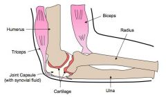
Label a diagram of a human elbow as an example of a synovial joint |
|
|
|
What is cartilage? What is its function? |
Tough, smooth tissues that covers the regions of the bone at the joint. Reduces friction in the joint, provides high tensile strength and support, absorbs compression |
|
|
What is synovial fluid? What is its function? |
Thick, viscous fluid found in the cavity of the synovial joints Reduces friction by providing lubrication in the joint, supplies oxygen and nutrients, removes CO2 and waste |
|
|
What is the joint capsule? What is its function? |
A tough ligamentous covering to the joint It seals the joint and holds in the synovial fluid and helps to prevent dislocation |
|
|
What is the radius and its function? |
Smaller forearm bone Lever attached to the biceps |
|
|
What is the ulna and its function? |
Longe forearm bone Lever connected to the triceps |
|
|
What is the biceps and its function? |
The muscle connected to the radius Contracts and causes flexion |
|
|
What is the triceps and its function? |
The muscle attached to the ulna Contracts and causes extension |
|
|
What are the muscles attached to bones called? |
Skeletal muscles |
|
|
What is the other name for skeletal muscles? Why are they called this? |
Striated muscles - they appear striped through a microscope |
|
|
What are striated muscles composed of? |
Bundles of muscle cells known as muscle fibres. |
|
|
What is the name of the plasma membrane that surrounds each muscle fibre? |
The sarcolemma |
|
|
Muscle fibres are much longer than typical cells, and have many nuclei. Why? |
Muscle cells are formed when embryonic muscle cells fuse together |
|
|
What are muscle fibres composed of? |
Many parallel elongated fibres called myofibrils |
|
|
What is the name of the reticulum that extends throughout the muscle fibre and wraps around each myofibril? |
The sarcoplasmic reticulum |
|
|
What is the function of the sarcoplasmic reticulum? |
It sends a signal to all parts of the muscle fibre to contract at the same time |
|
|
What does the sarcoplasmic reticulum store? |
Calcium |
|
|
What organelles are found between the myofibrils? |
Large numbers of mitochondria, providing the energy needed for contractions |
|
|
What is the name of the plasma membrane of the muscle cell? |
The sarcolemma |
|
|
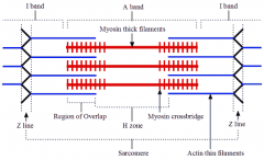
What gives striated muscle its striped? |
The alternating dark and light bands of myofibrils |
|
|
What is found in the centre of each light band of the myofibrils? |
A disc-shaped, dense protein structure known as the Z-line |
|
|
What is the part of the myofibril that lies between two Z-lines? |
The sarcomere - the functional unit of the myofibril |
|
|
What is the pattern of light and dark bands in sarcomeres due to? |
A precise and regular arrangement of 2 types of protein filament: - thin actin filaments - thick myosin filaments |
|
|
How are actin and myosin filaments arranged in muscles? |
- Actin filaments are attached to a Z-line at one end - Myosin filaments are interdigitated with actin filaments at both ends, occupying the centre of the sarcomere - Each myosin filament is surrounded by 6 actin filaments and forms cross bridges with them during muscle contraction |
|
|
Draw a labelled diagram of the structure of a sarcomere |
|
|
|
What happens during a muscle contraction? |
- myosin filaments pull action filaments towards the centre of the sarcomere - the sarcomere and length of muscle fibre is shortened - myosin heads bind to sites on the actin filaments, sliding the actin filaments along the myosin using ATP - the heads and binding sites are regularly spaced along the myosin and actin, so many cross bridges can form at once |
|
|
When a muscle is relaxed, what protein blocks the binding sites on actin? |
A regulatory protein called tropomyosin |
|
|
What happens when a motor neurone sends a signal to a muscle fibre to make it contract? |
The sarcoplasmic reticulum releases calcium ions |
|
|
What happens when the sarcoplasmic reticulum relates calcium ions? |
The ions bind to troponin, causing the tropomyosin to move and the binding sites on actin to be exposed. - myosin heads bind and swivel towards the centre of the sarcomere, moving the actin filaments a small distance |
|
|
What is necessary for the filaments to slide? |
ATP hydrolysis and cross-bridge formation
|
|
|
Why is ATP important in the sliding of muscle filaments? |
It is what causes the breaking of cross-bridges by attaching to the myosin heads, causing them to detach form the binding sites on actin
It provides the required energy for the myosin head to move outwards |
|
|
How does ATP provide the required energy for the myosin head to move outwards? |
ATP undergoes hydrolysis, into ADP and phosphate - this causes the myosin heads to change their angle - the heads are said to be 'cocked' in their new position as they are storing potential energy from ATP |
|
|
Where are new cross-bridges formed? |
Between the myosin heads and the binding site next to the one they were previously attached to |
|
|
What happens once the myosin head has bound to its new site? |
The energy stored when the myosin head was 'cocked' is released, and the head moves inwards towards the centre of the sarcomere, moving the actin filament. - this sequence of stages is repeated until the motor neurone stops sending signals |
|
|
What happens once the contraction is over?
|
The calcium ions are pumped back into the sarcoplasmic reticulum and tropomyosin blocks actin binding sites again - the muscle is relaxed |
|
|
What has been used to study the cyclic interactions in muscle contraction? |
Fluorescence |
|
|
What is osmolarity? |
The solute concentration of a solution |
|
|
Many animals are known as omoregulators. What does this mean? |
They maintain a constant internal solute concentration, even when living in marine environments with different osmolarities |
|
|
What animals are osmoreglators? |
All terrestrial animals, freshwater animals and some marine organisms like bony fish |
|
|
What are osmoconformers? |
Animals whose internal solute concentration tends to be the same as the concentration of solutes in the environment |
|
|
Arthropods have a circulating fluid. What is this fluid? |
Hemolymph - it combines the characteristics of tissue fluid and blood
|
|
|
When animals break down amino acids, the nitrogenous waste product is toxic and needs to be excreted. What forms is this waste product in in insects and mammals? |
Insects: uric acid Mammals: urea |
|
|
What body part do insects have to excrete uric acid? |
They have tubes that branch off from their intestinal tract called Malpighian tubules |
|
|
How do Malpighian tubules excrete uric acid? |
1. cells lining the tubules actively transport ions and uric acid from the hemolymph the tubules. (this draws water by osmosis from the hemolymph into the tubules) 2. the tubules empty their contents into the gut 3. in the hindgut most of the water + salts are reabsorbed. The nitrogenous waste is excreted with the faeces |
|
|
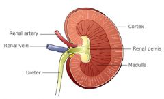
There are 5 main structures that make up the kidney. What are they? |
- cortex (outer layer) - medulla (mid layer) - pelvis of kidney (inner part) - renal artery - renal vein |
|
|
Draw and label a diagram of a kidney. |
|
|
|
What is the function of the kidney? |
It is responsible for removing unrequited/harmful substances from the blood. |
|
|
What substances are present in higher amounts in the renal artery than the renal vein? |
- toxins and other substances that are ingested and absorbed but not fully metabolised (e.g. retain pigments in beetroots and drugs) - excretory waste products including nitrogenous waste products, mainly urea |
|
|
What else is removed by the kidneys? |
- excess water, produced by cell respiration or absorbed from food in the gut - excess salt, absorbed from food in the gut |
|
|
How much of the plasma from blood flowing through kidneys is filtered? |
About 1/5 |
|
|
What other differences are between the composition of the blood in the renal artery and renal vein? |
- blood leaving the kidney by the renal vein is deoxygenated because kidney metabolism needs oxygen - renal vein has a higher partial pressure of CO2 is a waste product of metabolism - renal vein has less glucose |
|
|
Why should plasma proteins be of equal concentrations in the renal kidney and renal artery? |
Because they are not filtered by the kidney - plasma protein in the urine indicates abnormal function |
|
|
What is the basic functional unit of the kidney called? |
The nephron - there are many in the medulla of the kidney |
|
|
What are the parts of the nephron? |
1) The afferent arteriole 2) The glomerulus (inside the Bowman's capsule) 3) The efferent arteriole 4) The proximal convoluted tubule 5) The loop of Henle 6) The distal convoluted tubule 7) The collecting duct |
|
|
In which part of the kidneys is the pressure in capillaries particularly high and the permeability of their wall great? |
In the glomerulus - the volume forced out of the capillaries is about 100x greater than other tissues - the fluid forces out = glomerular filtrate |
|
|
Almost all proteins are retained in the capillaries of the glomerulus. What is the separation of different size particles called? |
Ultrafiltration |
|
|
What particles can pass through the walls of glomerulus capillaries? |
- those with a relative molecular mass below 65,000 atomic mass units - larger molecules passing through depends on their shape and charge |
|
|
What separates the capillaries in the glomerulus? |
Basement membrane |
|
|
What are the projections from the basement membrane of the glomerulus? |
Podocyte foot processes, which attach the podocytes (specialised epithelial cells) to the membrane. - the podocytes function as a barrier through which waste products are filtered from the blood |
|
|
There are 3 parts to the ultrafiltration system. What are they? |
1) Fenestrations 2) The basement membrane 3) Podocytes |
|
|
What are fenestrations? |
- between the cells in the wall of the capillary - about 100nm in diameter - they allow fluid to escape, but not blood cells |
|
|
What is the function of the basement membrane? |
- covers and supports the wall of the capillaries - it is made of negatively-charged glycoproteins, which form a mesh - prevents plasma proteins being filtered out |
|
|
What are podocytes? |
- they form the inner wall of the Bowman's capsule - they have extensions that wrap around the capillaries of the glomerulus - very narrow gaps between foot processes help prevent small molecules from being filtered out |
|
|
Where does glomerular filtrate flow? |
Into the proximal convoluted tubule |
|
|
What is the proximal convoluted tubule? |
The first part of of the nephron - where most of the glomerular filtrate is reabsorbed |
|
|
How are sodium ions absorbed in the proximal tubule? |
- they are moved by active transport from filtrate to space outside the tubule - then they pass to the peritubular capillaries - pump proteins are located in outer membrane of tubule cells |
|
|
How are chloride ions absorbed in the proximal tubule? |
- they are attracted from filtrate to space outside the tubule because of charge gradient set up by active transport of sodium ions |
|
|
How is glucose absorbed in the proximal tubule? |
- it is co-transported out of filtrate into fluid outside the tubule by co-transporter proteins in outer membrane of tubule cells - sodium ions move down concentration gradient from outside tubule into tubule cells, this provides energy for glucose to move at the same time to the fluid - the same process is used to reabsorb amino acids |
|
|
How is water absorbed in the proximal tubule? |
- pumping solutes out filtrates and into the fluid outside creates a solute concentration gradient, causing water to be reabsorbed from filtrate by osmosis |
|
|
There are 2 parts to the loop of Henle. What are they? |
1) the ascending limb 2) the descending limb |
|
|
What is the overall effect of the loop of Henle? |
To create a gradient of solute concentration in the medulla |
|
|
What happens in the descending limb of the loop of Henle? |
PERMEABLE TO WATER NOT TO IONS The increased solute concentration in the medulla causes water to be drawn out of the filtrate until equilibrium is met |
|
|
What happens in the ascending limb of the loop of Henle? |
PERMEABLE TO IONS NOT TO WATER Sodium ions are pumped out to the interstitial fluid in the medulla. - water is retained in the filtrate *the interstitial fluid is now hypertonic relative to the filtrate |
|
|
Why is the loop of Henle a countercurrent multiplier system? |
Countercurrent - fluid flows in opposite directions Multiplier - it causes a steeper solute concentration gradient to develop |
|
|
Why may the length of the loop of Henle vary between animals? |
The longer the loop of Henle, the more water will be reclaimed. Animals in dry habitats will have adapted to have a longer loop of Henle. - the medulla must become thicker to do this |
|
|
What is ADH? |
Anti-diuretic hormone - it is the hormone that helps to keep our blood dilute if we haven't consumed enough water
|
|
|
How does ADH work if blood is dehydrated? |
ADH signals for the walls of the distal convoluted tubule and the collecting duct to become permeable to water (through the use of aquaporins). Water exits the nephron, enters the medulla, and the urine becomes concentrated. |
|
|
What nitrogenous waste do marine mammals produce? |
Urea: they could produce ammonia directly (it can be easily diluted within the environment) but they produce urea because of their evolutionary history - amphibians release ammonia as larvae and urea after metamorphosis |
|
|
What is the benefit of uric acid? |
It is not water soluble so doesn't require water to be released
- useful for birds: they don't have to carry water for excretion so less energy is expended on flight |
|
|
What are the consequences of dehydration? |
- darker urine - low blood pressure due to low blood volume - increased heart rate - body temp. regulation issues: inability to sweat |
|
|
What are the consequences of over hydration? |
- dilution of blood solutes - swelling of cells due to osmosis - headaches - nerve function disrupted |
|
|
What are the two treatments for kidney failure? |
1. (Hemo) dialysis 2. Kidney transplant |
|
|
What happens during kidney dialysis? |
A steady flow of blood passes over an artificial semi-permeable membrane in the dialysis machine - the small waste products pass through the membrane (the larger ones cannot) - the purified blood is returned to the patient via a vein |
|
|
What is the benefit of a kidney transplant? What is the cost? |
+ results in greater independence of movement and freedom to travel compared to dialysis - the recipient's body could reject the organ |
|
|
What are the costs of dialysis? |
- takes several hours - carries the risks of infection or complications |
|
|
What can be tested for using urinary tests? |
Blood cells - indicates UTI (white blood cells) or kidney stone or tumour in the urinary tract (red) Glucose - diabetes Proteins - damage to the kidneys Drugs |
|
|
What does sexual reproduction involve? |
The development and fusion of haploid gametes |
|
|
What is spermatogenesis? |
The production of sperm (the male gamete) through meiosis |
|
|
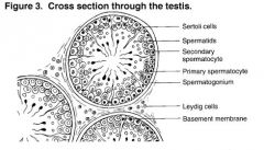
Where does spermatogenesis take place? |
In the testes |
|
|
What are the testes composed of? |
A mass of narrow tubes called seminiferous tubules |
|
|
What are the cells that fill the gaps between seminiferous tubules called? |
Interstitial cells |
|
|
The seminiferous tubules are also made of cells. What is the outer layer of cells called? |
The germinal epithelium |
|
|
What happens in the germinal epithelium? |
Sperm production - the most mature sperm are found near the fluid-filled centre of the seminiferous tubules |
|
|
What are the cells that have developed tails? |
Spermatozoa, abbreviated to sperm |
|
|
What are the other cells in the wall of the tubules called? |
Sertoli cells: they are large nurse cells that help with the production of sperm |
|
|
Outline the processes of spermatogenesis. |
1. 2n Germinal epithelium cells divide by mitosis to produce diploid cells 2. Diploid cells grow larger and are called primary spermatocytes 3. Primary spermatocyte carry out 1st division of meiosis to produce 2 secondary spermatocytes (n) 4. Secondary spermatocyte carry out 2nd meiosis division to produce 2 spermatids 5. Spermatids associate with Sertoli cells. They develop into spermatozoa 6. Sperm detaches from Sertoli cells and are carried out of the testes by fluid in seminiferous tubule |
|
|
What is oogenesis? |
The production of eggs (female gametes) through meiosis |
|
|
Where does oogenesis take place? |
In the ovaries |
|
|
Outline the process of oogenesis. |
1. Germ cells (2n) in the fetal ovary divide by mitosis to produce many 2nn germ cells called oogonia 2. Oogonia grow in the cortex until they are large enough and ready to go through meiosis (primary oocytes) 3. Primary oocytes go through 1st meiosis division, which is stopped in prophase when follicle cells surround the dividing oocyte (primary follicle - about 400,000 in newborn) 4. These follicles remain in this 1st stage of meiosis until the girl reaches puberty |
|
|
What happens at the start of each menstrual cycle? |
A small batch of primary follicles are stimulated to develop by FSH - usually only 1 goes on to become a mature follicle, containing a secondary oocyte |
|
|
What happens when the secondary oocyte begins to go through the 2nd meiotic division? |
It is released from the ovary - it will not complete the 2nd division unless the oocyte is fertilised |
|
|
Annotate a diagram of a seminiferous tubule |
|
|
|
Annotate a diagram of an ovary. |
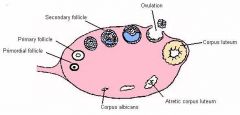
|
|
|
What are the components of an egg? |
- haploid nucleus - cytoplasm with droplets of fat - cortical granules - layer of gel composed of glycoproteins - layer of follicle cells - plasma membrane - 2 centrioles - 1st polar cell |
|
|
What are the components of a sperm? |
- acrosome - haploid nucleus - 1 centriole - plasma membrane - mid-piece - helical mitochondria - tail with microtubules and protein fibres |
|
|
What are the differences between spermatogenesis and oogenesis? |
- 4 male gametes produced, 2 female gametes produced from every germ cell - sperm cells are small with little cytoplasm, egg cells large with lots of cytoplasm - millions of sperm produced every day, 1 secondary oocyte ovulated each month |
|
|
What does fertilisation involve? |
1. Acrosome reaction 2. Fusion of plasma membrane of egg + sperm 3. Cortical reaction |
|
|
What is fertilisation? |
The combining of the male and female gametes to produce a zygote |
|
|
How are sperm stimulated to swim? |
By calcium ions in the vaginal fluids |
|
|
Where do the majority of fertilisations take place? |
In the fallopian tubes |
|
|
How do the sperm reach the fallopian tubes? |
They follow chemical signals produced by the egg |
|
|
What reaction occurs when the sperm reaches the egg? |
The acrosome reaction |
|
|
What is the purpose of the acrosome reaction? |
To allow the sperm to break through the layer of glycoproteins |
|
|
Outline the acrosome reaction. |
The acrosome in the head of the sperm releases hydrolytic enzymes onto the glycoprotein layer surrounding the egg (the zona pellucida) This digests the layer, allowing the sperm to force their way through the zona pellucida through vigorous tail beating |
|
|
What happens once a sperm has broken through the zona pellucida?
|
The first one that breaks through fuses with the egg's membrane (the membrane of the tip of the sperm has special proteins that can bind to the membrane of the egg) - the sperm's nucleus is released into the egg cell |
|
|
What reaction happens once the membranes have fused together? |
The cortical reaction |
|
|
Outline the cortical reaction. |
Cortical granules near the surface of the egg membrane are released by exocytosis - the chemicals in the granules combine with the glycoproteins in the zona pellucida to cross-link with each other - this creates a hard layer impermeable to other sperm - prevents polyspermy |
|
|
There are 2 types of fertilisation. What are they?
|
Internal External |
|
|
What does internal fertilisation ensure? |
Close proximity of the sperm and egg to ensure fertilisation takes place |
|
|
Most aquatic organisms rely on external fertilisation. What does this involve? |
Releasing the sperm and egg at a close proximity, in the water outside the female's body |
|
|
What risks are involved in external fertilisation? |
- predation - changes to the external environment (pH, pollution, temperature etc.) |
|
|
What happens after the gametes fuse to produce a zygote? |
The zygote divides by mitosis to form a 2-cell embryo - the 2 cells grow and replicate their DNA and undergo another cell division - this process continues, and the embryo moves along the fallopian tubes to the uterus |
|
|
What is the embryo called when it reaches 16-32 cells? |
The morula |
|
|
What is the embryo called when it reaches 100-128 cells? |
A blastocyst |
|
|
What does it mean when a blastocyst is formed? |
It is ready for implantation into the endometrium |
|
|
Describe the structure of the blastocyst. |
- inner cell mass that will become the body of the embryo - group of cells surrounding embryo called trophoblast that will become the placenta - fluid-filled cavity called the blastocoel - outer layer of cells that will become finger-like projections, allowing the embryo to penetrate the uterine wall during implantation |
|
|
What hormone does a human embryo make when it is implanted into the endometrium? |
HCG (Human chorionic gonadotropin) |
|
|
What is the role of HCG? |
It promotes the maintenance of the corpus luteum and prevents its disintegration - this allows for the continued production of progesterone |
|
|
What does progesterone do during pregnancy? |
It enriches the uterus with a thick lining of blood vessels and capillaries so it can sustain the growing foetus |
|
|
How can HCG protect the foetus during early pregnancy? |
It might repel the immune cells of the mother |
|
|
Humans are placental mammals. What other groups of mammals are there? |
Monotremes: lay eggs Marsupials: give birth to undeveloped offspring that develop inside a pouch |
|
|
Why is the placenta needed? |
Because the body surface area to volume ratio becomes smaller as the foetus grows |
|
|
From what does the placenta develop? |
The trophoblast layer of the blastocyst
|
|
|
What is the basic functional unit of the placenta? |
A finger-like piece of foetal tissue called a placental villus - these villi increase in number during pregnancy to cope with the increasing demands of the foetus |
|
|
How does the maternal and foetal blood exchange materials? |
Maternal blood leaves the arteries and enters the inter-villus space, where it pools and surrounds the placental villi. Foetal blood circulates in capillaries within villi and microvilli is very close to the surface Materials like oxygen, nutrients and vitamins diffuse into the foetal capillaries CO2 and waste diffuse out of foetal capillaries into the inter-villous space |
|
|
What do the cells that separate the maternal and foetal blood form? |
The placental barrier |
|
|
What starts to happen with the placenta after about 9 weeks? |
It starts to produce progesterone and oestrogen - the placenta produces enough of these steroids to maintain the pregnancy (the corpus luteum is no longer needed) - there is a risk of miscarriage at this stage |
|
|
During pregnancy, what hormone is inhibited by progesterone? |
Oxytocin |
|
|
What happens at the end of pregnancy? |
The foetus secretes hormone that signal the placenta to stop producing progesterone, oxytocin is therefore secreted |
|
|
What does oxytocin do at the end of pregnancy? |
Stimulates contractions of the muscle fibres in the myometrium (uterus) |
|
|
What happens when contractions begin? |
The contractions are detected by stretch receptors, which signal the pituitary gland to increase oxytocin secretion |
|
|
What is the effect of increased oxytocin secretion? |
It causes the contractions to become more frequent and more vigorous, causing more oxytocin secretion (positive feedback) |
|
|
What happens to the amniotic sac when contractions occur? |
It breaks, releasing the amniotic fluid |
|
|
How is the baby born? |
The relaxation of the muscle fibres in the cervix cause it to dilate, eventually allowing the contractions to push the baby out through the cervix and vagina. |
|
|
What happens to the placenta? |
It is expelled "afterbirth" - about 15 mins after baby is born |
|
|
Describe the correlation between animal size an development of young. |
In many cases, the longer the gestation period, the greater the mass and development at birth |
|
|
What are mammals that give birth to smaller, immature young called? |
Altricial species |
|
|
What are mammals called that give birth to mature offspring? |
Precocial species |