![]()
![]()
![]()
Use LEFT and RIGHT arrow keys to navigate between flashcards;
Use UP and DOWN arrow keys to flip the card;
H to show hint;
A reads text to speech;
122 Cards in this Set
- Front
- Back
|
What occurs in the nucleus |
ALL TRANSCRIPTION -replication and splicing too |
|
|
Where does translation occur |
All translation begins in the cytosol |
|
|
Cytosolic protein is translated in |
Start and finish in cytosol |
|
|
Which proteins finish translation in the rough ER |
1. Secreted protein in the body 2. transmembrane protein 3. Lysosomal protein (ER and Golgi "resident' protein) |
|
|
Lysosomal protein |
-Finish translation in rough ER -Stay in vesicle. Since digestive enzyme can not be free in cytosol -Is an acid hydrolase: Need acidic pH to work. Interior of vesicle is lower pH |
|
|
Signal Sequence |
Signal that directs (secreted/membrane-bound/lysosomal) proteins to finish translation in the Rough ER. Proteins in secretory pathway -They are hydrophobic |
|
|
For MCAT need to know about signal sequence: |
1. Who's got the signal sequence 2. Where in the protein (AA sequence) is the signal found 3. What ultimately happens to signal |
|
|
Signal Sequence in secreted and lysosomal proteins (where is is located and what happens) |
1. Signal is first few AA translated 2. Signal is removed on completion of translation |
|
|
Signal Sequence in membrane bound protein (where is is located and what happens) |
1. Can be anywhere in the AA seq 2. May appear several times 3. No removed. Remains as membrane bound part of protein |
|
|
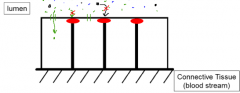
Signal sequence diagram for secreted protein |
Signal seq only good for holding protein down during trans. When finished - protein cut off from SS |
|
|
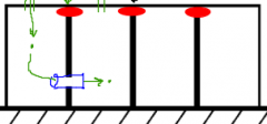
Signal sequence diagram for membrane bound protein |
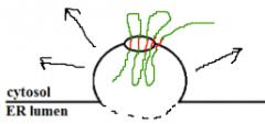
This vesicle will then fuse to outer membrane. Cytosol stays cytosol side. ER lumen becomes extracellular side |
|
|
Cholesterol in the membrane |
1. Stabilizes: normally phospholipids can shift all over but ones next to cholesterol are hydrogen bonded to it hydroxy group and cant move 2. Keeps it fluid: prevents fatty acid tails from packing up to closely together (making membrane more solid) |
|
|
Where are carbohydrates found in the plasma membrane |
Attached to the phospholipids or glycoproteins on the extracellular side. 1. help to identify cell 2. Help to increase receptor specificity |
|
|
Components of cell membrane |
1. Phosopholipds 2. Cholesterol 3. Proteins 4. Carbohydrates |
|
|
Electrolytes |
Free ions in solution produced as a result of dissolving ionic substances ex. NaCl = 2 ions produced |
|
|
Van't Hoff factor (i): |
Number of ions per molecule produced when the molecule dissolves in water -plays role in colligative properties. |
|
|
Colligative Properties |
Properties that depend on the number of solute particles but not their identity |
|
|
Types of colligative properties and what happens when you add particles |
Freezing point (depression- particles interfere w/ arrangement of crystal part. fomation) Vapor pressure (depression) Boiling point (elevated) Osmotic pressure |
|
|
Vapor pressure depression |
-Molecules of gas in equilibrium above the surface of a liquid (molecules of gas form pressure on liquid) -Particles in solution act like anchors that "hold" the solvent molecules down and prevent them from evaporating -can increase temp to soln to increase (more kinetic energy) |
|
|
Boiling point elevation |
Must increase temp to increase kinetic energy of solvent molecules to allow them to escape the anchors of solute holding them down |
|
|
Osmosis |
Movement of water - From its high concentration area to its low concentration area |
|
|
Hypertonic |
Have more particles than.... |
|
|
Hypotonic |
Have less particles than..... |
|
|
Isotonic |
Has same number of particles as.... |
|
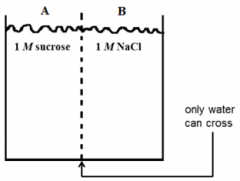
In what direction will osmosis occur? |
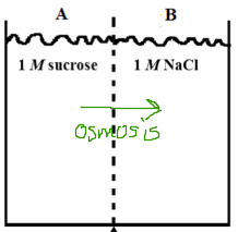
1 M sucrose = 1 mol particles 1 M NaCl = 2 moles ions |
|
|
Osmotic Pressure |
substitute this for particle concentration in a question |
|
|
What are red blood cells isotonic too? |
Physiological saline in lab 0.9% NaCl soln |
|
|
Things that can cross via simple diffusion |
(small/hydrophobic) -CO2 -Oxygen -Cholesterol -Steroid hormones -Lipids |
|
|
Types of facilitated transport proteins |
1. Pores: non-specific holes in membrane (size specific) Typically intracellular -mitochondria/nucleus 2. Channels: highly specific holes in membrane 3. Porters: Conformational change to move molecules across. (co transporter, uniporter, antiporter) |
|
|
Sodium/Potassium ATPase diagram |
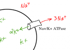
Cells also have potassium (K+) leak channel. |
|
|
Reasons for Na+/K+ ATPase |
1. Maintains osmotic balance (since water is flowing in) Ions moving out/ so water doesn't move in 2. Establishes electrical gradient (RMP = approx -70 mV) Inside neg 3. Sets up sodium gradient for secondary active transport (which indirectly uses ATP - primary uses ATP directly) |
|
|
Na+/glucose cotransporter |
Transports glucose (against gradient) and sodium (down gradient) -Runs on gradient set up my Na/K ATPase (Na is pumped out of cell) |
|
|
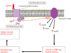
G-Proteins: Adenylyl Cyclase Diagram |
|
|
|
Three types of filaments in Cytoskeleton |
1. Microtubules 2. Microfilament 3. Intermediate filament |
|
|
Microtubules: Protein, diameter, and uses |
-Alpha and Beta tubulin -Large -Mitotic spindle, intracellular transport, cilia and flagella |
|
|
Cilia/flagella cross section |
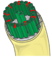
-Made with microtubules -"9+2" (nine pairs plus 2 singles) -tubules kept together with Dynein (contractile protein) |
|
|
Microfilament: Protein, diameter, and uses |
-Actin -Small -Muscle contraction, pseudopod formation, cytokinesis (contractile ring that separates cells) |
|
|
Intermediate Filament: Protein, diameter, and uses |
-Many different proteins (tend to be more stable- not as temporary as other types) -Medium diameter -Structural roles |
|
|
Desmosomes |
Cell Junction -general adhesive junction -two proteins that hold two cells together |
|
|
TIght Junctions |
Type of cell junction -Prevent unwanted molecules from getting in b/w cells to get to other side. -Seal lumens/ separate environments -Find tight junctions in the intestines/ brain capillaries |
|
|
Tight Junction Diagram |
|
|
|
Gap Juctions |
Cell-to-cell communication -Allow group of cells to work together. Ex. Cardiac muscle cells |
|
|
Gap Junction Diagram |
|
|
|
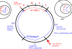
G1 Stage in Cell cycle |
Normal cell growth and activity -Very heavy regulation b/w G1 to S phase -G0 if you never leave G1 |
|
|
S stage in cell cycle |
"Synthesis" DNA replication -now committed to regulation |
|
|
G2 |
Growth -prep for mitosis -Check point b/w G2 and M (mitosis) |
|
|
Cell Cycle Diagram |
|
|
|
Goals of Prophase |
1. Break down nuclear membrane 2. Build mitotic spindle 3. Condense DNA |
|
|
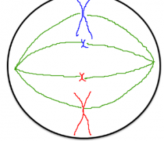
Metaphase |
When chromosomes align at cell center (random alignment) |
|
|
Goal of Anaphase |
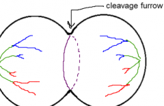
1. Separate sister chromatids 2. Begin cytokinesis (actin ring in the middle) |
|
|
Goal of telophase |
(reverse of prophase) 1. Rebuild nuclei 2. Break down spindle 3. De condense DNA 4. Finish cytokenesis |
|
|
Order of steps in Mitosis |
(I Pee on the MAT) Interphase Prophase Metaphase Anaphase Telophase |
|
|
What is cancer |
1. Mutation 2. Cell cycle unregulated 3. Divides out of control 4. Can migrate away and spread (metastasize) |
|
|
What are the two types of cancer genes? |
-Oncogenes -Tumor suppressor genes |
|
|
Proto-onocogene |
Gene that regulates the cell cycle (turns it on/off when necessary) |
|
|
Oncogene |
Mutated version of a proto-oncogene - permanently on. -uncontrolled cell division |
|
|
Tumor suppressor genes |
1. Code for protein that turn off cell cycle 2. Monitor genome of cells in the cell cycle. 3. If DNA damaged, initiate repair pathways 4. If DNA not repairable, then tumor suppressor proteins trigger apoptosis (not just cell lysis) |
|
|
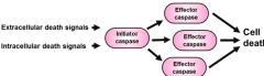
Capases and types |
Proteins that run apoptosis (proteases - cut on the C terminal side - C apases) 1. Activator capases 2. Effector capases |
|
|
Apoptosis Diagram |
|
|
|
Pathway of secreted proteins in cell |
ER -> Golgi -> Plasma membrane |
|
|
Job of nucleus |
-Contain and protect DNA -transcription -partial assembly of ribosomes (Replication, transcription, and splicing) |
|
|
Mitochondria |
Produce ATP via krebs and oxidative phosphorylation |
|
|
RER |
Location of synthesis/ modification of secretory, membrane-bound, and organelle proteins |
|
|
SER |
detoxification and glycogen breakdown in liver; steroid synthesis in gonads |
|
|
Golgi apparatus |
modification and sorting of proteins, some synthesis |
|
|
Lysosomes |
Contain acid hydrolases that digest various substances. |
|
|
Peroxisomes |
Metabolize lipids and toxins using H2O2 |
|
|
Heterochromatin |
densely packed chromatin |
|
|
Euchromatin |
More loosely packed - allow genes to be activated. |
|
|
Nuclear matrix |
AKA nuclear scaffold - provide structure |
|
|
Which molecules can pass into the nucleus |
Molecules smaller than 60 kilodaltons -Larger proteins need a nuclear localization sequence (contain a sequence of basic amino acids) |
|
|
Aminoacyl tRNA synthetase |
Enzymes (cytoplasm) that attach amino acids to their respective tRNAs. |
|
|
Targeting Signal |
Default target for proteins that go through the secretaory path is the plasma membrane. This signal is needed if a protein in that path needs to end up somewhere else (golgi, ER, lysosome) |
|
|
Localization signals |
Needed for proteins that are made in the cytoplasm but need to be sent to an organelle that is not part of the secretory path (nucleus, mitochondria, peroxisomes) |
|
|
Possible signals a protein may carry (review) |
-Signal sequence: signals protein to finish translation in ER -Localization signal: translated in cytoplasm but need to go to organelle -Transmembrane domain -Targeting signal: Part of secretory cycle but needs to go to golgi, ER, lysosome (since default is plasma membrane) |
|
|
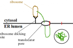
Signal Recognition Particle (SRP) |
Recognizes the signal sequence in a protein and binds to ribosome - ER has SRP receptors that dock the ribosome-SRP complex on cytoplasmic surface. |
|
|
On what type of proteins do you find disulfide bridges |
D bridges are found in extracellular proteins b/c cytoplasm is a reducing environment that changes cysteine to two cysteine. Which is why they are formed in the ER lumen (ER lumen is contiguous with extracellular space) |
|
|
Unidirectional travel of vesicles through the Golgi |
(from ER) Cis -> medial -> trans |
|
|
Regulated Secretory Pathway |
Specialized secretory cells (such as pancreatic cells, B-cells of immune system, ect) store secretory proteins in secretory vesicles and release them only at certain times. Different from constitutive secretory pathway which is unregulated (straight from Golgi to cell surface) |
|
|
Autophagy |
Self eating. Example is when a lysosome breaks down a damaged mitochondria by hydrolysis |
|
|
Phagocytosis |
Cell eating -when lysosomes degrade large particulate matter engulfed by the cell |
|
|
Macrophages |
of the immune system engulf bacteria and viruses - the organism ends up in phagocytic vesicle which will fuse with a lysosome |
|
|
Crinophagy |
lysosomal digestion of unneeded (excess) secretory products |
|
|
Acid hydrolases |
Enzymes responsible for degradation in lysosomes. They only function in acidic environment. pH = 5. pH of cytoplasm is 7.4 |
|
|
Perixisomes |
Contains enzymes thayt produce hydrogen peroxide (H2O2) as a by product. Essential for lipid breakdown. In liver they asssit with detoxification of drugs. They also contain catalase which converts H2O2 -> H2O and O2. |
|
|
Catalase |
Enzyme in peroxisomes which convert hydrogen peroxide by product - inactivates - H2O and O2. Protects cell from damage by peroxides or oxygen radicals. |
|
|
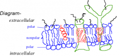
The three lipids contained in the plasma membrane: |
Phospholipids, glycolipids, and cholesterol |
|
|
Glycolipid |
membrane lipid consisting of a glycerol molecule esterified to 2 fatty acid chains and a sugar molecule -also have hydrophobic and hydrophilic regions |
|
|
Peripheral membrane proteins |
Not embedded in the membrane but rather stuck to integral membrane proteins, held by hydrogen bonding and electrostatic interactions |
|
|
Plasma membrane polarity meaning: |
inside face and outside face reamin different since lipids and proteins in membrane can diffuse laterally but can not flip-flop. Proteins anchored to cytoskeleton cannot move at all. |
|
|
Weak electrolyte |
Solutes that do not dissolve completely and remain ion-paired to some extent |
|
|
Van't Hoff (ionizability) factor |
Tells us how many ions one unit of substance will produce in a solution -Non-ionic that dont dissolve = 1 -NaCl - 2 -CaCl2 -> Ca2+ and 2 Cl- = 3 |
|
|
Osmosis |
Movement of water from low solute conc to region of higher solute concentration |
|
|
Osmotic Pressure |
Pressure it would take to stop osmosis from occuring |
|
|
Kinetic difference b/w simple diffusion and facilitated diffusion |
Simple: limited only by surface area Facilitated: exhibits saturation kinetics (transport proteins become saturated and therefore plateau in graph occurs) |
|
|
3 reasons the Na+/K+ ATPase is important: |
1. Maintain osmotic balance b/w inside/outside of cell 2. To establish resting membrane potential 3. To provide sodium (Na) concentration gradient used to drive secondary active transport. |
|
|
Note about active transport: |
If conditions change drastically a pump can run backwards -All active transporters are reversible -Also mV (RMP) would be more positive is no leaky K channels were in cell |
|
|
Ions with higher concentrations outside and inside cell |
More OUTSIDE: Na+, Cl-, Ca2+ More INSIDE: K+ |
|
|
Three types of endocytosis: |
1. Phagocytosis (nonspecific uptake - merges with lysosome) 2. Pinocytosis (cell drinking) 3. Receptor mediated endocytosis |
|
|
Endosome |
Vesicle formed during endocytosis since cytoplasm can't mix with extracellular fluid |
|
|
Receptor mediated Endocytosis |
Very specific - site is marked by pits coated with molecule clathrin (inside cell) and with receptors outside. -ex. uptake of cholesterol from blood (transported in lipoproteins) (clathrin - fibrous protein inside cell that associates with cytoplasmic portions of cell surface receptors that bind to lipoproteins) |
|
|
Atherosclerosis |
A build up of plaque on the walls of arteries - too much cholesterol in blood (accumulates in blood stream, sticking to inner walls of arteries) |
|
|
Signal Transduction |
When the binding of a ligand to its receptor triggers a response within the cell |
|
|
Ligand-gated ion channel |
(Type of signal-transducing cell surface receptor) -open an ion channel upon binding a particular neurotransmitter. -ex is ligand gated sodium channel on the surface of muscle cells - (ligand - conformation change - once open the massive influx of Na down its gradient - depolarizes cell - causes muscle to contract |
|
|
Catalytic Receptors |
(Type of signal-transducing cell surface receptor) -have an enzymatic active site on cytoplasmic side of membrane. Enzyme activity is initiated by ligand binding outside cell. -Usually involves protein kinase (modification of proteins with phosphates regulates activity) ex. insulin receptor |
|
|
G-linked protein recepetor |
Does not directly transduce signal, but transmits it into the cell with the aid of a second messenger. Chemical signal that relays instructions from cell surface to enzymes in cytoplasm -2nd messengers allow a much greater signal than receptor alone produces. -Hormone activates G protein linked receptor - which activates many G proteins - process continues. -ex. Cyclic AMP (cAMP) |
|
|
Cyclic AMP (cAMP) |
most important second messenger in G-protein-inked receptor -"Universal hunger signal" - 2nd messenger of hormones epinephrine and glucagon (glycogen and fat breakdown) |
|
|
cAMP process - review don't memorize |
Hormone activates G-linked receptor protein, which activates may G proteins -Each G protein activates many adenylyl cyclase enzymes, each cyclase makes lots of cAMP (from ATP) each cAMP activates cAMP-dPK, these phosphorylates many enzymes -End result is entire cell works towards same goal: energy mobilization |
|
|
Important to understand about cAMP |
-cAMP as a second messenger -signal transduction (binding outside triggers response inside of cell) -signal amplification |
|
|
Three proteins of cytoskeleton |
Microtubules, intermediate filaments, microfilaments -All composed of non-covalently polymerized proteins (quaternary protein structure) |
|
|
Microtubule Organizing Center (MTOC) |
Anchors growing microtubules (therefore can only extend from one side). Centrioles w/in MTOC (centrioles not essential but microtubules are for mitosis |
|
|
The aster |
The microtubules that radiate from the centrioles during mitosis. (star shape) -Microtubules connecting the chromosomes to aster are called polar fibers (and all together known as mitotic spindle) |
|
|
Function of microtubules |
-Mitosis -Mediate transport of substances wi/in cells -make up eukaryotic cilia and flagella (9+2) |
|
|
p53 |
Common tumor supressor gene - directs apoptosis |
|
|
Oxidative Stress |
-Is linked to cancer -Also component of immune system (broadly kill pathogens) |
|
|
Senescene |
Process of biological aging -telomeres shortening -cells become prone to apoptosis |
|
|
Order of apoptosis |
Dissemble cytoskeleton, break down nuclear membrane, break down genome, phagocytic digestion |
|
|
What molecule would not be involved in active transport? Vitamin D, bicarbonate, glucose, dipeptide? |
Vitamin D: fat soluble vitamin derived from cholesterol and can freely cross the membrane -All others are polar |
|
|
In oxidative phosphorylation - Name directions protons are moving down and against their gradient? |
AGAINST: Protons move out of matrix and into the inter-membrane space (requires energy) PASSIVE: From Inter-membrane space -> mitochondrial matrix (drives ATP formation) |
|
|
Large proteins, most notably albumin, are dissolved in the plasma and serve an important role in regulation of plasma volume.Reducing the amount of albumin to below-normal levels would most likely have which of the following effects? |
Movement of water from bloodstream into the tissues w/ resulting swelling, due to reduced osmotic pressure |
|
|
Note about BP elevation |
boiling point elevation is proportional to the van’t Hoff value for the solutes involved |
|
|
ER lumen corresponds to which compartments? |
Interior of Golgi = secretory vesicles = extra cellular environment |