![]()
![]()
![]()
Use LEFT and RIGHT arrow keys to navigate between flashcards;
Use UP and DOWN arrow keys to flip the card;
H to show hint;
A reads text to speech;
41 Cards in this Set
- Front
- Back
|
Question 1: What does the technique Positron Emission Tomography (PET) involve? |
Answer 1: It involves administering a radioactive isotope (e.g., oxygen-15) to the patient, thereby exposing the patient to a non-significant amount of ionizing radiation. |
|
|
Question 2: What is the difference between MRI and fMRI? |
Answer 2: MRI images the STRUCTURE of the human brain (i.e., shows you where the skull is, where the white matter is, etc). fMRI images the ACTIVITY of the human brain. |
|
|
Question 3: What does the "BOLD" in "BOLD fMRI" stand for? |
Answer 3: BOLD = Blood Oxygen Level-Dependent |
|
|
Question 4: How does BOLD fMRI allow us to measure changes in neural activity? |
Answer 4: Neural activity uses oxygen. When neurons fire, the brain increases blood flow to them. As the brain sends so much blood to the active area, the oxygen content of the blood INCREASES (i.e., neural activity UP, blood oxygen level UP). According to this logic, we compare the BOLD fMRI signals coming from the brain when 1) the subject performs the task to 2) when the subject either does nothing or performs a control task. Subtracting (2) from (1) reveals the areas of the brain that were preferentially activated by the task. |
|
|
Question 5: Assume your experiment has two conditions. In both, the observer saw exactly the same stimulus. In Condition 1, the observer tracks targets. In Condition 2, the observer passively views the same stimulus. You obtain BOLD fMRI signals for both conditions. Using those, how can you identify the brain areas involved in object tracking? |
Answer 5: If a brain area is involved in tracking objects, then it will be more active in Condition 1 than in Condition 2. Consequently, its BOLD fMRI activity will be greater in Condition 1 than in Condition 2. Thus, by subtracting the BOLD fMRI activity of Condition 2 from Condition 1, you can identify those brain areas involved in object tracking. |
|
|
Question 6: When will a proton absorb energy? |
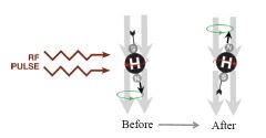
Answer 6: A proton will absorb energy ONLY at its resonance frequency. If the resonance frequency of the proton matches that of the radio frequency of the pulse, then the proton will absorb that energy. |
|
|
Question 7: What information is transmitted to the computer after the radio frequency pulse is turned off? |
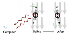
Answer 7: When the radio frequency pulse is turned off, the protons will remit their stored energy, generating radio frequency pulses. The computer decodes the pulses to create an image of the brain. |
|
|
Question 8: What are two crucial properties that allow us to use proton emissions to generate an image of the brain? |
Answer 8: 1. Not all of the protons' stored energy is emitted immediately - it takes take to flip back to their aligned states, and as they revert they emit a signal 2. The precession frequency of protons depends on the strength of the magnetic field - protons in different magnetic fields will have different precession frequencies. |
|
|
Question 9: How do radio frequency signals generated by a homogeneous magnetic field combine? |
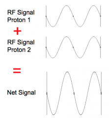
Answer 9: Coherent signals sum together to produce a large net signal. |
|
|
Question 10: How do radio frequency signals generated by an inhomogeneous magnetic field combine |
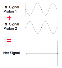
Answer 10: Incoherent signals cancel each other out to produce no net signal. |
|
|
Question 11: How is the precession rate of protons useful for measuring neural activity? |
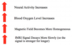
Answer 11: Since precession rate depends on the strength of the magnetic field, exciting the protons with an inhomogeneous magnetic field causes neighbouring protons and the RF signals they emit to be out of phase and cancel each other out. The more inhomogeneous the magnetic field, the faster different protons will become de-phased, and hence the faster the signal will decrease. Thus by measuring the rate at which the strength of the RF signals emitted by the tissue decreases, we can estimate how homogeneous the magnetic field is. |
|
|
Question 12: How does the fMRI excite just part of the brain? |
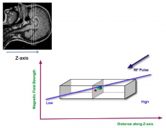
Answer 12: As protons will absorb radio frequency (RF) pulses only when the frequency of the pulse matches the proton's precession (i.e., resonance) frequency, by causing the magnetic field to vary linearly, we can cause the resonance frequency to vary throughout the brain. Thus, an RF pulse will excite only a slice of the brain – that slice of the brain where the resonance frequency of the protons matches the frequency of the RF pulse. |
|
|
Question 13: What shape is the RF signal emitted by our brain and how does that vary? |
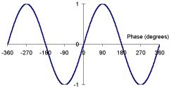
Answer 13: Sinusoidal. A sine wave varies in a periodic manner. |
|
|
Question 14: How do we determine the z-coordinate of the location of the brain area from which the BOLD signal is originating? |

Answer 14: We excite a slice of the brain to determine the z-coordinate. Slice-specific excitation: z-coordinate |
|
|
Question 15: How do we determine the x-coordinate of the location of the brain area from which the BOLD signal is originating? |
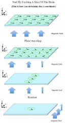
Answer 15: We arrange for the magnetic field to decrease along the x-axis for a SHORT duration. The phase of the precession now varies along the x-axis. By measuring the phase of the brain's RF pulse, we can determine the x-coordinate of the location from where it was emitted. This is known as phase encoding. Phase encoding: x-coordinate |
|
|
Question 16: How do we determine the y-coordinateof the location of the brain area from which the BOLD signal is originating? |

Answer 16: To determine the y-coordinate, we vary the magnetic field along the y-axis at readout. By listening to only a particular frequency, we can record signals of a particular y-coordinate. Frequency-specific readout: y-coordinate |
|
|
Question 17: What is BOLD fMRI? |
Answer 17: It is the measure of the oxygen level in the blood. |
|
|
Question 18: Why does one have to be careful when trying to infer neural activity from the BOLD signal? |
Answer 18: The BOLD signal is influenced, but not entirely determined, by the neural activity. |
|
|
Question 19: What is the biggest concern when trying to infer neural activity from the BOLD signal? |

Answer 19: The biggest concern is that there is a hemodynamic lag between the increase in neural activity and the increase in the BOLD signal. As seen in the image to the left, the peak BOLD response is delayed by about 8 seconds and continues for about 15 seconds after the stimulus has disappeared. Based on this hemodynamic response function, the maximum temporal resolution of the BOLD technique is about 2 seconds. This means that if two neural events occur within about two seconds of each other, the BOLD fMRI responses to the two events will become confused. |
|
|
Question 20: What is the upper limit of the spatial resolution achievable by the BOLD technique and what determines it? |
Answer 20: 1x1x1 mm^3 The upper limit is determined by how accurately the body can direct blood to the volume of the brain that needs it. If a given neuron "requests" blood, all neighbouring neurons within about 1mm of it will receive an increased blood flow. |
|
|
Question 21: What resolution do we scan at in a practical setting? |
Answer 21: We scan with voxels of 3x3x3mm^3 (at a lower resolution than 1x1x1 mm). |
|
|
Question 22: Why can we scan the same subject repeatedly in fMRI? |
Answer 22: This is because fMRI is non-radioactive and has no long-term effects on the subject. |
|
|
Question 23: What is a major safety concern of fMRI? |
Answer 23: Due to the presence of a very strong magnet (1.5T - 3T: 30,000 to 60,000 times the strength of the earth's magnetic field), any ferromagnetic object brought too close to the machine will literally fly into the magnetic field, destroying whatever is inside the magnet (e.g., the patient). |
|
|
Question 24: Due to the presence of the strong magnet, what type of patients cannot be brought into the scanning room? |
Answer 24: Depending on the metal, patients that have metal inside them may not be able to be brought into the scanning room. Similarly, patients with electronic implants (e.g., pacemaker) cannot be brought into the scanning room. |
|
|
Question 25: What are three other safety concerns of fMRI? |
Answer 25: 1. Claustrophobia - the space inside the machine is small. 2. Scanners make loud noises. Earplugs + headphones are normally used. 3. The scanner might have to be quenched. During a quench, the wire becomes resistive and therefore generates heat. The heat boils off the cryogenic fluid very quickly. If a large magnet undergoes a quench, the inert vapour formed by the evaporating cryogenic fluid can present a significant asphyxiation hazard to operators by displacing breathable air. |
|
|
Question 26: Which area of the brain did Kanwisher et al. (1997) find responded more strongly to faces than to objects, and what is it called? |
Answer 26: They found that an area of the fusiform gyrus responded more strongly to faces than objects, and this area is now referred to as the fusiform face area. |
|
|
Question 27: How did Kanwisher et al. (1997) show that this finding was not just a statistical fluke? |
Answer 27: They ran the same subject multiple times and got essentially the same result. This shows that the result is repeatable within a subject. By running a few other subjects, they also showed that the result is repeatable across subjects. |
|
|
Question 28: What control experiment did Kanwisher et al. (1997) need to run and why? |
Answer 28: While their results were repeatable within and across subjects, they could not rule out that they were due to something special about the control (non-face) stimuli. To test this Kanwisher et al. redid the experiment using different control stimuli - scrambled faces, houses, and hands. |
|
|
Question 29: What were the results of Kanwisher et al.'s (1997) control experiments? |
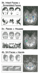
Answer 29: Regardless, of which control stimuli she used, she found that the fusiform face area always responded most strongly to faces. This strongly suggests that it was preferentially processing faces. |
|
|
Question 30: What are three common criticisms of fMRI? |
Answer 30: - The multiple comparisons problem - Non-independent sample selection - Over-interpretation of null results |
|
|
Question 31: What is the multiple comparisons problem? |
Answer 31: In fMRI one considers each voxel in turn and performs a t test to compare the activity in condition A versus the activity in condition B via a t test. The problem is that the more t tests that are performed, the greater the possibility of a false positive (i.e. the greater the probability of incorrectly rejecting a null hypothesis). This is known as the multiple comparisons problem: the more tests you do, the greater chance there is of reporting a false positive. |
|
|
Question 32: What is one way to correct for the multiple comparisons problem? |
Answer 32: One way of correcting for the multiple comparisons problem is to perform a Bonferroni Correction. That is, if you want there to be overall a 1% chance of reporting a false positive when you perform n t tests,then you perform each t test atthe 0.01/n level. |
|
|
Question 33: What is another way of avoiding the multiple comparisons problem? |
Answer 33: In order to avoid the multiple comparisons problem, many studies specify a region of interest (ROI) By averaging all the voxels within an ROI, they do just one t test per subject. Thus, they do not have to perform the Bonferroni correction so they are more likely to find a statistically significant result. |
|
|
Question 34: When is averaging the voxels in a ROI a valid method for analysing fMRI data? |
Answer 34: This is a valid method if the ROI is chosen in one scanning session but the data used for the t test is taken from a second scanning session. In other words, selection and testing must use different data sets (i.e. use different scanning sessions). |
|
|
Question 35: When does the non-independent sampling confound arise? |
Answer 35: A non-independent sampling confound occurs when a researcher uses the same scanning session to select the ROIs, and to test the ROIs. |
|
|
Question 36: Why is it important to test an ROI using a different scan to the one used to select it? |
Answer 36: The fundamental issue is that even if in reality two conditions are identical, the BOLD fMRI signal from each voxel will be slightly different in each condition because of random noise. To illustrate, suppose we form an ROI using voxels where the BOLD fMRI signal in Condition A happened (because of random noise) to be larger than that in Condition B. If we now do a t test on this ROI comparing its activity in the two conditions, we will of course find that the t test shows us that the ROI was more active in Condition A than in Condition B. This tells us absolutely nothing about how activity in this brain area differs in the two conditions, because this is how we defined the ROI in the first place. |
|
|
Question 37: What claim did Grill-Spector et al. make in her study? |
Answer 37: Grill-Spector et al. challenged the finding that the fusiform face area (FFA) specifically processes faces. She claimed that different regions of FFA were highly sensitive to different objects (not just faces; e.g. the car ROI responded very strongly to cars but not as strongly to faces, animals or sculptures). |
|
|
Question 38: Why is Grill-Spector's claim invalid? |
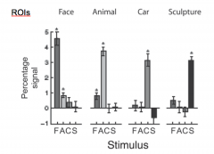
Answer 38: In order to select her ROIs, she identified which voxels in FFA responded maximally to each category (face, car, animal, sculpture), she then compared the BOLD fMRI activity generated when she presented each of the four category types compared to when she presented random noise. Thus, her sampling method was not independent. |
|
|
Question 39: What study did Baker et al. do to demonstrate the statistical problems inherent with the Grill-Spector et al.study? |
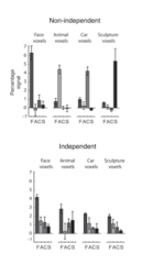
Answer 39: Baker et al. redid the study using the same flawed technique, and obtained the same results. They also did the analysis with voxels that were not in the brain, and again got the same results. Baker et al. then choose the ROIs using the data from one scanning session and tested the ROIs using data from a second scanning session - the effect then disappeared. |
|
|
Question 40: What can be concluded for voxels in which there is no statistically significant difference between the 2 conditions? |
Answer 40: A finding of no statistically significant difference is a finding of a "null" result - not much can be concluded from this! It could be that for these voxels there really was a difference between the two conditions but that the statistical tests were not sensitive enough to detect it. |
|
|
Question 41: What is the problem with over-interpreting null results? |
Answer 41: The failure to prove that a given brain area is involved in a particular task does not prove that the brain area is not involved in the task. Thus, when you report that a set of brain areas are involved in a given task, you should always acknowledge that there might be a whole lot of other brain areas involved in the task but that your statistical tests were not sensitive enough to detect their involvement. |