![]()
![]()
![]()
Use LEFT and RIGHT arrow keys to navigate between flashcards;
Use UP and DOWN arrow keys to flip the card;
H to show hint;
A reads text to speech;
25 Cards in this Set
- Front
- Back
|
Myoglobin basic structure |
-Contains a spherical red heme group in the center of Fe(II) - made up of eight helices |
|
|
Oxo-myoglobin Fe(II) group bonds |
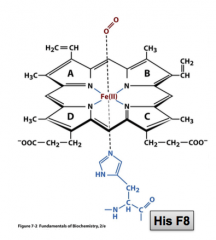
- Bound to the four Nitrogen of the heme group - One molecule of O2 can bind - Also bound to His F8 |
|
|
The Myoglobin heme complex structure |
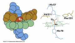
The heme group is packed between hydrophobic side chains of Val E11 and Phe CD1 ** when O2 is bound to the Fe(II), it also interacts with the NE2 of the imidazole of HisE7 |
|
|
What is YO2? |
Stands for the fractional oxygen saturation: the amount of O2 bonding sites occupied by oxygen Mathematically: Y = pO2 / (pO2 + K), where K = [ O2][Mb]/[MbO2] |
|
|
P50 |
The partial pressure of O2 (pO2) when saturation (Y) =0.5 **pO2 in venous blood is about 30 torr. 760 torr =1atm |
|
|
Myoglobin O2 binding curve |
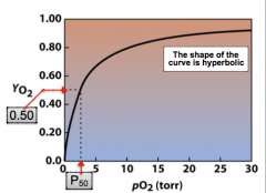
The shape of the curve is hyperbolic |
|
|
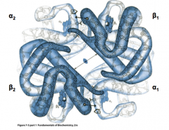
Hemoglobin general information |
The oxygen transporter in the blood of most animals, usually embedded in RBC's - consists of four myoglobin-like subunits - two Hb chains are called a chains, which have the same amino acid sequence - there are also two B chains that have the same sequence |
|
|
"Cooperativity" in hemoglobin |
Refers to the cross-talk between the four subunits of hemoglobin -increases the efficiency of O2 transport from lungs to other organs - responsible for the sigmoidal binding curve |
|
|
Deoxy versus Oxo Hemoglobin |
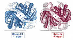
Deoxy Hb = T state Oxy Hb = R state - in oxy state, the central site gets smaller - salt bridges in the T state are broken in the R state |
|
|
O2 binding to heme in Hemoglobin |
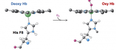
1. Fe move into the plane of heme, which becomes planar when the O2 binds 2. NO oxidation or reduction of Fe(II) occurs 3. Movement of His F8 acts like a trigger 4. Protons released due to change in pKa of groups in salt bridges |
|
|
Changes in the heme groups upon oxygenation: "the trigger" |
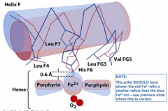
Upon oxygenation: the movement of the Fe2+ into the plane of the heme group,and the greater planarity of the heme after this move, is the “trigger” of the T-state (Deoxy-Hb) to R-state (Oxy-Hb) transition |
|
|
Deoxy versus Oxy Hemoglobin absorbance |
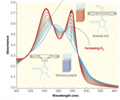
As more O2 binds, the visible spectrum shifts from blue to red. At 580nm, you can measure how much has been oxygenated |
|
|
O2 binding in Hemoglobin versus Myoglobin |
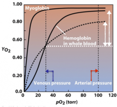
Hb picks up O2 in lung and delivers to tissue much more efficiently. - Hb is much less saturated (has more room for O2 to bind) than Mb at venous pO2 |
|
|
The Hill Equation |
The equation used to describe the sigmoidal curve of Hb-O2 binding. The larger the Hill coefficient, the steeper the S-shaped O2 binding plot.
|
|
|
The Bohr Effect |
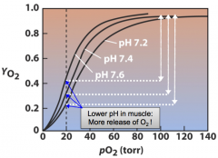
Describes the delivery of O2 from a region of higher pH (the lungs) to lower pH (the tissue) |
|
|
The Allosteric Effect |
The binding of one ligand at one site affects the binding ofanother ligand at another site. This often, but not necessarily always, requires interactionsbetween subunits of oligomeric proteins. |
|
|
The Symmetric model of allosteric proteins |
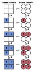
Assumes that each subunit can exist in either T or R. and the the ligand can bond to either conformation ** The key point is that hemoglobin cannot adopt an intermediate conformation.The tetramer is either in the deoxy (T) state or in the oxy (R) state. |
|
|
The Sequential Model of allosteric proteins |
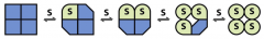
Main difference is that it assumes that symmetry does not need to be maintained and that subunits can adopt multiple conformations |
|
|
BGP (D-2,3-Bisphosphoglycerate): what it is and where it binds |
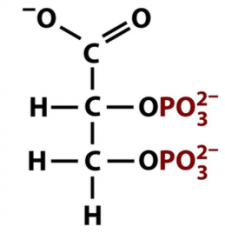
At high altitude the [BGP] in blood increases from ~4mM to ~8mM - binds at the central cavity between the B-chains in deoxy Hb. Does NOT bind to Oxy Hb |
|
|
Effects of BPG |
The net effect of BGP is to decrease Hb's affinity for O2, which increases the efficiency of O2 delivery significantly How?: increase in [BPG] causes the P50 of Hb to increase from 26 to 31 torr. |
|
|
Hemocyanin |
The oxygen binding protein in blue blood of mollusks and arthropods - much larger than hemoglobin -O2 binding site is based on Cu, not Fe(II) Mollusk hemocyanin: gigantic cylinders with 10-20 subunits per cylinder Arthropod hemocyanin: hexameters with D3 dihedral symmetry (6 subunits/hexamer) |
|
|
O2 binding center in hemocyanin (v general) |
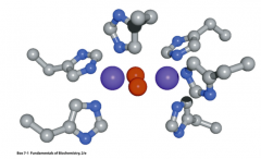
Oxygen (red) bound using two Cu(I) ions (purple) liganded by six histidines.And all embedded in a totally different protein environment than in hemoglobin. |
|
|
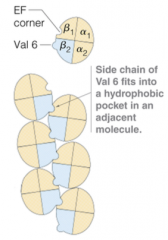
Sickle Cell Anemia |
Makes RBC's rigid and jagged, making it extremely challenging to pass through capillaries - The prime event is the point mutation fromGlu6 in HbA to Val6 in the HbS β-chain. -Heterozygotes with 40% HbS still have normal RBCs. - Common in South Africa because it confers immunity to Malaria
|
|
|
B1 to B2 mutation in Sickle Cell Anemia |
Deoxy HbS tetramers start to form fibers in an uncontrolled manner. Under deoxy conditions, these RBCs cannot pass trough capillaries |
|
|
Myoglobin and Hemoglobin differences: (subunits and hemes bonded) |
Hemoglobin has four subunits and bond 4 heme groups Myoglobin has one subunit and binds one heme group |