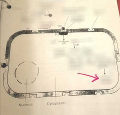
![]()
![]()
![]()
Use LEFT and RIGHT arrow keys to navigate between flashcards;
Use UP and DOWN arrow keys to flip the card;
H to show hint;
A reads text to speech;
136 Cards in this Set
- Front
- Back
|
Special Senses |
1. Olfaction 2. Gustation 3. Audition 4. Vision |
|
|
How does a special sense occur? |
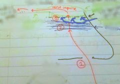
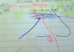
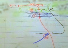
A receptor receives a stimulus which initiates a nerve impulse. A sensory nerve takes the nerve impulse to the CNS where it is interpreted |
|
|
Receptors |
1. Chemoreceptors 2. Nociceptors 3. Thermoreceptors 4. Mechonoreceptors 5. Photoreceptors 6. Propioreceptors |
|
|
Types of mechonoreceptors? |
Tactile Baroreceptor |
|
|
Tactile receptor |
Simulated by touch Ex Meissner's Corpuscles |
|
|
Baroreceptor |
Simulated by pressure Ex Pacinian Corpuscles |
|
|
Olfaction |
Smell |
|
|
Gustation |
Taste |
|
|
Audition |
Hearing |
|
|
Chemoreceptors |
Stimulated by chemical compounds Ex taste buds |
|
|
Nociceptor |
Stimulated by pain |
|
|
Thermoreceptor |
Stimulated by temperature |
|
|
Mechanoreceptor |
Stimulated by mechanical forces |
|
|
Photoreceptor |
Stimulated by light Ex cones and rods |
|
|
Propioreceptor |
Stimulated by body position |
|
|
Requirements for a substance to be smelled |
1. Substance must be volatile 2. Substance must bevwater soluble 3. Substance must be lipid soluble |
|
|
Volatile |
Capable of entering into a gas |
|
|
Why is a substance's volatility important for olfaction? |
It allows the substance to penetrate the nostrils |
|
|
Why is a substance's water solubility important for olfaction? |
It allows it to penetrate the mucous layer |
|
|
Why is a substance's lipid solubility important for olfaction? |
It allows it to stimulate the olfactory hairs/olfactory nerves |
|
|
Medial gyrus of the temporal lobe |
|
|
Olfactory tract |
|
|
Olfactory bulb |
|
|
Cribriform plate (ethmoid bone) |
|
|
Olfactory hairs/ nerves |
|
|
Mucous layer |
|
|
Nostril |
|
|
Adaptation (in relation to olfaction) |
To get used to the smell of something |
|
|
Of all the special senses, ___ is the most powerful for memory |
Olfaction |
|
|
Mastication |
Chewing |
|
|
Saliva |
Dissolves chemicals in the food that we eat so they can stimulate the taste buds |
|
|
Papilla |
Connective tissue Elevations on the surface of the tongue that contain taste buds (with one exception) |
|
|
Types of papilla |
1. Filiform 2. Fungiform 3. Circumvallate 5. Foliate |
|
|
Filiform |
Do not contain taste buds Most numerous type Give surface of the tongue texture |
|
|
Fungiform |
Mushroom- like shape |
|
|
Circumvallate |
Largest papilla Located on posterior 1/3 of tongue In ^ arrangement |
|
|
Foliate |
"Fish gills" Located on lateral edges of the tongue |
|
|
Primary tastes |
1. Sweet (tip) 2. Sour 3. Salt 4. Bitter 5. Umami 6. Water |
|
|
Myopia |
When the refractive power of the cornea and lens is too great relative to the length of the eye (eye is too long) Aka nearsightedness |
|
|
Hyperopia |
Caused when cornea is too flat or lens has too little refractive power relative to the length of the eye (eye is too short) Aka farsightedness |
|
|
Nearsightedness |
Ability to see close but not distant objects |
|
|
Farsightedness |
Ability to see distant but not close objects |
|
|
Tinnitus |
Phantom sound sensations, such as ringing in the ears Can be caused by hearing loss |
|
|
Otitis Externa |
Infection of the outer ear canal often caused by water remaining in the ear after swimming, creating a moist environment for bacterial growth Aka swimmers ear |
|
|
Cranial nerves involved in taste |
1. VII Facial Nerve 2. IX glossopharyngeal 3. X Vagus |
|
|
VII Facial Nerve |
Anterior 2/3 of tongue |
|
|
IX glossopharyngeal |
Posterior 1/3 of tongue |
|
|
X Vagus |
Throat |
|
|
Audition |
Sound waves are converted to vibrations, which are converted to a nerve impulse |
|
|
Ear wax |
Helps to moisten the auditory canal Traps dust and pathogens entering the canal |
|
|
The middle ear is typically a __ filled cavity |
Air |
|
|
Otitis Media |
Middle ear infection Infection starts in throat and travels up the eustachian tube
Most common in young children |
|
|
Cause of higher frequency of otisis media in children? |
Their eustachian tubes are shorter and more horizontal |
|
|
Ear tubes |
Treatment for otitis media Inserted into the tympanum Drains fluid from the middle ear |
|
|
Dynamic Equilibrium |
Maintaining balance while the head is moving |
|
|
Static Equilibrium |
Maintaining balance while the head is motionless |
|
|
What affects volume? |
The stronger the vibration of the tympanum, the greater the volume |
|
|
Conjunctiva |
Membranous covering on the anterior surface of the eye, not including the surface of the cornea. Secretes a lubricating fluid |
|
|
Conjunctivitis |
Pink eye Treated with antibiotic otic drops |
|
|
Iris |
Colored portion of the eye Made of smooth muscle |
|
|
What gives the iris color? How does it work? |
Melanin More = brown Middle = green Little = blue |
|
|
Pupil |
A hole in the center of the iris Admits light |
|
|
Circular fibers |
Contract in order to make the pupils smaller |
|
|
Radial Fibers |
Contract to make the pupil larger |
|
|
Pupil |
|
|
Radial fibers |
|
|
Iris |
|
|
Photoreceptors |
Receive light Located in the retina |
|
|
Types of photoreceptors |
Cones Rods |
|
|
Process of vision |
Light hits rods and cones Creates a nerve impulse Nerve impulse travels to the optic chiasma To the optic tracts To the thalamus To the posterior tip of the occipital lobe |
|
|
Cataract |
The lens loses transparency |
|
|
Glaucoma |
Over production of aqueous humor or inadequate drainage If left untreated, can cause blindness |
|
|
Astigmatism |
Abnormal curvature of the cornea and/or the lens |
|
|
Presbyopia |
"Old age farsightedness" Caused by the lens losing its ability to accommodate |
|
|
Accommodation |
The ability of your lens to change shape when looking at far to near objects Near vision: lens becomes bulbous Far vision: lens becomes flat |
|
|
Macular Degeneration |
Deterioration of the macula lutea and the fovea centralis |
|
|
Lacrimal glands |
Secretes tears, which drain into the nasolacrimal duct and into the nasal cavity |
|
|
Layers of the eye |
Fibrous tunic Vascular tunic Nervous tunic |
|
|
Fibrous tunic |
Connective tissue structures: Sclera Cornea |
|
|
Vascular tunic |
Rich blood supply Choroid Ciliary body Iris |
|
|
Nervous tunic |
Composed of neurons Retina |
|
|
Sclera |
"White of the eye" Provides shape Protects inner eye Composed of dense connective tissue |
|
|
Cornea |
"Window of the eye" Admits and refracts light Responsible for 75% of refraction |
|
|
Choroid |
Provides nutritive blood supply to the retina and absorbs light |
|
|
Ciliary body |
Involved in accommodation of the lens |
|
|
Iris |
Regulates the amount of light that enters the pupil and thus the eye |
|
|
Retina |
Image formation takes place here Light is received and transformed into nerve impulses which go to the optic nerve |
|
|
Anterior cavity |
Contains aqueous humor Contains Anterior chamber Posterior chamber |
|
|
Anterior chamber |
Posterior to the cornea, anterior to the iris |
|
|
Posterior chamber |
Posterior to the iris, anterior to the lens |
|
|
Canal of schlemm |
Located at the junction of the cornea and sclera Drains aqueous humor |
|
|
Aqueous humor |
Continually produced Maintains the shape of the eye Keeps choroid and retina together Refracts light |
|
|
Posterior cavity |
Contains vitreous humor Posterior to the lens |
|
|
Vitreous humor |
Helps maintain the shape of the eyeball Keeps the choroid and retina together Refracts light Not continually produced |
|
|
Lens |
Refracts light |
|
|
Optic disc |
"Blind spot" Where the optic nerve enters the back of the eye No image formation takes place here |
|
|
Refraction |
The bending of light rays as they pass through transparent media |
|
|
Convergence |
Medial movement of the eyes on an object as it approaches the face |
|
|
Macula lutea |
The yellow spot on the back of the eye Located in the center of the retina |
|
|
Fovea Centralis |
The depression in the center of the macula lutea Contains cones only |
|
|
Cones |
Stimulated by bright light Gives best visual acuity Color vision |
|
|
Rods |
Stimulated by dim light Poor visual acuity Colorless vision Most densely concentrated in the peripheral regions of the retina |
|
|
Pinna |
Aka auricle Collects sound waves |
|
|
External auditory meatus |
Directs sound waves to the tympanic membrane |
|
|
Tympanic membrane |
Sound waves cause it to vibrate and these vibration are transmitted to the ossicles |
|
|
Ossicles (in order according to use in audition) |
Malleus Incus Stapes |
|
|
Eustachian tubes |
Aka auditory tubes Functions to equalize pressure on either side of the tympanic membrane |
|
|
Oval Window |
The stapes fit into this. Separates the middle and inner ear |
|
|
Semicircular canals |
Tracts within the bony labyrinth |
|
|
Ampulla |
Sac like dilations at the base of the canals Part of the bony labyrinth |
|
|
Vestibule |
Connects the semicircular canals with the cochlea Part of the bony labyrinth |
|
|
Cochlea |
Contains the organ of corti Part of the bony labyrinth |
|
|
Semicircular ducts |
Part of the membranous labyrinth within the semicircular canals Contains the cristae |
|
|
Cristae |
Receptors for dynamic equilibrium |
|
|
Utricle and saccule |
Parts of the membranous labyrinth within the vestibule Contains the macula |
|
|
Macula |
Receptors for static equilibrium |
|
|
Cochlear duct |
Part of the membranous labyrinth within the cochlea Aka scala media Contains the organ of corti |
|
|
Organ of Corti |
Receptor for hearing |
|
|
Vestibulocochlear nerve |
Cranial nerve IIX Aka auditory nerve |
|
|
Endolymph |
Fluid inside the membranous labyrinth |
|
|
Perilymph |
Fluid surrounding the membranous labyrinth |
|
|
Physiology of Hearing |
1. Sound waves collected by pinna => external auditory meatus 2. Waves strike tympanic membrane 3. Vibrations from tympanic membrane travel to malleus, then incus, then stapes 4. Vibrating stapes pushes the oval window in and out 5. Movement from oval window creates fluid waves and increases pressure in the perilymph of the scala vestibuli 6. Increase in pressure makes the vestibular membrane to push inward, increasing the pressure on the endolymph of the cochlear duct 7. Basilar membrane gives under pressure and bulges into the scala tympani 8. Vibrations of the basilar membrane cause the hair cells of the Organ of Corti to strike the factorial membrane, leading to the generation of nerve impulses in the vesribulocochlear nerve 9. Impulse => medulla => inferior colliculi of the midbrain => thalamus => superior gyrus of the temporal lobe for interpretation 10. Pressure in scala tympani pushes the perilymph toward round window, causing it to bulge back in the inner ear. Helicotrema transmits fluid waves of the scala vestibuli b into the scala tympani. This ceases the stimulation of the Organ of Corti by stopping the fluid waves |
|
|
Conduction Deafness |
Caused by damage to your tympanum and/or the ossicles |
|
|
Nerve Deafness |
Damage to the cochlea and/or the auditory nerve |
|
|
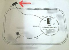
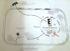
Steroid hormone Passes through target cell membrane |
|
|
The hormone enters the cell nucleus and combines with the nuclear receptor, forming a steroid-protein complex and causing gene activation |
|
|
Gene activation causes the formation of mRNA, a copy of that gene |
|
|
mRNA Contains information needed to make a specific protein Meets with a ribosome within the cell |
|
|
New protein molecule Leads to the desired action of the hormone |
|
|
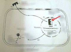
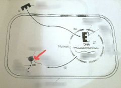
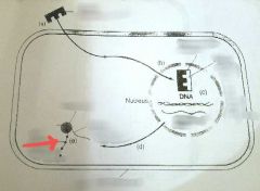
Anime, peptide, or protein hormone molecule |
|
|
Hormone combines with the cell membrane receptor |
|
|
Adenylate cyclase An enzyme, activated by the hormone receptor combination |
|
|
Adenylate cyclase causes ATP to be converted into cAMP (cyclic AMP) |
|
|
cAMP leads to the activation of an inactive protein already present within the cell |
|
|
Activated protein causes the desired action or cellular changes of the hormone |
|
|
Circular fibers |