Reading...
![]()
Play button
![]()
Play button
![]()
Use LEFT and RIGHT arrow keys to navigate between flashcards;
Use UP and DOWN arrow keys to flip the card;
H to show hint;
A reads text to speech;
31 Cards in this Set
- Front
- Back
|
PICTURE
Frontal belly of occipitofrontalis, Orbicularis oculi, Zygomaticus major, Orbicularis oris, Temporalis, Occipital belly of occipitofrontalis, Masseter |
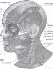
Frontal belly of occipitofrontalis, Orbicularis oculi, Zygomaticus major, Orbicularis oris, Temporalis, Occipital belly of occipitofrontalis, Masseter
|
|
|
PICTURE
Sternocleidomastoid, Teres major, Deltoid (cut), External oblique, Internal oblique, Transversus abdominis (seen through a window cut in the internal abdominal oblique), Rectus abdominis, External oblique, Latissimus dorsi, Biceps brachii, Pectoralis major, Deltoid, Trapezius, Platysma |
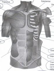
Sternocleidomastoid, Teres major, Deltoid (cut), External oblique, Internal oblique, Transversus abdominis (seen through a window cut in the internal abdominal oblique), Rectus abdominis, External oblique, Latissimus dorsi, Biceps brachii, Pectoralis major, Deltoid, Trapezius, Platysma
|
|
|
PICTURE
Deltoid, Triceps brachii, Infraspinatus |
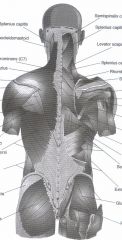
Deltoid, Triceps brachii, Infraspinatus
|
|
|
PICTURE
Flexor carpi radialis, Brachioradialis, Extensor digitorum |
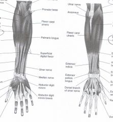
Flexor carpi radialis, Brachioradialis, Extensor digitorum
|
|
|
PICTURE
Adductor longus, Gracilis, Sartorius, Vastus medialis, Gluteus medius, Tensor fasciae latae, Vastus lateralis, Rectus femoris, Semitendinosus, Semimembranosus, Gluteus maximus, Long head of biceps femoris |
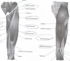
Adductor longus, Gracilis, Sartorius, Vastus medialis, Gluteus medius, Tensor fasciae latae, Vastus lateralis, Rectus femoris, Semitendinosus, Semimembranosus, Gluteus maximus, Long head of biceps femoris
|
|
|
PICTURE
Fibularis longus or Peroneus longus, Tibialis anterior, Extensor digitorum longus, Soleus |
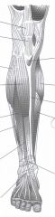
Fibularis longus or Peroneus longus, Tibialis anterior, Extensor digitorum longus, Soleus
|
|
|
PICTURE
Medial head of gastrocnemius, Lateral head of gastrocnemius |
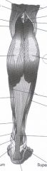
Medial head of gastrocnemius, Lateral head of gastrocnemius
|
|
|
PICTURE
Posterior or Dorsal median sulcus, Anterior or Ventral median fissure, Central canal, Anterior gray horns, Posterior gray horns, Lateral gray horns, Anterior white columns, Posterior white columns, Lateral white columns, Spinal nerve, Dorsal root, Dorsal root ganglion, Ventral root |
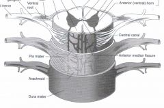
Posterior or Dorsal median sulcus, Anterior or Ventral median fissure, Central canal, Anterior gray horns, Posterior gray horns, Lateral gray horns, Anterior white columns, Posterior white columns, Lateral white columns, Spinal nerve, Dorsal root, Dorsal root ganglion, Ventral root
|
|
|
PICTURE
Cerebral hemisphere, Diencephalon, Brain stem, Cerebellum |

Cerebral hemisphere, Diencephalon, Brain stem, Cerebellum
|
|
|
PICTURE
Sulcus, Olfactory bulb, Olfactory tract |
Sulcus, Olfactory bulb, Olfactory tract
|
|
|
PICTURE
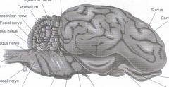
Optic nerve, Midbrain, Pons, Abducens nerve, Medulla oblongata, Hypoglossal nerve, Spinal cord, Accessory Nerve, Vagus nerve, Glossopharyngeal nerve, Vestibulocochlear nerve, Facial nerve, Trigeminal nerve, Trochlear nerve, Oculomotor nerve, Optic tract, Optic chiasma, Olfactory bulb |
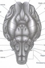
Optic nerve, Midbrain, Pons, Abducens nerve, Medulla oblongata, Hypoglossal nerve, Spinal cord, Accessory Nerve, Vagus nerve, Glossopharyngeal nerve, Vestibulocochlear nerve, Facial nerve, Trigeminal nerve, Trochlear nerve, Oculomotor nerve, Optic tract, Optic chiasma, Olfactory bulb
|
|
|
PICTURE
Cerebral hemispheres, Thalamus, Corpora quadrigemina, Pineal gland |
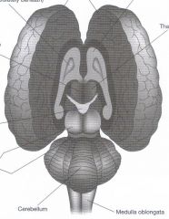
Cerebral hemispheres, Thalamus, Corpora quadrigemina, Pineal gland
|
|
|
PICTURE
Cervical spinal nerves, Thoracic spinal nerves, Lumbar spinal nerves, Sacral spinal nerves, Filum terminale, Cauda equina, Conus medullaris, Lumbar enlargement, Dura mater and Arachnoid, Cervical enlargement |
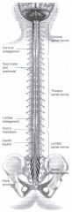
Cervical spinal nerves, Thoracic spinal nerves, Lumbar spinal nerves, Sacral spinal nerves, Filum terminale, Cauda equina, Conus medullaris, Lumbar enlargement, Dura mater and Arachnoid, Cervical enlargement
|
|
|
PICTURE
Pineal gland, Superior colliculus, Inferior colliculus, Arbor vitae (white matter), Pituitary gland, Infundibulum, Optic chiasma, Hypothalamus, Corpus callosum |
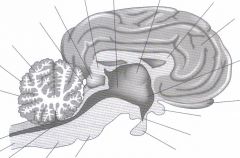
Pineal gland, Superior colliculus, Inferior colliculus, Arbor vitae (white matter), Pituitary gland, Infundibulum, Optic chiasma, Hypothalamus, Corpus callosum
|
|
|
PICTURE
V1 - Optical Division (upper face), V2 - Maxilliary Division (mid face), V3 - Maxilliary Division (lower face). C2 - Occipital Protuberance, C3 - Supraciavicular Fossa, C4 - Acromiociavicular Joint, C6 - Thumb, C7 - Middle Finger, C8 - Little Finger, T1 - Medial Antecubisal Fossa, T2 - Apex of Axillia. L1 - Upper Anterior Thigh, L2 - Mid Anterior Thigh, L3 - Medial Femoral Condyle, L4 - Medial, L5 - Dorsum 3rd MTP Joint, S1 - Lateral Heel, S2 - Popliteal Fossa, S3 - Ischial Tuberosity, S5 - Parianal Area |
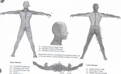
V1 - Optical Division (upper face), V2 - Maxilliary Division (mid face), V3 - Maxilliary Division (lower face). C2 - Occipital Protuberance, C3 - Supraciavicular Fossa, C4 - Acromiociavicular Joint, C6 - Thumb, C7 - Middle Finger, C8 - Little Finger, T1 - Medial Antecubisal Fossa, T2 - Apex of Axillia. L1 - Upper Anterior Thigh, L2 - Mid Anterior Thigh, L3 - Medial Femoral Condyle, L4 - Medial, L5 - Dorsum 3rd MTP Joint, S1 - Lateral Heel, S2 - Popliteal Fossa, S3 - Ischial Tuberosity, S5 - Parianal Area
|
|
|
PICTURE
Sensory Cell Bodies, Ganglion, Sensory Fibers, Recepter Effector, Spinal Nerve, Motor Fibers, Ventral Root, Motor Cell Bodies, Anterior or Ventral median fissure, Central Canal - Contains Cerebrospinal Fluid, Gray Matter, White Matter, Posterior or Dorsal Median Sulcus, Dorsal Root |
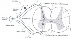
Sensory Cell Bodies, Ganglion, Sensory Fibers, Recepter Effector, Spinal Nerve, Motor Fibers, Ventral Root, Motor Cell Bodies, Anterior or Ventral median fissure, Central Canal - Contains Cerebrospinal Fluid, Gray Matter, White Matter, Posterior or Dorsal Median Sulcus, Dorsal Root
|
|
|
PICTURE
Effector, Motor Neuron, Sensory Neuron, Interneuron |
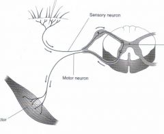
Effector, Motor Neuron, Sensory Neuron, Interneuron
|
|
|
Pupillary Reflex
|
An autonomic nrevous system reflex that controls the diameter of the pupils in response to a increase in the level of light
|
|
|
Reflex: A grade of 0
|
Partner shows no response
|
|
|
Reflex: A grade of 1
|
Partner has a reflex response although it is only a slight response
|
|
|
Reflex: A grade of 2
|
Partner has a strong, visible response. This is considered a normal response
|
|
|
Reflex: A grade of 3
|
Partner has a very strong respose
|
|
|
Reflex: A grade of 4
|
Referred to as a clonus response. Partners muscles will exhibit repeated contractions. Abnormal
|
|
|
Glabellar Reflex
|
Primitive reflex. Parner closes eyes. Gently tap the center of thier forehead
|
|
|
Jaw Jerk Reflex
|
Tapping mandable with hammer will cause jaw to close
|
|
|
Biceps Reflex
|
Tapping the inner elbow at the biceps tendon will cause arm to raise
|
|
|
Triceps Reflex
|
Tap the triceps tendon just above its insertion with the narrow end of the reflex hammer. Muscle will contract
|
|
|
Brachioradialis Reflex
|
Tapping the brachioradialis tendon will cause contraction of that muscle
|
|
|
Patellar Reflex
|
Your lower leg will rise up when the bottom of knee cap is touched
|
|
|
Calcaneal Reflex
|
Foot lifts when calcaneal muscle is tapped
|
|
|
Plantar Reflex
|
Tuck your toes in when someone draws an L shape on the bottom of your foot
|