![]()
![]()
![]()
Use LEFT and RIGHT arrow keys to navigate between flashcards;
Use UP and DOWN arrow keys to flip the card;
H to show hint;
A reads text to speech;
274 Cards in this Set
- Front
- Back
|
1. Regulators/Conformers
|
-Regulators use internal control mechanisms to moderate internal change in the face of external, environmental fluctuation
-Conformers conform ***Animals may regulate some environmental variables while conforming to others |
|
|
Homeostasis 1
|
Maintenance of a 'steady state' regardless of external environment
-humans regulate pH, body temp, glucose concentration |
|
|
Homeostasis 2
|
•For a given variable,fluctuations above or below a set point serve as a stimulus; these are detected by a sensor and trigger a response
•The response returns the variable to the set point -Regulation can be positive or negative |
|
|
negative feedback loop
|
helps return a variable to a normal range
build up of the end product shuts the system off |
|
|
Thermoregulation
|
process of maintaining body temperature
-Endothermic animals generate heat by metabolism; birds and mammals are endotherms. -Ectothermic animals gain heat from external sources. |
|
|
Vasoconstriction vs. vasodilation
|
Dilation: When you're hot, blood flow increases and heat exchange increases - more sweat and cooling
Constriction: blood flow decreases to conserve heat |
|
|
Cooling by Evaporation
|
•Sweating or bathing moistensthe skin, helping to cool an animal down.
•Panting increases the coolingeffect in birds and many mammals. •Birds have no sweat glands for cooling. |
|
|
Human Thermoregulation
|
Hypothalamus is the thermostat - will induce vasodilation or shivering
Fever creates vasoconstriction and shivering to raise body temp |
|
|
Bioenergetics
|
The overall flow and transformation of energy in an animal
|
|
|
Metabolic Rate
|
The amount of energy an animal uses an a unit of time
|
|
|
metabolic rate
|
can be determined by an animal's heat loss or the amount of oxygen used or CO2 released
|
|
|
Basal Metabolic Rate
|
the metabolic rate of an endotherm at rest at a “comfortable” temperature.
|
|
|
Standard Metabolic Rate
|
The metabolic rate of an ectotherm at rest at aspecific temperature.
|
|
|
Body mass vs. BMR - Important
1gram of mouse uses more energy than an elephant |
•The higher metabolic rateof smaller animals leads to a higher oxygen delivery rate, breathing rate,heart rate, and greater (relative) blood volume, compared with a larger animal.
mouse BMR is 20x higher than the elephant BMR. If an elephant has a mouse BMR it will have to eat ~20x times more food. Instead of 330 lbs. of food, the San Diego zoo would need 6600 lbs. of food per elephant per day!• |
|
|
2. Animal Nutrition
|
–Herbivores eat mainly plants and algae
–Carnivores eat other animals –Omnivores regularly consume animals as well as plants or algae Influences how digestive tract is adapted |
|
|
An animal’s diet must supply...
|
1. Chemical energy, which is converted into ATP to power cellular processes 2. Organic building blocks, such as organic carbon and organic nitrogen, to synthesize a variety of organic molecules 3. Essential nutrients, which are required by cells and must be obtained from dietary sources |
|
|
Essential Nutrients
|
-Essential Amino Acids
-Essential Fatty Acids -Vitamins-Minerals |
|
|
Essential Amino Acids
|
Animals require 20 and can make about half of them - The rest must come from food
Most plant proteins are incomplete in Amino acid composition What about vegans? |
|
|
Essential Fatty Acids
|
Animals can synthesize most of the fatty acids they need
a few must be obtained directly from diet and are unsaturated fish, eggs, broccoli |
|
|
Vitamins
|
Fat and water soluble organic molecules needed in small amounts
easy to get but critical lack of vitamins can be severe sometimes - Scurvy |
|
|
Minerals
|
Simple inorganic nutrients needed in small amounts
eating too much of some minerals can upset homeotic balance |
|
|
Undernutrition
|
Too few calories
Stages: 1.Use up stored fat and carbohydrates 2. Break down its own proteins 3. Lose muscle mass 4. Suffer protein deficiency 5. Die or suffer irreversible damage |
|
|
Malnourishment
|
long term absence from the diet of an essential nutrient
Deficiencies inessential nutrients can cause deformities, disease, and death |
|
|
Digestion (Chemical and Mechanical)
|
Process of breaking down food into molecules that can absorbed
•Mechanical digestion, including chewing, increases the surface area of food. •Chemical digestion splits food into small molecules that can pass through membranes; these are used to build larger molecules. |
|
|
Intracellular digestion
|
Food particles engulfed by phagocytosis
|
|
|
Ingestion
|
Advantage of digestive tube:
Specialized regions can break food down better |
|
|
Suspension Feeders and Filter Feeders
|
Important: Bivalve filter-feeding mollusks remove large quantities of suspended material from the water
|
|
|
Substrate Feeders
|
•Substrate feeders are animals that live in or on their food source•
Important: rootworm is a corn pest nicknamed the"billion dollar bug problem" |
|
|
Fluid Feeders
|
•Fluidfeeders suck nutrient-rich fluid from a living host
Important: Mosquito bites transmit Malaria. In 2010 an estimated 219 million cases of malaria occurred worldwide and 660,000 people died. |
|
|
Bulk Feeders
|
•Bulkfeeders eat relatively large pieces of food•
|
|
|
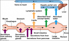
Mammalian Digestive System
|
Alimentary Canal and accessory glands
|
|
|
Digestion
|
First stage is mechanical - takes places in oral cavity
salivary glands deliver saliva for lubrication, enzymes |
|
|
Oral Digestion
|
Teeth break down food into smaller pieces which are exposed to amylase - initiating breakdown of glucose polymers
Saliva also contains mucus, a viscous mixture of water, salts, cells, and glycoproteins Epiglotis swings shut to close trachea |
|
|
Carbohydrate digestion in mouth
|
Amylase breaks down polysaccharides into maltose and smaller polysaccharides - disaccharides left alone
Amylase helps know immediately if something has sugar in it |
|
|
peristalsis
|
Food is pushed along by peristalsis, rhythmiccontractions of muscles in the wall of the canal•
|
|
|
Chemical digestion in stomach
|
stores food and secretes gastric juice (HCl and pepsin) that converts a meal to acidic chyme
gastric juice has a pH of about 2 - kills bacteria and denatures proteins Chief cells secrete inactive pepsinogen, which is activated to pepsinprotease that degrades protein |
|
|
Production of Gastric Juice
|
1. Pepsinogen and HCl ions (from parietal cells) secreted into lumen
2. HCl converts pepsinogen into pepsin (low pH) 3. pepsin activates more pepsinogen, starting chain reaction (positive feedback loop) |
|
|
Protection
|
mucous keeps bacteria from dissolving stomach walls
sphincters in the stomach keep everything contained - acidic chyme then moves on to small intestine bad sphincter = heartburn |
|
|
Stomach adaptations
|
Cows have multiple stomachs to keep digesting grass - vomit and redigest to extract all nutrients
|
|
|
Small Intestine
|
Enzymatic digestion is completed as peristalsis moves the chyme and digestive juices along the small intestine
Sugars broken down into monosaccharides Proteins into amino acids nucleic acids broken down to into molecules |
|
|
Pancreas
|
Produces proteases trypsin and chymotrypsin that are activated in the lumen of theduodenum
Its solution is alkaline and neutralizes the acidic chyme. The pancreas also has an endocrine component: Insulin-Glucagon. |
|
|
Liver
|
Bile is made in the liver and stored in the gallbladder
In the small intestine, bile aids in digestion and absorption of fats |
|
|
Form/Function of Small Intestine
|
The small intestine has a huge surface area, due to villi and microvilli that are exposed to the intestinal lumen
|
|
|
Nutrient Absorption
|
Villi and microvilli direct nutrients into capilaries which direct nutrients to the hepatic portal vein
|
|
|
Hepatic Portal Vein
|
-The hepatic portal vein carries nutrient-rich blood from the capillaries of the villi to the liver, then to the heart
-The liver regulates nutrient distribution, interconverts many organic molecules, and detoxifies many organic molecules |
|
|
Absorption of Fat
|
Fats break down into fatty components in small intestine lumen, diffuse through membrane, and recombine - also cholesterol, phospholipids, and proteins
|
|
|
More fat absoption
|
Chylomicron - lipoprotein particles that consist of triglycerides (85–92%), phospholipids (6–12%), cholesterol (1–3%), and proteins (1–2%).[1] They transport dietary lipids from the intestines to other locations in the body.
|
|
|
Lacteal
|
A lacteal is a lymphatic capillary that absorbs dietary fats in the villi of the small intestine. Taken into lymphatic system
Directs to large veins to the heart |
|
|
Large Intestine
|
Colon is connected to the small intestine
Absorption of many nutrients that were broken down cecum (first part of large intestine) has an extension called the appendix - minor role in immunity |
|
|
Colon
|
Major function is to recover water that entered alimentary canal
houses bacteria (e.g. E.Coli) that live on undigested organic material - produce some vitamins feces becomes more solid as it moves through colon |
|
|
Cecum
|
Aids in fermentation of plant material
Herbivores have large cecum/colon and a short small intestine Carnivores have small cecum/long small intestine |
|
|
Herbivores
|
Many herbivores have fermentation chambers, where mutualistic microorganisms digest cellulose
bacteria in the stomachs break down cellulose e.g. cow |
|
|
Mutualistic Adaptations
|
•The coexistence of humans and many bacteria involves mutualistic symbiosis
•Some intestinal bacteria produce vitamins; intestinal bacteria also regulate the development of the intestinal epithelium and the function of the innate immune system |
|
|
Regulation of Energy Storage
|
•The body stores energy-rich molecules that are not needed right away for metabolism
•Excess energy is stored in fat in adipose cells •When fewer calories are taken in than expended, the human body expends liver glycogen first, then muscle glycogen and fat |
|
|
Eating and Hormones
|
Hunger is regulated by hormones which change based on hunger and if you're eating
•Hormones regulate long-term and short-term appetite by affecting a “satiety center”in the brain |
|
|
Ghrelin
|
Hormone secreted by stomach wall - triggers feelings of hunger before a meal
|
|
|
Insulin & PYY
|
Insulin from pancreas Used to breakdown sugars in the blood |
|
|
Leptin
|
produced by adipose (fat) tissue, also suppresses appetite and plays a role in regulating body fat levels
|
|
|
See Slide 70
|
|
|
3. Gas Diffusion/Exchange
|
•A gas diffuses from aregion of higher partial pressure to a region of lower partial pressure
•Partial pressure is the pressure exerted by a particular gas in a mixture of gases Gas exchange occurs across specialized respiratory surfaces |
|
|
Trachae
|
-The tracheal system of insects consists of tiny branching tubes that penetrate the body
-The respiratory and circulatory systems are separate |
|
|
Gas Exchange Considerations
|
•Gas diffusion increases with surface area and decreases with distance.
•Respiratory surfaces are built to maximize the surface area and have the blood as close to the air/water as possible. (lungs are thin) E.g. gills have large area and small distance |
|
|
Countercurrent Exchange
(Cocurrent max is 50%) |
Since substances flow from high to low pressure, the flow of water across a gill allows maximum O2 to be absorbed and CO2 to be diffused through the water (~100%)
As the water flows down the gradient, it will release the maximal amount of oxygen and absorb the maximum amount of CO2 |
|
|
O2 in water
|
O2 has low solubility in water as compared to in air
If the partial pressure of gas in water is greater than in the atmosphere, the gas ‘boils’ out of the water. Fish suffocate on land because the gills collapse (lose surface area) |
|
|
Lungs
|
Infolding of the body surface
•The circulatory system (open or closed) transports gases between the lungs and the rest of the body. •The size and complexity of lungs correlate with an animal’s metabolic rate. |
|
|
Bird lungs
|
Birds inhale and exhale twice - clean and dirty air don't mix - posterior and anterior air sacs hold air around lungs
flying demands a lot of O2 and energy - their system supplies a lot of O2: small animal with high BMR per gram (breath continuously, almost like a gill) |
|
|
Mammalian System
|
Air is warm and humid from nose
mucus traps particles gas exchange occurs in alveoli |
|
|
Alveoli
|
Have lots of capillaries close to the O2 rich air
Lack cilia and are susceptible to contamination Surfactants, lower surface tension, coat the alveoli surface Preterm babies lack them treated with artificial ones |
|
|
Circulatory System
|
Has a fluid, heart, and interconnecting vessels
•The circulatory system connects the fluid that surrounds cells with the organs that exchane gases, absorb nutrients, and dispose of wastes. |
|
|
Open vs. closed systems
|
Insects have open systems using hemolymph
Advantage of closed: pressure allows heart to overcome gravity and pump all over |
|
|
Single Circulation
|
Found in only fishes.
Mode of circulation: Blood passes only once through the heart to supply once to the body. Less efficient: one pump has to supply the whole body - lower pressure |
|
|
Double Circulation
|
Found in amphibians, reptiles, birds and mammals.
More efficient as blood flows at higher pressure, especially in birds and mammals, which increases the rate of food and oxygen supply to the cell and also rapid removal of wastes from them. |
|
|
Amphibian Circulation
|
3-chambered heart where O2 and CO2 rich blood mix - great thing for them
Allows blood flow to the lungs to be shutoff when underwater •The 1 ventricle pumps blood into a forked artery that splits the ventricle’s output into the pulmocutaneous circuit and the systemic circuit |
|
|
Reptilian Circulation (Double)
|
Have 2 atria and a ventricle which is partially divided by a septum
Blood can bypass the lungs when reptiles are dormant and don't need much O2 - pumping blood to lungs requires mad energy. 2 systemic aortas direct blood out |
|
|
Arteries vs. Veins
|
Arteries and veins are distinguished by the direction of blood flow, not by O2 content
Artery=away from heart vein=towards heart |
|
|
Animal and Bird Circulation
|
-Mammals and birds have a four-chambered heart with two atria and two ventricles
•Oxygen-rich blood does not mix with oxygen-poor blood in the heart. -Mammals and birds are endotherms and require more O2 than ectotherms Valves keep blood flowing the correct way |
|
|
Blood flow part 1
|
1. CO2 rich Blood comes in from superior and inferior vena cavas into right atrium
2. right ventricle 3. pulmonary artery 4. capillaries of the lungs 5. pulmonary vein |
|
|
Blood flow part 2
|
6. Left atrium
7. Left ventricle 8. Aorta/arteries 9. Capilaries in the rest of the body 10. Veins to vena cava |
|
|
Heart Valves
|
Atrioventricular - self explanatory
Semilunar - Ventricles to arteries |
|
|
Heart Cycles
|
1. Atrial and ventricular diastole(filling) - atrioventricular valves open - semilunar valves closed
2. Atrial systole(pumping to ventricle) and ventricular diastole (filling) 3. Atrial and ventricular systole (contracting/pumping) semilunar valves open - atrioventricular valves closed |
|
|
Cardiac Output
|
Heart rate/pulse - beats per minute
Stroke volume - amount of blood pumped in a single contraction Cardiac output - volume of blood pumped into the system per minute, depends on above |
|
|
Heart rate control
|
Sympathetic Nervous system: speeds up the pacemaker of the heart
Parasympathetic nervous system: slows down the pacemaker of the heart Pacemaker is also controlled by hormones |
|
|
Structure of Blood Vessels
|
Arteries (lots of muscle) /veins have (from outer to inner) connective tissue, smooth muscle, endothelium
|
|
|
EPETHELIAL TISSUE
|
One of the four main types of tissue - lines cavities and surface structures of the body
|
|
|
Capillary Bed
|
Networks of capilaries where chemical exchange takes place between the blood and interstitial fluid - HUGE surface area
Control of temperature |
|
|
Blood Flow
|
Capilaries: high area, low speed allows for maximum exchange. Because of high resistance and cross-sectional area
Veins/Arteries: high speed, low area Pressure: High in arteries, lowest in veins |
|
|
Respiratory Pigments
|
Proteins which transport oxygen
hemoglobin is contained in erythrocytes (red blood cells) |
|
|
Hemoglobin
|
A single hemoglobin molecule can carry FOUR O2 molecules in 4 heme groups
When an O2 molecule binds, it changes the shape to make it more O-affinative |
|
|
Hemoglobin Dissociation Curve
|
PO2 is highest in the lungs, coinciding with maximum Hemoglobin O2 saturation.
Lower PO2 = lower hemoglobin O2 As it goes further down, O2 is released - it doesn't release a lot at tissue at rest - can give a lot to muscles exercising |
|
|
CO2 to hemoglobin
|
CO2 reacts with water in the body to make bicarbonate, lowering blood pH
|
|
|
Bohr Shift
|
CO2 produced during cellular respiration lowers blood pH and decreases the affinity of hemoglobin for O2; this is called the Bohr shift
|
|
|
Fetal Hemoglobin
|
Has a stronger affinity for oxygen at a given PO2, so it can snatch O2 from the mother
|
|
|
Homeostasis in circulation
|
1. Blood pH falls due to buildup of CO2 from exercise
2. Medulla receives signal from sensors in carotid artery 3. rate and depth of breathing increased until blood pH is raised to normal |
|
|
PO2/CO2
|
PO2 starts high on inhale, then drops to ~40% after capillary bed exchange
PCO2 remains at about 40% throughout most carbon dioxide in the blood is carried as bicarbonate in buffer system, converted via carbonic acid in lungs or blood - co2 also more soluble in blood than o2 |
|
|
4. Osmoregulation/Excretion
|
-osmoregulation - regulation of solute concentration and balancing of water
- excretion - removal of nitrogenous metabolites and other wastes |
|
|
osmolarity
|
solute concentration
***To make a side more dilute, you can either move solute out OR water in hypo/hyper |
|
|
osmolarity 2
|
isoosmotic - concentrations are equal - water will cross equally in both directions
hypo/hyper - solute will move from more saturated to less saturated |
|
|
mobilizing solute
|
can be used to equilibrate solutions
|
|
|
Water balance
|
blank
|
|
|
Freshwater Fish
|
Do not drink water
large volume of urine, less concentrated than body fluids principle problem is keeping water out and salt actively transported in |
|
|
Saltwater fish
|
Drinks a lot water
Principle problem is keeping water in |
|
|
Land mammals
|
Drink only a little water and intake salt
|
|
|
Nitrogenous Wastes
|
Among the most significant wastes are nitrogenous breakdown products of proteins and nucleic acids
Some animals convert toxic ammonia (NH3) to less toxic compounds prior to excretion |
|
|
Ammonia
|
Most toxic, released by most aquatic animals
Needs lots of water, can be released across the whole body |
|
|
Urea
|
•The liver of mammalsand most adult amphibians converts ammonia to the less toxic urea that requires less water than ammonia
|
|
|
Uric Acid
|
•Uric acid is relatively nontoxic and does not dissolve readily in water, secreted as a paste with little water.
Birds, reptiles, land snails Most energy intensive to make |
|
|
Key processes of Excretory Systems
|
–Filtration: Filtering of body fluids
–Reabsorption: Reclaiming valuable solutes –Secretion: Adding non essential solutes and wastes from the body fluids to the filtrate –Excretion: Processed filtrate containing nitrogenous wastes, released from the body |
|
|
Juxtamedulary Nephron Units
|
1. Glomerulus (Bowman's Capsule)
2. Proximal Tube 3. Loop of Henle (Descending and Ascending) 4. Distal Tubule 5. Collecting Duct |
|
|
1. Glomerulus (Bowman) (cortex)
|
First part of collection - blood plasma is filtered through capillaries which flow through the GLOMERULUS
Filtrates contains salts, glucose, amino acids, vitamins, nitrogenous wastes, and other small molecules then flows into the Bowman's Capsule to begin processing |
|
|
2. Proximal Tube (cortex)
|
•Reabsorption of ions, water, and nutrients
•Molecules are transported actively and passively from the filtrate into the interstitial fluid and then capillaries •Toxic materials are actively secreted into the filtrate •Materials to be excreted become concentrated in the proximal tubule without changing osmolarity |
|
|
3a. Descending Loop of Henle (medulla)
|
Water leaves by osmosis - filtrate becomes more concentrated with urea and NaCl
Permeable to water |
|
|
3b. Ascending Loop of Henle (medulla)
|
Salt, NOT water, is able to diffuse out at the bottom due to concentration gradient
At the top, salt is actively transported out as interstitial fluid becomes less saturated Filtrate becomes more dilute without taking water from interstitial fluid! |
|
|
4. Distal Tubule
|
Filtrate entering the tubule is MORE dilute than body fluids Absorption of a LOT of water •The distal tubule regulates the K+ and NaCl concentrations of body fluids•The controlled movement of ions contributes to pH regulation Nephron Cortex |
|
|
5. Collecting Duct
|
•The collecting duct carries filtrate through the medulla to the renal pelvis - maximum saturation at the bottom
•One of the most important tasks is reabsorption of solutes and water •Urine is hyperosmotic to body fluids Aquaporins are added here if blood volume/pressure is low |
|
|
The countercurrent multiplier system involving the loop of Henle maintains a high salt concentration in the kidney
|
shorter collecting duct = more dilute urine
|
|
|
Adaptations to reduce water loss
|
Desert animal concentrate urine
Longer loops of Henle would make more concentrated urine |
|
|
Role of ADH
|
When blood osmolarity increases, hypothalamus signals posterior pituitary to release ADH (Vasopressin)
ADH creates aquaporins in the collecting duct to trigger reabsorption of water in epithelium |
|
|
Diabetes
|
any of various abnormal conditions characterized by the secretion and excretion of excessive amounts of urine.
Diabetes Mellitus: sweet urine |
|
|
Diabetes Insipdus
|
Rare condition where kidneys can't conserve water due to a lack of ADH production
Severe dehydration |
|
|
Vasopressin Case
|
Aquaporin-2 is the vasopressin-responsive water channel in the collecting duct. In congenital nephrogenic diabetes insipidus, the kidney does not respond to vasopressin. Ninety percent of these patients carry a mutation in the gene coding for the vasopressin V2 receptor located on the X chromosome. Autosomal recessive and dominant forms of nephrogenic diabetes insipidus that are caused by mutations in the aquaporin-2 gene have now been described.
|
|
|
The Renin-Angiotensin-Aldosterone System (RAAS)
|
•A drop in blood pressure near the glomerulus causes the juxtaglomerular apparatus (JGA) to release the enzyme renin
•Renin triggers the formation of the peptide angiotensin II |
|
|
Angiotensin II
|
Raises blood pressure and decreases blood flow to the kidneys - constricts arterioles
–Stimulates the release of the hormone aldosterone from adrenal cortex |
|
|
Aldosterone
|
from adrenal cortex: increases blood volume and pressure
|
|
|
RAAS function 1
|
1. Stimulus: low blood volume or blood pressure e.g. from dehydration or blood loss
2. JGA releases renin, triggering the formation of angiotensin I (also released from liver) 3. ACE converts angiotensin I to II |
|
|
RAAS function II
|
-Angiotensin 2 stimulates adrenal gland to release aldosterone. Also constricts arterioles to increase blood pressure
-Aldosterone triggers more absorption of Na and H2O in distal tubules, increasing blood volume |
|
|
ACE inhibitors
|
•ACE inhibitors areusually used to treat hypertension because they reduce the production of Angiotensin II, and thus causes dilatation of blood vessels, which results in lower blood pressure.
|
|
|
Kidney Pathology
|
•Poor regulation of solutes in blood•
•Compromised fluid balance• •Accumulation of toxic waste |
|
|
5. Hormones/Endocrine System
|
Long distance messaging, transported by blood, affect many organs, involuntary response, response may be short or long lived
|
|
|
Endocrine Signaling
|
Hormone travel in the blood
|
|
|
Paracrine
|
Hormone is released into the neighborhood, doesn't need to travel through blood
|
|
|
Neuroendocrine
|
specialized neurosecretory cells secrete molecules called neurohormones that travel to target cells via the bloodstream
|
|
|
Autocrine
|
Cell signals itself
Might be important in cancer - |
|
|
Receptors
|
Critical - if a cell doesn't have the right receptor, it can't receive the message
|
|
|
Phermones
|
Members of the same animal species sometimes communicate with pheromones, chemicals that are released into the environment
|
|
|
Endocrine vs. Exocrine
|
•Endocrine glands secrete hormones directly into surrounding fluid.•
•These contrast with exocrine glands, which have ducts and which secrete substances onto body surfaces or into cavities.•
Exo: Sweat glands and gastric are exocrine, hypothalamus is endocrine |
|
|
Endocrine and Exocrine Pancreas
|
•The exocrine pancreas, a gland behind the stomach, can raise the pH in the duodenum and secretes digestive enzymes. The pancreas has clusters of endocrine cells called pancreatic islets that produce glucagon and produce insulin in response to high blood glucose levels |
|
|
Classes of Hormones and their solubility
|
Polypeptides (water soluble)
Amines from Amino Acids (both) Steroid Hormones (Lipid) If it's lipid soluble, it won't want to travel in the water of the blood.Water soluble peptides need a mechanism to be released from the cell but will travel easily in the blood,can't get into the target cell |
|
|
Steroids
|
Can easily diffuse into blood, bind to a carrier protein to travel around
Can easily diffuse into cell membrane Receptors are inside the cell |
|
|
Water-soluble (proteins)
|
Diffused into blood via vesicles, can easily travel in blood
Receptors for water soluble hormones MUST be on cell membrane Signal must travel to the nucleus via signal cascade/second messengers |
|
|
Pathway for water-soluble hormone
|
Binding on membrane surface activates signal transduction pathway
|
|
|
Pathway for lipid-soluble hormones
|
•Steroids, thyroid hormones, and the hormonal form of vitamin D enter target cells and bind to protein receptors in the cytoplasm or nucleus. |
|
|
Multiple effects of Hormones
|
Same hormone can have different effects on cells via different receptors or signal transduction pathways
Ex: epinephrine decreases blood flow to stomach, increases blood flow to skeletal muscles |
|
|
Hormone Feedback Regulation
|
•A negative feedback loop inhibits a response by reducing the initial stimulus, thus preventing excessive pathway activity•
•Positive feedback reinforces a stimulus to produce an even greater response• |
|
|
Positive Feedback Example (Oxytocin)
|
Stimulus: Suckling leads to release of oxytocin from posterior pituitary (neurohormone)
Travels to mammary glands and triggers release of milk and further oxytocin production |
|
|
Hypothalamus
|
Receives information from the nervous system and initiates responses through the endocrine system
|
|
|
Pituitary Gland
|
Attached to the hypothalamus - divided into two parts: Anterior and Posterior
|
|
|
Posterior Pituitary (neuroendocrine)
|
Functions as an extension of the hypothalamus
axons of hypothalamus go into PP. Stores hormones produced by the neurosecretory cells of the hypothalamus and releases them when triggers |
|
|
Posterior Hormones
|
ADH: Kidney tubules - neurohormone
Oxytocin: Mammary Glands, uterine muscles Both act directly on non-endocrine tissues |
|
|
Anterior Pituitary
|
Basically a separate organ
Anterior endocrine hormones are controlled from neurohormones of the hypothalamus Ex: PRL (Prolactin releasing hormone) goes from hypothalamus to the anterior pituitary - prolactin released |
|
|
Some Anterior Hormones
|
FSH/LH - testes or ovaries
TSH - Thyroid ACTH - Adrenal cortex Prolactin - Mammary Glands MSH - Melanocytes GH - liver, other tissues, bones |
|
|
Thyroid Hormones
|
-Triiodothyronin (T3), with three iodine atoms
–Thyroxine (T4), with four iodine atoms - The Thyroid gland also produces other hormones: Calcitonin (C-Cells)and PTH (Parathyroidgland): Ca+ metabolism |
|
|
Thyroid Regulation
|
A hormone can trigger the release of a cascade of hormones, the last activates the non-endocrine target cell
|
|
|
Thyroid Process
|
Stimulus: Thyroid hormone Levels drop
1. hypothalamus secretes TRH into anterior pituitary causing... 2. Anterior pituitary secretes TSH 3. TSH stimulates thyroid to secrete T3 and T4. Negative feedback loop - hypothalamus turns off TRH after T3/T4 levels return to normal |
|
|
Disorders of the Thyroid
|
Hypothyroid: too little T3/4 leads to weight gain, lethargy, cold intolerance
Hyperthyroid: too much leads to high temperature,sweating, weight loss, irritability, and high blood pressure |
|
|
Malnutrition and Thyroid
|
can alter thyroid function - Insufficient dietary iodine leads to an enlarged thyroidgland, called a goiter.
T3/T4 carry 3 and 4 ions of iodine respectively |
|
|
Adrenal Glands
|
Located on top of the kidneys, divided into medulla and cortex
|
|
|
Adrenal Medulla
|
Secretes catecholamines
• epinephrine(adrenaline) & norepinephrine(noradrenaline) Flight or fight reflex triggered by nerves via spinal cord |
|
|
Catecholamines
|
•The release of epinephrine and norepinephrine occurs in response to involuntary nerve signals
–Trigger the release of glucose and fatty acids into the blood –Increase oxygen delivery to body cells –Direct blood toward heart, brain, and skeletal muscles and away from skin, digestive system, and kidneys |
|
|
Adrenal Cortex Hormones
|
Release corticosteroids in response to long term stress
Humans produce glucocorticoids and mineralocorticoids |
|
|
Steroids of adrenal cortex
|
•Glucocorticoids, such as cortisol, influence glucosemetabolism and the immune system
Mineralocorticoids, such as aldosterone, affect salt and water balance Aldosterone is part of RAAS system |
|
|
6 - Animal Reproduction
|
Moving on
|
|
|
Asexual reproduction
|
•Fission: separation of a parent into two or more individuals of about the same size
•Fragmentation: breaking of the body into pieces, which develop into adults •Budding: new individuals arise from outgrowths of existing ones •Parthenogenesis:is the development of a new individual from an unfertilized egg |
|
|
Sexual Reproduction
|
Creation of offspring with a sperm and egg --> zygote
Sexual species have half as many offspring as asexual ones - nearly all euks still sexual |
|
|
Advantages of sexual reproduction
|
–An increase in variation in offspring, providing an increase in the reproductive success of parents in changing environments
–An increase in the rate of adaptation –A shuffling of genes and the elimination of harmful genes from a population |
|
|
Variations of Sexual Reproduction
|
Hermaphrodites - each individual is male/female
sex reversals |
|
|
Gametes define sex
|
Male: motile, small
Female: small, immotile |
|
|
Review
|
Meiosis reduces number of chromosomes (n)
fertilization restores 2n |
|
|
Fertilization
|
External: eggs are shed by female and fertilized by sperm in the external environment
Internal: sperm are deposited in or near the reproductive tract and fertilization occurs therin |
|
|
Coordination of Fertilization
|
Successful fertilization depends on coordinating sperm availability with mature eggs.
Ovulation is the release of mature eggs at the midpoint of a female cycle. Other coordinators include pheromones, courtship dances, etc |
|
|
Cortical Reaction
|
Fusion of egg and sperm initiates reaction which forms a fertilization envelope, preventing further fertilization and polyspermy
Calcium wave is the rxn |
|
|
Internal fertilization
|
•Fertilization depends on mechanisms that help sperm meet eggs of the same species.
•requires behavioral interactions and compatible copulatory organs. •is typically associated with production of fewer gametes but the survival of a higher fraction of zygotes. |
|
|
Gamete Production/Delivery
|
Gametes are produced in gonads and systems include other organs to carry, nourish, and protect the gametes and embryo
|
|
|
Spermatogenesis
|
1. spermatogonium - 2n germ cells
2. primary spermatocyte - 2n 3. secondary spermocyte - n 4. early spermatid - differentiation 5. sperm cell released into lumen after maturation |
|
|
Ovaries
|
•Each ovary contains many follicles, which consist of a partially developed egg, called an oocyte, surrounded by support cells
•Once a month, an oocyte develops into an ovum (egg) by the process of oogenesis• |
|
|
Oogenesis
|
1. Begins in embryo with formation of oogonium
2. divide into primary oocyte - stop in prophase of meiosis I 3. Starting at puberty, eggs finish meiosis I and ovulation occurs 4. Secondary oocyte in mature follicle is arrested at metaphase of meiosis II |
|
|
Ovulation
|
1. Puberty begins when FSH stimulates a follicle to fully mature and release seconday oocyte
2. oocytes only finish meosis II if there is fertilization 3. Corpus leutem secretes estradiol and progesterone |
|
|
Hormonal Control of Female Reproduction
|
Cyclically regulated
Changes in the uterus are called menstrual cycle Changes in ovaries are ovarian cycle |
|
|
Ovarian Cycle
|
1. The sequential release of FSH and LH stimulates follicle growth
2. The follicular phase ends at ovulation, and the secondary oocyte is released. 3.After ovulation the corpus luteum secretes progesterone and estradiol. |
|
|
Hormones in Ovarian Cycle
|
1. GnRH released from hypothalamus, triggers release of FSH and then LH from anterior pituitary
2. Estradiol is slowly released from growing follicle - inhibits more FSH or LH 3. surge in estradiol causes LH surge via GnRH 4. Corpus luteum secretes progesterone and estradiol, preventing further ovulation 5. If there is no fertilization, hypothalamus begins releasing GnRH again |
|
|
Menstrual (Uterine) Cycle
|
1. After ovulation, estradiol and progesterone stimulate thickening of endometrium in the uterus in preparation for implantation
2. If there is no implantation, the endometrium is shed in menstruation |
|
|
Post-fertilization
|
1. Fertilization occurs in the fallopian tube
2. Embryo begins to rapidly cleave into many small cells - overall diameter does not change 3. Cleavage continues until embryo becomes blastocyst 4. blastocyst implants in endometrium 7 days after conception |
|
|
Blastocyst
|
-inner cell mass is called the embryo the outer layer(trophoblast) grows into placenta |
|
|
7. Development
|
asdlkfjsdf
|
|
|
Twins
|
-1 embryo splits: monozygous twins, genetically identical, one placenta
-2 distinct embryos: dizygotic twins, genetically distinct, two placentas |
|
|
Morphogenesis
|
After cleavage, the rate of cell division slows and thenormal cell cycle is restored
•Morphogenesis, the process by which cells occupy their appropriatelocations, involves –Gastrulation, rearranges the cells of a blastula into a three-layeredembryo, called a gastrula
–Organogenesis, the formation of organs |
|
|
Gastrulation
|
•The three layersproduced by gastrulation are called embryonic germ layers
–The ectoderm forms the outer layer –The endoderm lines the digestive tract –The mesoderm partlyfills the space between the endoderm and ectoderm •Each germ layercontributes to specific structures in the adult animal |
|
|
Process of Gastrulation
|
cells infold into the embryo, creating a distinct blastopore (anus) and the three layers
Gastrulation occurs AFTER implantation in the uterus (trophoblast) |
|
|
Organogensis
|
•various regions ofthe germ layers develop into rudimentary organs
•Neurulation, the formation ofprimitive CNS structures. - Cells form a neural plate which rolls into the neural tube, becomes brain and spinal cord |
|
|
Cytoplasmic determinants
|
•Determination is the term used todescribe the process by which a cell or group of cells becomes committed to aparticular fate•
•Differentiation refers to theresulting specialization in structure and function |
|
|
P Granules (fate mapping)
|
Germ cells are the specialized cells that give rise to sperm or eggs. •Complexes of RNA and protein are involved in the specification of germ cell fate. Called P Granules in C. Elegans Note: fate mapping = killing early cells and seeing what happens when they develop |
|
|
***P Granules *****
|
•P granules are distributed throughout the newly fertilized egg and move to the posterior end before the first cleavage division
•With each subsequent cleavage, the P granules are partitioned into the posterior-most cells •P granules act as cytoplasmic determinants, fixing germ cell fate at the earliest stage of development |
|
|
Implications
|
“C. elegans oocytes have no inherent polarity and fertilization itself does not polarize the embryo. About 30 minutes after fertilization, however, the symmetry of the one-cell embryo is broken when the anterior-posterior axis is established.”
|
|
|
Cell Potentiality
|
•Totipotent cell: can develop into all the possible cell types:fertilized egg.•
•Pluripotent cell: can develop into some cells: committed germ cell (ex: gastrulation) As embryonic cells acquire distinct fates, they influence each other’s fates by induction |
|
|
Spemann and Mangold
|
•transplanted tissues between early gastrulas and found that the transplanted dorsal lip triggered as second gastrulation in the host
•The dorsal lip functions as an organizer of the embryo body plan, inducing changes in surrounding tissues to form notochord, neural tube, and so on. |
|
|
Implications of Spemann and Mangold
|
Signaling molecules from the organizer induce the host cell to adopt different fates
Organizer is paracrine! |
|
|
Inductive Signaling
|
•Inductive signals play a major role in pattern formation, development of spatial organization
•The molecular cues that control pattern formation are called positional information, tells a cell where it is with respect to the body axes
•It determines how the cell and its descendents respond to future molecular signals |
|
|
Limb formation
|
Begin as limb buds
AER: Apicalectodermal ridge - where limb forms ZPA: Zoneof polarizing activity -indicates posterior
Tissue transplantation experiments support the hypothesis that the ZPA produces an inductive signal that conveys positional information indicating “posterior” |
|
|
8. Stem Cells
|
Must be able to differentiate into one other cell type and turn into another stem cell
|
|
|
Types of stem cells
|
Embryonic - have greater potential, can basically do anything - can reproduce indefinitely
Adult - can generate multiple cell types - used to replace non-regenerating cells as needed |
|
|
Embryonic Stem Cells
|
Undifferentiated cells derived from a preimplantation embryo….. that are capable of dividing without differentiating fora prolonged period in culture, and are known to develop into cells and tissuesof the three primary germ layers.
|
|
|
Embryonic Stem Cell power
|
1.Most changes to the genome during development are reversible.
2.There is something in the Egg that resets the adult nucleus and allows it to start development again. Seen in nuclear transfer cloning: skin cell nucleus put into an egg cloned! |
|
|
Cloning
|
Same technique of implanting an adult nucleus into an egg was used to clone Dolly
Was a very inefficient process - takes a lot of cells and stuff to work Started with 277, got 1 |
|
|
Reprogramming
|
Could remove nucleus from egg, implant adult cell that you wanted to get embryonic stem cells with the genetic material of the adult
Oocyte reprograms the nucleus |
|
|
Fusion
|
Can fuse an adult cell with an ES cell
Use stem cell regulator genes to force a differentiated cell into an embryonic-like state Same factors in the egg are also in ES's It works! Introduction of 4 genes into a fibroblast |
|
|
Critereon to make stem cells
|
1.Are they similar to embryonic stem cells?
2.Do they divide and self renew like embryonic stem cells? 3.Do they express the same set of genes? 4.Are they able to differentiate into different tissues like stem cells do?• |
|
|
1.Are they similar to embryonic stem cells?
2. Do they divide too? |
1. They divided a lot and produced more stem cells - tracked with GFP
2. Yes they divided, not as fast but still |
|
|
3. Do they divide and self-renew like embryonic stem cells?
|
Use of nanog to show that the induced cells expressed similar gene activity to original cells
Fxb was much worse than ES Nanog about the same |
|
|
4.Are they able to differentiate into different tissues like stem cells do?
|
Injected into mice - formed teratomas - have all three germ layers
|
|
|
iPS germ line
|
contribute to all germ layers even germ cells. Thus, they are truly pluripotent!
|
|
|
9. Neurons
|
Axon traveling = electrical
synapse = chemical |
|
|
Structure of a neuron
|
Most organelles in the cell body
Dendrites: highly branched extensions - receive signals Axon: extension that transmits signals (very long) Synapse: Junction between dendrite and axon Glia: nourish and insulate neurons (myelin) |
|
|
Membrane Potential
|
•Every cell has a voltage (difference in electrical charge) across its plasma membrane called a membrane potential
•The resting potential is the membrane potential of a neuron not sending signals •Changes in membrane potential act as signals, transmitting and processing information |
|
|
Ion Concentrations
|
Potassium: More concentrated inside -
Sodium: More concentrated outside Chlorine: More concentrated outside Ion channels allow ions to diffuse without using ATP |
|
|
Charge
|
Any net movement of ions carries the charge with them, generating a membrane potential/voltage across the membrane
K+ channels are always open, maintaining an outflow and a negative charge in the cell An equilibrium is established between inflow of Na+ and outflow of K+ |
|
|
Na+/K+ Pump
|
Uses ATP for active transport - allows cells to maintain a concentration gradient that is different from the surrounding area
Effect is to make the interior negatively charged and the outside positively - changes electrical equilibrium(3 + out, 2 + in) Driving source of the charge |
|
|
Ion Channel Diversity
|
Nongated
Voltage gated Chemically Gated |
|
|
Non-gated ion channels
|
Responsible for membrane resting potential
Always open: K+ diffuses down its gradient to the outer chamber: creates negative charge –Negative charge (Cl–) builds up in the inner chamber **At equilibrium, both the electrical and chemical gradients are balanced together |
|
|
Electrochemical Equilibrium
|
Higher solute concentration: Negative
Lower solute concentration: Positive Again: At equilibrium, both the electrical and chemical gradients are balanced TOGETHER K wants to go out, but the charge can only get so negative before it pulls backwards |
|
|
Equilibrium Potential
|
The membrane voltage for a particular ion at equilibrium •The equilibrium potential of K+(EK)is negative (-90V), while the equilibrium potential of Na+ (ENa) is positive |
|
|
Potential of Neurons
|
•Every cell has avoltage (difference in electrical charge) across its plasma membrane called a membrane potential
•The resting potential is the membranepotential of a neuron not sending signals •Changes in membrane potential act as signals, transmitting and processing information |
|
|
Resting Potential
|
The concentration of KCl is higher in the inner chamber and lower in the outer chamber |
|
|
Increasing K permeability
|
When gated K+channels open, K+ diffuses out, making the inside of the cell more
negative hyperpolarization, an increase in magnitude of the membrane potential |
|
|
Increasing Na Permeability
|
When Na+channels open and Na+ diffuses into the cell
Depolarization, a reduction in the magnitude of the membrane potential Depolarization triggers the action potential |
|
|
Voltage-gated Channels
|
Voltage-gated channels are responsible for generation and propagation of the action potential, the outgoing signal from the neuron.
voltage gated channels are triggered if membrane potential passes a particular level - opening some channels makes even more open: positive feedback Only open for a certain amount of time, then go inactive |
|
|
Generation of Action Potentials
|
1. Cell is in resting state, ungated channels open to allow K+ to flow freely and maintain - charge
2. A stimulus opens some gated Na channels, allowing membrane depolarization - IF it reaches threshold, action potential is triggered |
|
|
Action Potential phases
|
1: Rising - depolarization opens most Na voltage channels, K voltage channels closed - influx of Na makes interior of membrane positive
2. Falling: most Na channels closed, K voltage channels open to make cell negative again 3. Undershoot: All Na channels close, some K still open - membrane returns to resting state |
|
|
Conduction of action potentials
|
As an action potential is created in one cell, it will depolarize a neighboring region of the membrane - you can sense your neighbor
The process is repeated along the axon. Currents across the membrane cause the action potential to be propagated along the length of the axon |
|
|
Myelin Sheath
|
•In vertebrates, axons are insulated by a myelin sheath, which causes an action potential’s speed to increase
•Myelin sheaths are made by glia— oligodendrocytes in the CNS and Schwann cells in the PNS •Action potentials in myelinated axons jump between the nodes of Ranvier in a process called saltatory conduction |
|
|
Multiple Sclerosis
|
In MS, myelin becomes damaged, affecting neural conductivity
|
|
|
Synapses (Chemically-gated)
|
At the terminal end of an axon, the presynaptic neuron makes neurotransmitters and packages them into vesicles
The arrival of the action potential opens Ca voltage gated channels. Ca goes in, vesicles go out |
|
|
Process of synapse firing
|
1. Action potential arrives and destabilizes membrane
2. depolarization opens Ca voltage cated channels, Ca flows in 3. Elevated Ca causes vesicles to fuse to membrane, releasing neurotransmitter to synapse 4. neurotransmitter binds to ligand gated ion channels, passing on signal |
|
|
Neurotransmitters
|
•There are more than100 neurotransmitters, belonging to five groups: acetylcholine, biogenicamines, amino acids, neuropeptides, and gases
•A single neurotransmitter may have more than a dozen different receptors |
|
|
Receptors
|
Ionotropic - Acetylchloride (direct and fast)
Metabotropic - movement of ions through the channel depends on metabolic steps (indirect, slow) |
|
|
Metabotropic Receptor
|
•Binding of a neurotransmitter to a metabotropic receptor activates a signal transduction pathway in the postsynaptic cell involving a second messenger that then opens
•Compared toligand-gated channels, the effects of second-messenger systems have a slower onset but last longer |
|
|
Termination of the Signal
|
Enzymatic breakdown in the synaptic cleft
Reuptake by presynaptic neurons Diffusion |
|
|
Postsynaptic Potentials
|
•Direct synaptictransmission involves binding of neurotransmitters to receptors in the postsynaptic cell•
•Neurotransmitter binding causes ion channels to open, generating Excitatory (towards threshold) or Inhibitory(away from threshold) postsynapticpotential |
|
|
Temporal Summation
|
Two excitatory postsynaptic potentials from ONE axon come together to trigger an action potential
|
|
|
Spatial Summation
|
Two excitatory postsynaptic potentials from TWO axons come together to trigger an action potential
Summation can also occur with inhibitory potentials |
|
|
Lecture 10 - Nervous Systems
|
Brain:
Gray matter: Cell bodies White matter: myelinated axons |
|
|
Nerves
|
•bundlesthat consist of the axons of multiple nerve cells.
|
|
|
More than neurons
|
•Glia havenumerous functions to nourish, support, and regulate neurons
–Embryonicradial glia form tracks along which newly formed neurons migrate –Astrocytesinducecells lining capillaries in the CNS to form tight junctions, resulting in a blood-brainbarrier andrestricting the entry of most substances into the brain |
|
|
Ganglia
|
segmentally arranged clusters of neurons
|
|
|
Examples of Glia
|
Schwann Cells in PNS, Oligodendrocytes in CNS
|
|
|
Astrocytes
|
Glia that form the blood-brain barrier
|
|
|
Peripheral Nervous System
|
Afferent Neurons bring information in for processing in CNS
Efferent neurons send out signals |
|
|
Efferent Components
|
Autonomic Nervous System
Motor System - skeletal muscle contractions |
|
|
Autonomic System
|
involuntary
Made of sympathetic, parasympathetic, and enteric divisions |
|
|
Sympathetic
|
Nerves that stimulate something
Mostly in the spine System makes you ready to fight or flight |
|
|
Parasympathetic
|
Mostly in the brain
Rest and digest response |
|
|
Sensory Reception
|
1. The receptor can be an afferent neuron - stimulus makes neuron fire
2. Sensory neuron secretes a neurotransmitter which triggers an afferent neuron -The post synpaticneuron (Afferent) must have the correct receptor. The reception of theneurotransmitter would trigger a depolarization via Na+ |
|
|
Not all neurons depolarize when activated
|
Photoreceptors in the eye
When it's inactive, sodium channels are open, cell is depolarized, and glutamate is constantly sent as a neurotransmitter When active, cell becomes hyperpolarized and stops sending neurotransmitter |
|
|
Photoreceptors
|
Cones and Rods
|
|
|
Cones
|
Sensitive to red, green, and blue
dichromat people are lacking in one of the cones |
|
|
PNS transmits information via afferent nerve to the brain
|
Happens in brain in the primary somatosensory cortex
|
|
|
Perception
|
Brain's construction of stimuli
Stimuli from other sensory receptors travel as action potentials along dedicated neural pathways brain distinguishes stimuli from different receptors based on the area in the brain where they arrive |
|
|
Central Nervous System
|
•Specificbrain structures are particularly specialized for diverse functions•
•Thesestructures arise during embryonic development As we develop, telnecephelon turns into most of the brain |
|
|
Cerebrum
|
Cerebrum is the biggest structure - comes from telencephelon
essential for awareness, speech, language, cognition, memory, consciousness |
|
|
How to study the brain
|
Measure activity - fMRI
analyze injuries make lesions in monkeys |
|
|
Broca's area
|
disorder there causes aphasia (speech disorder)
huge lesion in a part of the cortex |
|
|
Limbic System
|
•Generationand experience of emotions involve many brain structures including theamygdala, hippocampus, and parts of the thalamus
•Thestructure most important to the storage of emotion in the memory is the amygdala, amass of nuclei near the base of the cerebrum |
|
|
Location Specialization
|
In the somatosensory cortex and motorcortex, neurons are arranged according to the part of the body that generatesinput or receives commands
|
|
|
Primary Motor Cortex
|
The largest and most important parts of the body (hands, face, eyes, taste, etc)
If there is an injury in one part, you will have a big problem in that area, but would be okay elsewhere - depends on the injury OUTPUT |
|
|
Somatosensory Cortex
|
•Somatosensoryreceptors provide information about touch, pain, pressure, temperature, and theposition of muscles and limbs
INPUT |
|
|
Nervous Disorders
|
ALS: small injury, motor neurons gone = paralysis
Others are Alzheimer, Parkinson, addiction Many disorders are genetic - high similarity in identical twins (schizophrenia) |
|
|
Modulating the Network
|
•Thebrain’s reward system rewards motivation with pleasure
•Some drugsare addictive because they increase activity of the brain’s reward system •Addictive drugsenhance the activity of the dopamine pathway |
|
|
Cocaine/Amphetamines
|
Prevent removal of dopamine from synaptic cleft
-Thelonger the neurotransmitter is around, the longerand more active downstream neurons will be. Keep you happy bykeeping dopamine in synapses longer |
|
|
Nicotine
|
Stimulates dopamine-releasing VTA Neuron
Effect is to secrete more dopamine in the system Same consequence as cocaine |
|
|
Opium/Heroin
|
Decrease activity of inhibitory neurons
Reward/response system is more active, other things gofaster and make you happy Neurons release dopaine forlonger |
|
|
Neural Plasticity
|
describes the ability of the nervoussystem to be modified after birth
|
|
|
Learning
|
Learning isthe modification of behavior based on specific experiences.
•Habituation –simplest form of learningForm of nonassociativelearning – decrease in response to stimulus due to repetition |
|
|
Imprinting
|
•Imprinting is abehavior that includes learning and innate components and is generallyirreversible
•It isdistinguished from other learning by a sensitive period• Asensitive period is a limited developmental phase that is the only time whencertain behaviors can be learned |
|
|
Associative learning
|
•In associativelearning,animals associate one feature of their environment with another
E.g. Classical conditioning - anarbitrary stimulus is associated with a reward or punishment |
|
|
Operant conditioning
|
•Conditioning is atype of associativelearning inwhich an animal learns to associate one of its behaviors with a reward orpunishment
•Itis also called trial-and-error learning |
|
|
•Thequestions will focus on topics cover in lectures and recitations.Thequestions in recitations are good examples of the type of questions I ask
|
THE END!
|