![]()
![]()
![]()
Use LEFT and RIGHT arrow keys to navigate between flashcards;
Use UP and DOWN arrow keys to flip the card;
H to show hint;
A reads text to speech;
86 Cards in this Set
- Front
- Back
|
Action Potential |
electrochemical signals sent by the nervous system |
|
|
Soma |
central cell body of neuron that contains the nucleus; site of all biosynthesis including neurotransmitters |
|
|
What are neurons with one/multiple dendrites called? |
one: bipolar multiple: multipolar |
|
|
How do action potential travel down a neuron? |
Dendrites receive the signal and transmit them to the soma. Then, axons carry the action potential away from the cell body |
|
|
Axon Hillock |
Integrates the signal for transmission |
|
|
Neuron Picture |
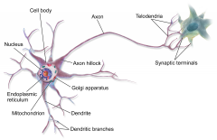
|
|
|
Resting membrane potential |
-70mV |
|
|
What helps establish the resting potential? |
Sodium pottasium pump |
|
|
What does it mean that neuron cells are polarized at resting potential? |
Negative on the inside and positive on the outside of the membrane |
|
|
Depolarization |
A change in the membrane potential from resting membrane potential of -70 to a less negative or even positive potential Causes an action potential |
|
|
Repolarization |
After depolarization occurs, repolarization returns the membrane potential back to normal |
|
|
What do voltage gated sodium channels do? |
they are located in the plasma membrane of the axon. They all sodium ions to flow down their gradient into the cell when the membrane potential changes. This allows depolarization to occur to +35mV and allows an action potential to propagate through the axon. The sodium ions flow down the interior of the axon and slightly depolarize the next section of the membrane. Then the process starts over again and continues until the action potential propagates all the way through the axon |
|
|
Threshold potential |
-50 mV cause voltage gated sodium channels to open |
|
|
What happens during repolarization? |
1.) After the depolarization due to voltage gated sodium channels, the voltage gated sodium channels inactivate 2.) Voltage gated potassium channels open slowly and stay open for a longer period of time causing hyperpolarization by allowing extra potassium to leave the cell causing the interior of the cell to be negative again (-90 mV) 3.) Sodium potassium pump returns cell to resting potential |
|
|
Myelin |
an insulating sheath that surrounds the axons of many neurons does not allow ions to exit or enter the neuron so depolarization cannot occur speeds up action potential by making it jump from one node of Ranvier to the next |
|
|
Schwann cells |
type of glial cell located in the peripheral nervous system; create the myelin sheath |
|
|
nodes of Ranvier |
periodic gaps in the myelin sheath that is concentrated with voltage gated sodium and potassium channels that allow depolarization and repolarization to occur |
|
|
Saltatory conduction |
jumping of action potential from node to node |
|
|
glial cells |
specialized, non neuronal cells that provide structural and metabolic support to neurons |
|
|
Oligodendrocytes |
type of glial cells located in the central nervous system; create myelin sheath |
|
|
Other types of glial cells |
1.) astrocytes: in central nervous system; guide neuronal development; regulate synaptic communication 2.) Microglia: in CNS; remove dead cells 3.) Ependymal cells: produce and circulate cerebrospinal fluid |
|
|
All Action Potentials are of the same size |
yuh |
|
|
Equilibrium potential for sodium and potassium |
Sodium: +50 mV Potassium: -90 mV |
|
|
Are there more sodium or potassium channels in the membrane? |
the equilibrium potential for potassium is -90 mV. Since the resting membrane potential is -70 mV, this shows that there are more potassium channels than sodium
|
|
|
Refractory period |
After an action potential occurs, there is a period of time that another one cannot occur. this is the refractory period |
|
|
2 phases of the refractory period |
1.) absolute refractory period: neuron cannot fire action potential no matter how strong depolarization is because the voltage gated sodium channels have been inactivated (they only activate again once resting membrane potential has been reached) 2.) Relative refractory period: neuron can be induced to transmit action potential because voltage gated sodium channels are active again after hyperpolarization, but requires a greater depolarization; this is because the neuron has been hyperpolarized (more negative than resting potential) by the voltage gated potassium channels that cause the neuron to have a potential of -90 mV |
|
|
Synapse |
Junction between axon terminus of one neuron and dendrites, soma, or axon of another neuron |
|
|
2 types of synapse |
1.) electrical synapse: occurs when cytoplasm of 2 cells are joined by gap junctions; action potential spreads directly from one cell to other 2.) Chemical synapse: found at the end of axons where they meet their target cell; action potential here is converted to chemical signal |
|
|
Synaptic knob |
End of the axon |
|
|
How is a an action potential transmitted across a chemical synapse? |
1.) action potential reaches the synaptic knob 2.) presynaptic membrane is depolarized and opens voltage gated calcium channels 3.) calcium moves into presynaptic cell and causes exocytosis of neurotransmitter in secretory vesicles 4.) neurotransmitter binds to receptor proteins on postsynaptic membrane 5.) ligand gated ion channels open after neurotransmitter binds to it and alters the membranes polarization 6.) if threshold voltage is reached, action potential is initiated |
|
|
Acetylcholine |
neurotransmitter in the neuromuscular junction |
|
|
Excitatory neurotransmitter |
neurotransmitter that causes depolarization in the postsynaptic membrane and allows an action potential to occur |
|
|
Inhibitory neurotransmitter |
causes hyperpolarization which drives the membrane potential farther away from the threshold needed for action potential |
|
|
Temporal summation |
presynaptic neuron fires action potentials very rapidly; the excitatory or inhibitory post synaptic potentials pile up and the additive effect will allow depolarization or hyperpolarization |
|
|
Spatial summation |
ALL EPSP and IPSP from all synapses on postsynaptic membrane are added at a given moment in time |
|
|
Efferent neurons |
carry info from CNS to appropriate glands/organs |
|
|
Afferent neurons |
carry info toward CNS |
|
|
Peripheral Nervous system |
includes axons, dendrites, and cell bodies that are not part of central nervous system Somatic: voluntary movement (if you need to or want to) of skeletal muscle; excitatory only Autonomic: involuntary processes such as digestion, circulation etc; excitatory or inhibitory |
|
|
Division of Autonomic system |
Sympathetic: fight or flight parasympathetic: rest and digest |
|
|
Central nervous system |
Brain and spinal cord |
|
|
Where are most neuronal cell bodies found? |
in the Central nervous system; they bunch together in the CNS to form nuclei |
|
|
What are cell bodies bunched together outside of the CNS called? |
Ganglia |
|
|
3 divisions of the brain |
1.) Hindbrain (rhombencephalon) 2.) Midbrain (mesencephalon) 3.) Forebrain (prosencephalon) |
|
|
Hindbrain |
1.) medulla: located below pons; area of brain that connects to spinal cord; regulates vital autonomic functions such as blood pressure and digestion; contains respiratory rhythmicity center 2.) pons: located above medulla; coordinates balance 3.) cerebellum: located behind medulla; coordinates complex movements; pons and cerebellum receive info from vestibular apparatus in ear which monitors acceleration and position due to gravity |
|
|
Midbrain |
located above pons; relays visual and auditory information; contains Reticular Activating System which controls arousal and wakefulness |
|
|
Forebrain |
1.) diencephalon: thalamus and hypothalamus; thalamus is located above hypothalamus and contains processing centers for sensory info and relays info between spinal cord cerebral cortex. hypothalamus is located above midbrain and controls emotions, hormones, and serves as the primary link between nervous and endocrine system by controlling the pituitary gland 2.) Telencephalon: Cerebrum which controls memory, planning, higher learning etc Basal Nuclei: regulation of motor movement Limbic System: has amygdala and hippocampus Amygdala controls emotions; hippocampus controls memory and learning |
|
|
Left side of brain vs Right side |
Left side: controls motor functions of right side of body; responsible for speech and logic Right side: controls motor functions of left side of body; responsible for music and creativity |
|
|
Corpus Callosum |
a thick bundle of axons that connects the two hemispheres of the brain |
|
|
Cerebrum |
the main part of the brain that consists of both hemispheres |
|
|
Cerebral cortex |
1.) frontal lobe: initiate all voluntary movement; helps in complex reasoning skills and problem solving 2.) Parietal lobe: involved in touch and taste sensations 3.) Temporal lobes: involved in sound and smell sensations; involved in short term memory, language comprehension 4.) Occipital lobe: process visual sensation |
|
|
Brocas area |
speech production |
|
|
Wernickes area |
language comprehension |
|
|
Limbic system |
located between cerebrum and diencephalon; important in emotion and memory and learning; includes amygdala and hippocampus |
|
|
Cranial nerves |
there are 12 pairs of cranial nerves that convey sensory and motor information to and from the brainstem |
|
|
Spinal nerves |
There are 31 pairs of spinal nerves that convey information to and from the spinal cord |
|
|
Vagus Nerve |
a cranial nerve; part of the parasympathetic system; |
|
|
Anatomy of Somatic System |
1.) All somatic motor neurons innervate skeletal muscle cells, use acetylcholine, and have their soma in the brain stem or front part of spinal cord
2.) All somatic sensory neurons have a long dendrite that goes from a sensory receptor to the soma which is in the dorsal root ganglion. The axon goes into the spinal cord and the first synapse is in the CNS |
|
|
Anatomy of Autonomic System |
1.) contains preganglionic and postganglionic neurons. The preganglionic neuron sends and axon to the postganglionic neuron which send an an axon to an organ or gland. 2.) All autonomic preganglionic neurons release acetylcholine; all parasymathetic postganglionic also release acetylecholine; all sympathetic postganglionic release norepinephrine |
|
|
Sympathetic vs. Parasympathetic system |
Sympathetic: preganglionic neurons are in thoracic and lumbar region of spinal cord; preganglionic axon is short; ganglia is far from target; postganglionic axon is long; usually releases norepinephrine Parasympathetic: preganglionic neurons are in brainstem and sacral spinal cord; preganglionic axon is long; ganglia is close to target; post is short; releases acetylcholine |
|
|
Four properties that sensory receptors need to communicated to CNS |
1.) stimulus modality (type of stimulus) 2.) Stimulus location 3.) Stimulus intensity 4.) Stimulus duration |
|
|
Structure of outer ear |
contains auricle or pinna and external auditory canal |
|
|
Structure of middle ear |
contains ossicles which are three small bones known as malleus, incus, and stapes |
|
|
Structure of inner ear |
contains cochlea, semicircular canals, utricle and saccule |
|
|
What do the semicircular canals, utricle and saccule do? |
they help with balance |
|
|
Eustachian tube |
passage from back of throat to middle ear that equalizes pressure on both sides of the ear drum |
|
|
Organ of Corti |
the basilar membrane and tectorial membrane; primary site at which auditory stimuli is detected |
|
|
How is sound converted to hearing in the ear? |
Sound waves travel through the outer ear and cause tympanic membrane to vibrate. This causes the bones of the middle ear to vibrate; then, the stapes, the last bone of the middle ear causes vibrations on the oval window; this causes perilymph and endolymph which are fluids in the cochlea to vibrate; this causes the basilar membrane to vibrate which is in the cochlea; this causes hair cells to move against the tectorial membrane; this causes neurotransmitters to release and stimulates dendrites of afferent neurons; nerve impulses are created |
|
|
Oval window |
membrane that divides inner ear from middle ear |
|
|
Pitch (frequency) |
distinguished by different regions of the basilar membrane vibrating; low frequency is at the apex of the cochlea while high frequency is detected near the oval window |
|
|
How can you determine the source of where a sound is coming from? |
based on the difference detected between two ears; if the sound is coming from your right, your right ear will receive the sound waves slightly sooner and more intensely than your left |
|
|
Loudness of sound |
based on amplitude of vibration; more vibration will cause more frequent action potentials |
|
|
Vestibular complex |
made of the three semicircular canals, the utricle, the saccule, and the ampullae all of these things contain endolymph and hair cells which detect rotational acceleration of the head |
|
|
Reflex Arc |
Does not involve the brain for motor output BUT signals still go to the brain |
|
|
Interneuron |
Small neuron in spinal cord that is Always inhibitory |
|
|
Monosynaptic Reflex |
Always the excitatory reflex because there is only one synapse |
|
|
Disynaptic Reflex |
Always inhibitory because there are two synapses due to an interneuron |
|
|
Cones in eye |
for color vision 3 types of cones: red, green, blue that work together to produce color Cones are located in the FOVEA mah nigga |
|
|
Rods in eye |
for black and white vision Located in periphery |
|
|
How the eye works |
No light: photoreceptors are depolarized (sodium channels are open) and either stimulate or inhibit the bipolar cell Light: photoreceptor is hyperpolarized and either inhibits or stimulates the bipolar cell |
|
|
2 types of hormones |
Peptide hormones and steroid hormones |
|
|
Peptide hormones |
1.) Poplypeptide made in the ER 2.) Hydrophillic (can travel in blood) 3.) Bind to cell surface receptors 4.) Use second messenger systems 5.) stored in vesicles 6.) Rapid response |
|
|
Steroid Hormones |
1.) Made of cholesterol 2.) Made my smooth ER 3.) Hydrophobic (travel using proteins) 4.) Bind to nuclear/cytoplasmic receptors 5.) Use transcription factors and regulate gene expression 6.) Slow response |
|
|
Pituitary |
1.) Controlled by hypothalamus 2.) Connected to hypothalamus by the Infundibullum 3.) Anterior Pituitary (controlled by hormones from the hypothalamus via a portal system) 4.) Posterior Pituitary (Controlled by Axons that extend down from hypothalamus) |
|
|
Portal System |
Two consecutive Capillary beds |
|
|
Anterior Pituitary Hormones |
FSH LH ACTH TSH Prolactin I(Nothing) GH |
|
|
Posterior Pituitary |
Oxytocin ADH (Vasopressin) |