![]()
![]()
![]()
Use LEFT and RIGHT arrow keys to navigate between flashcards;
Use UP and DOWN arrow keys to flip the card;
H to show hint;
A reads text to speech;
126 Cards in this Set
- Front
- Back
|
Nuclear Envelope- has how many membranes?
Has what, that enclose what? |
Double(2) Pores that enclose Nucleus |
|
|
Chromatin does what? |
Diffuses Threads, Containing DA |
|
|
The Nucleolus does what? |
Produces Subunits of Ribosomes |
|
|
ENDOPLASMIC RETICULUM contains what three things inparticular? |
Rough ER, Smooth ER, Ribosomes |
|
|
Describe Rough ER, and its Function. |
IT is Studded with Ribosomes, and Processes Proteins. |
|
|
Smooth ER. Lacks what? and what is its function? |
Lacks RIbosomes and Synthesizes LIPID Molecules. |
|
|
What are RIbosomes? Where are they located usually in the cell? |
Particles that out Protein Synthesis. Around the Rough ER in the ENDOPLASMIC RETICULUM. |
|
|
What is a Mitochondrion? |
An Organelle that carries out cellular Respiration, and produces ATP Molecules. |
|
|
Describe the Mitochondrion location of the cell and its shape. |
Below the SMOOTH ER, and is Bean shaped. |
|
|
What are Polyribosomes? |
String of Ribosomes that simultaneously synthesize proteins. |
|
|
Describe what the Golgi Apparatus does. |
Processes/packages/ and Secretes Modified Cell Products. (The UPS/Amazon of the Cell) |
|
|
What is the Function of the CYTOSKELETON? |
Maintains Cell shape and assits movement of cell parts. |
|
|
What are MICROTUBLES? |
Cylinders of Protein molecules |
|
|
What 4 cell parts are MICROTUBULES present in? |
Cytoplasm/Centrioles/Cilia/ and Flegella |
|
|
What are INTERMEDIATE FILAMENTS? |
Protein fibers that provide support and Strength. |
|
|
How many types of Filaments are there? What are they? |
2/ INTERMEDIATE/ACITIN |
|
|
What is an ACITIN FILAMENT? What is its Function? |
ACITIN FILAMENTS are Protein Fibers They play a role in MOVEMENT of CELLs and ORGANELLES. |
|
|
What are CENTRIOLES? What Protein in in a Centriole? |
Help with CELL DIVISION Short Cylinders of MICROTUBULES. |
|
|
What is a CENTROSOME? What Protein is in a CENTROSOME? |
ORGANIZING CENTER that contains PAIRS of CENTRIOLES. MICROTUBULES |
|
|
What is a VESICLE? |
MEMBRANE BOUND, SAC that STORES/TRANSPORTS substances. KEYS (STORAGE/TRANSPORT) |
|
|
What is a LYSOSOME? What type of ORGANELLE is it? |
A VESICLE that DIGESTS MACROMOLESCULES/CELL PARTS KEYS(remember) (Fat Boy that eats everything and digests MACROMOLECULES) |
|
|
CYTOPLASM is what? |
A SEMIFLUID MATRIX outside the NUCLEUS and CONTAINS (most) ORGANELLES of the CELL. |
|
|
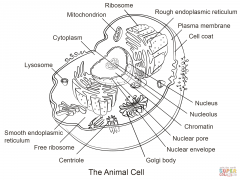
Name all 16 major cell parts and their location. |
|
|
|
What is a Tissue? |
Specialized CELLS of the SAME type that perform a common function in the Body. |
|
|
Name all 4 Major types of Tissues Families. |
CONNECTIVE/ MUSCULAR / NERVOUS / EPITHERLIAL |
|
|
DESCRIBE CONNECTIVE tissues. |
DIVERSE in Structure, and Function. All types of CONNECTIVE tissue have 3 SIMILAR COMPONENTS. (Specialized, Ground Substance, and Protein Fibers) |
|
|
How many types of CONNECTIVE FIBERS are there? What are they? |
3 WHITE COLLAGEN, RETICULAR, YELLOW ELASTIC |
|
|
How many types of CONNECTIVE tissue are there? What are they? |
6 Types FIBEROUS LOOSE DENSE CARTILAGE LYMPH BONE and a special type called ADIPOSE |
|
|
SUPPORTIVE types of connective tissue include what? |
CARTILAGE and BONE |
|
|
FIBEROUS CONNECTIVE tissue has how many forms? What are they? |
2 Forms LOOSE/ DENSE |
|
|
Both FIBEROUS and DENSE connective tissues have cells called what? (hint FIBRO-.....) |
Fibroblasts |
|
|
FIBROBLASTS are located close are away from each other? |
AWAY (seperated) |
|
|
What are FIBROBLASTS seperated by? What FIBERS do the substance contain? |
jellylike Gland substance WHITE COLLAGEN and YELLOW ELASTIC fibers |
|
|
What is the Jelly LIKE (ground/Gland) substance that seperates FIBROBLASTS? |
MATRIX |
|
|
LOOSE FIBROUS CONNECTIVE tissues include what 2 types of connective tissue? |
AREOLAR and RETICULAR |
|
|
LOOSE FIBROUS CONNECTIVE tissues support what? |
the EPITHELIUM and INTERNAL ORGANS |
|
|
What examples of Organs are LOOSE FIBROUS connective tissues found in? |
LUNGS/ ARTERIES / BLADDER |
|
|
LOOSE FIBROUS CONNECTIVE tissues allow organs to do what? |
EXPAND |
|
|
LOOSE FIBROUS CONNECTIVE TISSUES form what? Around what? |
Protective covering/ encasing internal organs, muscles, blood vessels, nerves. |
|
|
ADIPOSE TISSUE is what kind of special tissue? |
LOOSE CONNECTIVE |
|
|
(loose connective) ADIPOSE TISSUES do what?(function is?) |
Cells Enlarge and Store fat |
|
|
ADIPOSE TISSUES have and EXTRA what? |
MATRIX |
|
|
Describe (loose connective) ADIPOSE TISSUE. |
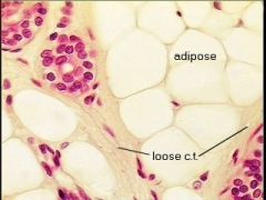
Yellow color |
|
|
What is the yellow color in loose connective ADIPOSE TISSUE? What is the black dot in between the yellow cells? |
Fat Nucleus |
|
|
ADIPOCYCTES are what? |
The Fat filled sigular cells that make up ADIPOSE TISSUE.(yellow color) |
|
|
(loose connective) ADIPOSE TISSUE/ADIPOCYTES, do what? |
ENLARGE and STORE FAT. Also has a little EXTRA MATRIX (adipocytes) that Release HORMONES called LEPTIN which REGULATES APPETITE control centers in the brain. |
|
|
(loose connective) ADIPOSE tissue primarily found where? |
Beneath the skin AROUND KIDNEYS / SURFACE of HEART. |
|
|
DENSE FIBROUS CONNECTIVE TISSUE contains what? |
Many COLLAGEN FIBERs packed together |
|
|
DENSE FIBROUS CONNECTIVE tissue has more specific functions than what other CONNECTIVE tissue? |
LOOSE CONNECTIVE TISSUE |
|
|
DENSE FIBROUS connective tissue in found where? |
TENDONS and LIGAMENTS |
|
|
TENDONS do what? |
CONNECT muscles to bone |
|
|
LIGAMENTS do what? |
CONNECT bones to other bones and joints. |
|
|
SUPPORTIVE CONNECTIVE TISSUE includes what 2 key keys parts of the body made of CONNECTIVE TISSUE? |
CARTILAGE and BONE |
|
|
SUPPORTIVE CONNECTIVE tissues have what function? |
SUPPORT / SHAPE / PRODECTION |
|
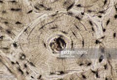
This a picture of what kind of SUPPORTIVE CONNECTIVE TISSUE? What is the large black circle in the middle of ring? The thin retangular pieces through out and around the ring is called what? The smaller round black parts are called what? |
BONE Central Canal Lamella Lacunae |
|
|
CARTILAGE is more flexible that bone and consists of what? |
small chambers called LACUNAE |
|
|
CARTILAGE lacks what? |
direct BLOOD FLOW supply |
|
|
How many types of CARTILAGE are there? What are their names? |
3 HYALINE / ELASTIC / FIBROCARTILAGE |
|
|
Which kind of CARTILAGE is MOST common? |
HYALINE |
|
|
HYALINE CARTILAGE collagen fibers are described how? |
fine |
|
|
HYALINE CARTILAGE contains what? |
fine COLLAGEN FIBERS |
|
|
HYALINE CARTILAGE's appearance is described how? |
glassy translucent |
|
|
HYALINE CARTILAGE is found where? |
NOSE / ends of BONES / RIBs / RESPIRATORY PASSAGES |
|
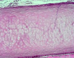
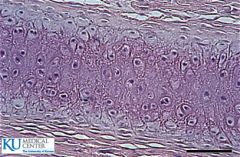
This picture is of what kind of (supportive connective tissue) CARTILAGE? |
HYALINE |
|
MORE IMAGES of HYALINE CARTILAGE (supportive connective tissue)
|
HYALINE |
|
|
ELASTIC CARTILAGE(supportive connective tissue) is found where in the body? |
OUTER EAR |
|
|
ELASTIC CARTILAGE(supportive connective tissue) more elastic what other CARTILAGE? |
HYALINE |
|
What kind of (supportive connective tissue) CARTILAGE is shown in the picture? |
ElASTIC |
|
What kind of (supportive connective tissue) CARTILAGE is this? |
HYALINE |
|
What kind of (supportive connective tissue) CARTILAGE is this? |
FIBROCARTILAGE |
|
|
FIBROCARTILAGE has STRONG COLLAGEN FIBERS contained in what? |
MATRIX |
|
|
FIBROCARTILAGE is found where in the body? |
DISKS between VERTEBRE and KNEE JOINTS |
|
|
FIBROCARTILAGE in found in the DISKS in the VERTEBRE and KNEE JOINTS because it and do what? |
WITHSTAND TENSION and PRESSURE |
|
|
This type of CONNECTIVE tissue is the MOST RIGID of the CONNECTIVE tissues. |
BONE |
|
|
BONE has a soft or hard MATRIX? |
HARD |
|
|
BONE MATRIX is made up of what? |
INORGANIC SALTS / CALCIUM SALTS / OSTEO(BLASTS) / OSTEO(CLASTS) |
|
|
What to OSTEOBLASTS and OSTEOCLASTS form? |
The MATRIX |
|
|
How many types of BONE are there? What are they? |
LONG BONE SHORT BONE FLAT BONE IRREGULAR BONE SESMOID BONE |
|
|
What are the 8 components of CONNECTIVE TISSUE? |
ADIPOSE TISSUE / GROUND SUBSTANCE / ELASTIC FIBER / BLOOD VESSELS / STEM CELL / COLLAGEN FIBER / FIBROBLASTS / RETICULAR FIBER / WHITE BLOOD CELL / |
|
|
How many types of BONE are there? What are they? |
2 COMPACT / SPONGY / |
|
|
The SHAFT and LONG BONE are made up of what kind of BONE? |
COMPACT |
|
|
COMPACT BONE consists of what? |
CYLINDRICAL STRUCTURAL UNITS CALLED OSTEONS |
|
|
What a OSTEON? |
fundamental functional unit of much compact bone that are CYLINDRICAL structures
|
|
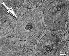
What is this picture of? |
OSTEON |
|
|
The BLANK of a OSTEON is surrounded by what? |
CENTRAL CANAL RINGS of HARD MATRIX |
|
|
BONE CELLS are located where? |
LACUNAE |
|
|
Where are LACUNAE located? |
BETWEEN RINGS of MATRIX |
|
|
SPONGY BONE surrounds what? |
MARROW CAVITY |
|
|
Describe SPONGY BONE |
OPEN / BONY / LATTICE WORK / BONY BARS / PLATES |
|
|
SPONGY BONE is designed for what? |
STRENGTH / SUPPORT |
|
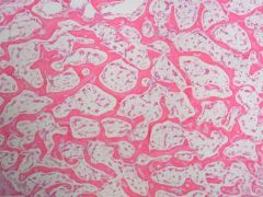
THIS is a picture of what kind of BONE? |
SPONGY BONE |
|
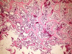
This is a picture of what kind of BONE through the Microscope? |
SPONGY BONE |
|
This a picture of what kind of BONE? |
COMPACT BONE |
|
|
There are how many types of FLUID CONNEC TIVE TISSUE? What are they? |
3 types RED BLOOD CELLS / WHITE BLOOD CELLS / LYMPH |
|
|
BLOOD consists of what elements? |
FORMED ELEMENTS / and PLASMA |
|
|
What is BLOOD's function? |
TRANSPORT of NUTRIENTS and OXYGEN to INTERSTITIAL FLUID |
|
|
What is INTERSTITIAL FLUID (or also called EXTRACELLULAR FLUID / TISSUE FLUID)? |
BATHES the body's CELLS and REMOVES CARBON DIOXIDE and other wastes. |
|
|
Besides BATHING CELLS and removing CARBON DIOXIDE, what else does blood do? |
DISTRIBUTES HEAT and plays a role in in FLUID, ION, and PH BALANCE. |
|
|
What are RED BLOOD CELLS and what do the do? |
SMALL, BICONCAVE, DISK SHAPED CELLS, WITH OUT a NUCLEI. |
|
|
What is another name for RED BLOOD CELLS? |
ERYTHROCYTES |
|
|
What give RED BLOOD CELLS its red PIGMENT? |
HEMOGLOBIN |
|
|
What is HEMOGLOBIN? |
they have 4 units and are composed of the PROTEIN GLOBIN and a IRON CONTAINING HEME. |
|
|
HEME helps do what? |
TRANSPORT OXYGEN |
|
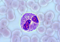
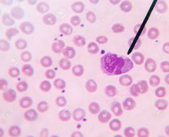
What type of cell is this? |
BLOOD CELL |
|
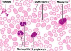
What kind of cells are in the picture? |
WHITE BLOOD CELL / RED BLOOD CELL |
|
|
WHITE BLOOD cells other wise known as LEUKOCYTES, Constist of what? |
HAVE a NUCLEUS / PROTECT BODY / |
|
|
Blood PLATELETS are not complete cells. TRUE of FALSE? |
TRUE |
|
Describe what the BLOOD PLATELETS look like in this picture. |
the small dark spots(looks like ink blots) |
|
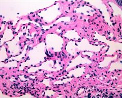
What kind of tissue is this? |
LOOSE FIBROUS CONNECTIVE |
|
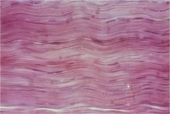
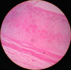
What kind of tissue is this? |
DENSE FIBROUS CONNECTIVE |
|
|
LYMPH is what type of tissue? |
FLUID CONNECTIVE |
|
|
What are the chractaristics of LYMPH? |
FAINTLY YELLOW / SURROUNDS TISSUES / CONTAINS WHITE BLOOD CELLS / |
|
|
LYMPHATIC VESSELS do what? |
Transport LYMPH |
|
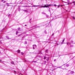
What type of TISSUE is in this picture? What specific family of tissue is this? |
LYMPH / FLUID CONNECTIVE |
|
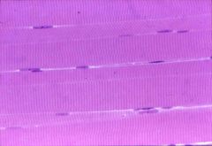
What type of tissue is this? |
SKELETAL MUSCULAR TISSUE |
|
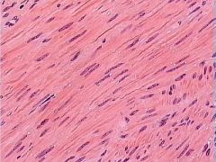
What kind of TISSUE is this? |
SMOOTH MUSCLE TISSUE |
|
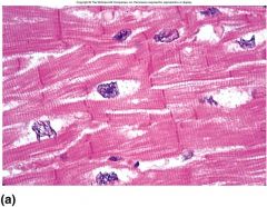
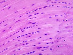
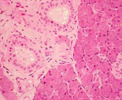
What type of tissue is this? |
CARDIAC MUSCLE TISSUE |
|
|
EPITHELIAL TISSUE is separated how many different shape of cells? What are they? |
3 SQUAMOUS / CUBOIDAL / COLUMNAR |
|
|
There are how many types of LAYERING of the EPITHELIUM? What are they? |
3 PSEUDOSTRATIFIED / STRATIFIED / SIMPLE |
|
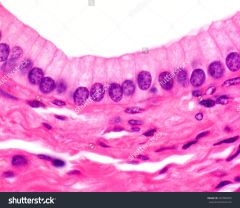
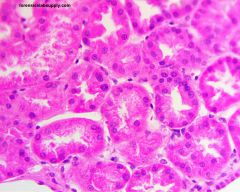
What type of EPITHELIUM is this? |
SIMPLE COLUMNAR (looks like teeth with roots) |
|
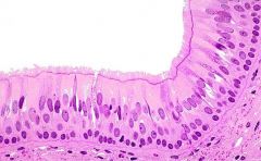
What type of EPITHELIUM is this? |
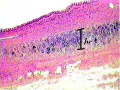
PSEUDOSTRATIFIED CILIATED COLUMNAR (COLUMNARS have COLUMNS)( Rows of cells close together.) |
|
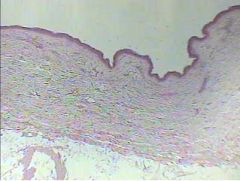
What type of EPITHELIUM is this? |
STRATIFIED SQUAMOUS (from far away it looks like a beach or shore) |
|
What Type of EPITHELIUM is this? |
Stratified SQUAMOUS (looks similar to Mitochondria or jalepanos) |
|
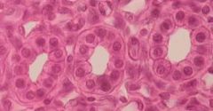
What type of EPITHELIUM is this? |
SIMPLE CUBOIDAL (donuts inside of donuts) |
|
What type of EPITHELIUM is this? |
STRATIFIED CUBOIDAL ( the (CUBES) are there and there is spacing and stretching.) The key is the CUBE or "donut" shape. |
|
|
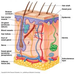
LIST all parts of HUMAN SKIN ANATOMY |
|