![]()
![]()
![]()
Use LEFT and RIGHT arrow keys to navigate between flashcards;
Use UP and DOWN arrow keys to flip the card;
H to show hint;
A reads text to speech;
174 Cards in this Set
- Front
- Back
|
Living things: |
• utilize materials and energy • grow and develop • respond to their environment • reproduce and pass their traits to offspring • evolve (change slowly) in response to their environment • Are made up of cells |
|
|
There are ___#_____ living species on Earth today |
over 10 million |
|
|
Robert Hooke |
• Created the name: Cell (like a prison cell) • 17th century man • Observed structure of cork (was looking at cellulose walls of dead plant cells) (1635-1703) overlap with Leeuwenhook |
|
|
Cells were not seen until _______ when _________. |
the 17th century, when the microscope was invented |
|
|
Antoine van Leeuwenhook |
• Built over 200 microscopes • First person to observe single living cells (1623-1732) overlap with Hooke |
|
|
Cells are _____________ of all ________ _________. |
the universal building blocks of all living tissues. • Cell Theory • every tissue is made up of cells |
|
|
Cell Theory |
• Cells are the universal building blocks of all living tissues • Matthias Schleiden (1804-1881) • Theodor Schwann (1810-1882) |
|
|
Matthias Schleiden |
Cell Theory: Cells are the universal building blocks of all living tissues |
|
|
Theodor Schwann |
Cell Theory: Cells are the universal building blocks of all living tissues |
|
|
Can life be spontaneously created? |
(spontaneous generation?) No. • Louis Pasteur (1860) – meat broth in open flask --> gone grey (bacteria grew in it, even after heating) – Plugged top (heated) --> no bacteria (either b/c no air or no things able to go in it) – gave it air, but prevented things from getting in --> no bacteria ••• living organisms that grew in the broth came from outside (spores on dust, rather than spontaneously generated within the broth) ••• Life must always come from another living thing •••All cells arise from pre-existing cells |
|
|
Louis Pasteur |
(1860) – meat broth in open flask --> gone grey (bacteria grew in it, even after heating) – Plugged top (heated) --> no bacteria (either b/c no air or no things able to go in it) – gave it air, but prevented things from getting in --> no bacteria ••• living organisms that grew in the broth came from outside (spores on dust, rather than spontaneously generated within the broth) ••• Life must always come from another living thing ••• All cells arise from pre-existing cells |
|
|
Where does the diversity of life come from? |
• All species are derived from variants of earlier species by selection of the fittest – Charles Darwin (1809-1882) – The Theory of Evolution (1838) |
|
|
The Theory of Evolution |
1838 • All species are derived from variations of earlier species by selection of the fittest • Charles Darwin (1809-1882) |
|
|
Charles Darwin |
(1809-1882) • Each island: different food source, different finches • Each finch specialized for the type of food on that island (size beaks) |
|
|
Origin of life...(when was it) – How do we know this? – What's implied by this fact? |
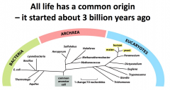
How do we know this? • DNA sequences of all organisms are related • The basic chemistry of all cells is similar • The fundamental processes are similar Implication of this fact: • What you learn about one type of cell will likely be true in other cell types ••• These are basic laws that govern cells ••• How does a single cell exist, duplicate, and become another? |
|
|
Why is it important to learn about cells? |
1. Our curiosity • What are cells made of? • How do they function? • How do new cells arise from pre-existing cells? • How can we grow from a single cell to an animal with 200 different cell types? + All diseases result from altered cell function |
|
|
All diseases result from ______________. What type of diseases? (list 3) |
• Hereditary diseases (genetic) • Environmental diseases (asthma) • Pathogenic diseases (mosquito) |
|
|
**Questions from Lecture 1: be able to answer: 1. What is the molecular composition of cells? 2. What is the internal organization of cells? 3. How are cells propagated? (reproduced) 4. How does a single cell develop to become an organism |
3. Cell division |
|
|
Cells ____________________ in isolation |
Do not exist in isolation • life in communities |
|
|
Unicellular organisms |
• use cell signaling to: – interact with their environment (cues) – are part of communities – b/c individual cells need to coordinate cell division |
|
|
cell signaling: |
• used by unicellular organisms – to interact with environment – are part of communities • used by multicellular organisms – to coordinate cell behaviors – regulate gene expression to generate different types of cells that perform different functions (ex. neurons and skeletal muscle: different fxns) |
|
|
Multicellular organisms |
• use cell signaling to coordinate cell behaviors • regulate gene expression to generate different types of cells that perform different functions – neurons and skeletal muscle: different from each other • originate from just one cell (zygote) – sexual reproduction – meiosis – inheritance – fertilization ••• Behavior and structure depend on DNA (instructions are in DNA) ••• How do these cells become different from each other? --> gene expression |
|
|
How do cells of multicellular organisms become different from each other? |
gene expression • can generate different types of cells that perform different functions |
|
|
nucleotides |
-the set of four monomers strung together in different sequences to convey information -they make the long DNA polymer chains |
|
|
protein |
-translated from RNA (DNA to RNA to protein) -structurally supports and dictates the appearance and behavior of a cell -built from amino acids -20 types of amino acids to make proteins (different combinations) |
|
|
central dogma |
transcription and translation, by which the information in genes flows into proteins: DNA → RNA → protein. Transcription is the synthesis of an RNA copy of a segment of DNA |
|
|
genome |
-the entire sequence of nucleotides in an organism's DNA |
|
|
extracellular matrix |
-dense material often made of protein fibers embedded in a polysaccharide gel -closely pack or separate the cells -fibrous proteins form gel-like structure that helps bind cells together to form tissues -- these proteins are secreted by the cells (ex. collagen) |
|
|
internal membranes |
-the membranes surrounding organelles |
|
|
transmission electron microscope |
-transmits a beam of electrons as opposed to light |
|
|
scanning electron microscope |
-scatters electrons off the surface of the sample, so is used to look at the surface detail of cells and other structures |
|
|
prokaryotes |
-spherical, rodlike, small a.k.a. bacterium -mostly single-celled organisms, but some join in chains -very diverse (live in different habitats) |
|
|
mitochondria |
-harness the energy from the oxidation of food molecules (sugars) to produce ATP -consumes O2 and releases CO2 -- called cellular respiration -organelles that generate energy in eukaryotic cells -might have evolved from aerobic bacteria that took to living inside the anaerobic ancestors of today's eukaryotic cells |
|
|
photosynthesis |
-using energy from sunlight to produce organic molecules from CO2 -in plant cells, chloroplasts: have evolved from photosynthetic bacteria |
|
|
prokaryotes |
-Traditionally classified as 2 domains: bacteria and the archaea -now bigger and more elaborate than bacteria and archaea -some independent living others multicellular -all have a nucleus |
|
|
nucleus |
-enclosed within 2 concentric membranes that form the nuclear envelope -contains molecules of DNA (in chromosomes, before a cell divides -- densely packed) |
|
|
DNA |
-found in both eukaryotic and prokaryotic cells -extremely long polymers that encode the genetic information of the organism |
|
|
chloroplasts |
-large green organelles found in cells of plants and algae -more complex than mitochondria: possess stacks of membranes containing chlorophyll -carry out photosynthesis: trap sunlight energy in chlorophyll and manufacture sugar molecules and release O2 -contain their own DNA like mitochondria -reproduce by diving in two -may have evolved from photosynthetic bacteria engulfed by a eukaryotic cell |
|
|
endoplasmic reticulum |
-maze of interconnected spaces enclosed by a membrane -most cell-membrane components and exports are made here -large areas of the ER have ribosomes attached to the cytosolic surface and are called rough ER: ribosomes actively synthesize proteins, which are delivered into ER lumen or ER membrane -smooth ER lacks ribosomes (scarce in most cells, but highly developed for functions in others): site of steroid hormone synthesis (adrenal gland) and alcohol detoxification (liver) |
|
|
golgi appartus |
-stacks of flattened membrane-enclosed sacs -modifies and packages molecules made in the ER -receives proteins and lipids from the ER and modifies and dispatches them to other parts of cell |
|
|
lysosomes |
-small, irregularly shaped organelles in which intracellular digestion occurs, releasing nutrients from ingested food particles -break down unwanted molecules for recycling or excretion from the cell -large and small molecules constantly being broken down and remade -will degrade worn-out organelles and macromolecules and particles taken into the cell by endocytosis -materials must first pass through series of compartments called endosomes before reaching lysosome |
|
|
peroxisomes |
-small, membrane-enclosed vesicles that provide a safe environment for a variety of reactions in which hydrogen peroxide is used to inactivate toxic molecules |
|
|
transport vesicles |
-move materials between one membrane-enclosed organelle and another -formed by the membranes -moderate the exchange of materials between the ER, golgi, lysosomes and outside the cell -- by pinching off from the membrane of one organelle and fusing with another |
|
|
endocytosis |
-when, at the surface of the cell, portions of the plasma membrane tuck inward and pinch off to form vesicles that carry material captured from the external medium into the cell -animal cells can engulf very large particles or even foreign cells by endocytosis think "engulfing" opposite: exocytosis |
|
|
exocytosis |
-when vesicles from inside the cell fuse with the plasma membrane and release their contents into the external medium -most of the hormones and signal molecules that allow cells to communicate with each other are secreted from cells by this process opposite: endocytosis |
|
|
cytosol |
-a concentrated aqueous gel of large and small molecules -site of many fundamental chemical reactions -site of early steps in the breakdown of nutrient molecules -site where most proteins are made by ribosomes -part of the cytoplasm that is not contained within intracellular membranes -if we were to strip the plasma membrane from a cell and remove all the membrane-enclosed organelles and nucleus, we'd get the cytosol |
|
|
cytoplasm (structure) |
-is crisscrossed by long, fine filaments, which are anchored at one end or radiate from a central site (not just a structureless soup of chemicals) -- this system is called the cytoskeleton |
|
|
cytoskeleton |
-system of protein filaments -composed of 3 major filament types: 1. actin filaments a) thinnest type b) serve as a central part of the machinery responsible for muscle contraction c) abundant in all eukaryotic cells but occur in especially large numbers inside muscle cells 2. microtubules a) thickest filaments in the cytosol b) in dividing cells, become reorganized to help pull the duplicated chromosomes in opposite directions (distribute them equally) 3. intermediate filaments b) serve to strengthen the cell -Attach together w/ other proteins to form a system of girders, ropes, and motors that gives cell mechanical strength, shape, mobility -can assemble and disappear quickly |
|
|
motor proteins |
-use the energy stored in ATP to carry organelles and proteins across the protein ropes |
|
|
eukaryotes |
-organisms whose cell has a nucleus -maybe the eukaryotic cell was a predator that fed on other cells -a eukaryotic cell carries out thousands of different chemical reactions (mutually incompatible), for different purposes (these must be segregated) ex. some reactions make glucose, others break down glucose etc. |
|
|
protozoans |
-actively motile organisms (single-celled microorganisms / eukaryotes) that can prey upon and swallow other cells -not all protozoans are predators -- they can be photosynthetic or sedentary -anatomy: sensory bristles, photoreceptors, beating cilia, stalk-like appendages, mouthparts, stinging darts, musclelike contractile bundles -can be as versatile and intricate as multicellular organisms |
|
|
2 ways procaryotic cells and eucaryotic cells isolate specific chemical reactions |
1. both can aggregate (form into a group) the different enzymes required to catalyze a particular sequence of reactions into large, multicomponent complexes 2. (many eukaryotes) confine different metabolic processes, and the proteins req. to perform them, within different membrane-enclosed compartments |
|
|
membrane-enclosed organelles |
-principal membrane-enclosed compartments of eukaryotic cells |
|
|
protein sorting |
-the transfer process, in which a unique set of proteins from each compartment have to be transferred selectively from the cytosol, where they are made, to the compartment in which they are used -transfer depends on signals built into the amino acid sequence of the proteins |
|
|
vesicles |
-small membrane-enclosed sacs -pinch off from one compartment, move through the cytosol, and fuse with another compartment in a process called vesicular transport |
|
|
vesicular transport |
-vesicles pinch off from one compartment, move through the cytosol, and fuse with another compartment |
|
|
cytosol |
-in both prokaryotes and eukaryotes (cells) -surrounded by plasma membrane -Eukaryotic cells have internal membranes |
|
|
membrane-enclosed organelles |
- eukaryotes (cells) -surrounded by cytosol, which is surrounded by plasma membrane -Eukaryotic cells have internal membranes |
|
|
nucleus |
-most prominent organelle in eukaryotes -surrounded by a double membrane called nuclear envelope -communicates with the cytosol via nuclear pores that perforate the envelope -outer nuclear membrane is continuous with the membrane of the ER |
|
|
Evolution of cells w/ membrane-enclosed organelles |
-today's eukaryotic cells probably could not have occurred without the development of internal membranes 1. nuclear membranes and membranes of the ER, golgi, endosomes...etc. are believed to have originated by invagination (turned inside out) of the plasma membrane 2. organelles communicate in and outside cell through small vesicles that bud off from the organelles and fuse with another 3. (mitochondria and chloroplasts) evolved from bacteria (prokaryote) engulfed by eukaryotes |
|
|
membrane invagination |
what?? |
|
|
endomembrane system |
-the membranes and the organelles they enclose |
|
|
glycosidic bonds |
-covalent bonds that can link monosaccharides to form larger carbohydrates |
|
|
condensation reaction |
-when a molecule of water is expelled as a bond is formed between an -OH group on one sugar and an -OH group on another -2 molecules are joined together to make a larger molecule with the loss of water -the bonds created by condensation reactions can be broken by the reverse process: hydrolysis |
|
|
hydrolysis |
-reaction involving the breaking of a bond in a molecule using water |
|
|
long-term stores of glucose held in reserve: -for animals? -for plants? |
glycogen in animals starch in plants |
|
|
cellulose |
-a polysaccharide of glucose that constitutes the chief part of the cell walls of plants chitin - found in insect exoskeletons and fungal cell walls (also a polysaccharide) |
|
|
glycoproteins |
-proteins with a sugar attached to them -many functions: immune system, communication, reproduction -attached to ER: called N-linked sugars -attached to the Golgi: called O-linked sugars -surfaces of most cells are decorated w/ sugar polymers that belong to these glycoproteins |
|
|
glycolipids |
-lipids with a sugar (carbohydrate) attached -function to maintain stability of the membrane and facilitate cellular recognition -surfaces of most cells are decorated w/ sugar polymers that belong to these glycolipids |
|
|
fatty acid + function? |
-has 2 chemically distinct regions: 1 long hydrocarbon chain (hydrophobic, unreactive), 1 carboxyl (-COOH) group (behaves like an acid) -becomes ionized in solution (hydrophilic, reactive) -function: concentrated food reserve in cells; can be broken down to produce energy (glucose) -stored in the cytoplasm (as triacylglycerol: molecules made of 3 fatty acid chains + 1 glycerol) -formation of membranes (from phospholipids) -almost always covalently linked to other molecules by carboxylic acid group -fatty acids w/ both hydrophobic and hydrophilic regions are called amphipathic -found in animal fats as well as plant oils -when a cell needs energy, fatty acid chains can be released from triacylglycerols and broken down into 2-Carbon units (glucose) |
|
|
hydrophobic |
-nonpolar molecules that repel the water molecules |
|
|
hydrophilic |
-molecules forming ionic or hydrogen bond with water molecule |
|
|
amphipathic |
-fatty acids w/ both hydrophobic and hydrophilic regions |
|
|
saturated vs. unsaturated hydrocarbons |
-saturated: has no double bonds between carbon atoms; contains highest number of hydrogens -unsaturated: one or more double bonds between carbons; double bonds create kinks, which interfere with ability to pack molecule together in a solid mass |
|
|
phospholipid |
-lipids that compose of the cell membranes -is amphipathic: both hydrophobic and hydrophilic on structure -can form lipid bilayers |
|
|
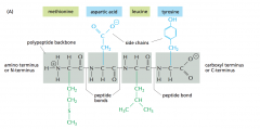
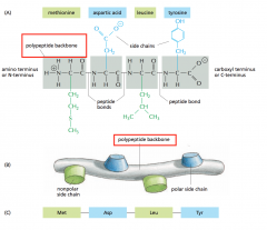
amino acids |
-molecules containing a carboxylic acid group and amino group attached to same carbon -building blocks for proteins -covalent linkage between 2 amino acids in a chain is called a peptide bond |
|
|
peptide bond |
-covalent linkage between 2 amino acids in a chain -formed by condensation reactions that link amino acids |
|
|
polypeptide always... |
has an amino (NH2) group at one end and a carboxyl (COOH) group at the other end |
|
|
nucleoside |
a molecule made of a nitrogen-containing ring compound linked to a 5-carbon sugar (ribose or deoxyribose) -nucleotide is a nucleoside sporting one or more phosphate groups attached to its sugar |
|
|
nucleotide |
- a nucleoside sporting one or more phosphate groups attached to its sugar -pyrimidines (cytosine, thymine, uracil) derive from 6-membered pyrimidine ring purine compounds (guanine, adenine) bound to a second, five-membered ring fused to the six-membered ring -carries of energy ATP -function: storage and retrieval of biological info; building blocks for construction of nucleic acids: long polymers in which nucleotide subunits are covalently linked by the formation of a phosphodiester bond betw. phosphate group on sugar of nucleotide 1 and hydroxyl group on sugar of nucleotide 2 |
|
|
nucleic acids |
-long polymers in which nucleotide subunits are covalently linked by the formation of a phosphodiester bond betw. phosphate group on sugar of nucleotide 1 and hydroxyl group on sugar of nucleotide 2 |
|
|
RNA |
ribonucleic acids -bases A, G, C, U -occurs in cells in form of a single-stranded polynucleotide chain -short-term carrier of molecular instructions |
|
|
DNA |
-hydroxyl at the 2' position of the ribose carbon ring is replaced by hydrogen -bases A,G,C,T (T similar to U) -form of double-stranded or double helix -long-term repository for hereditary info |
|
|
macromolecules |
-polymers that are constructed by covalently linking small organic molecules (monomers/subunits) into long chains |
|
|
subunits of the polymer chain are in a... |
sequence / particular order |
|
|
proteins types of proteins + functions? |
-the main building blocks from which cells are assembled -constitute most of the cell's dry mass -provide cell shape and structure -execute lots of functions (b/c of high # of different shapes) -made froma long chain of amino acids held together by covalent peptide bonds, and their amino acid chains are called polypeptide chains -in a type of protein, amino acids are in a unique order or amino acid sequence, same from one molecule of protein to the next (ex. molecule of human insulin has same amino acid sequence as every other molecule of human insulin) -some carry messages from cell to cell -some act as motors that propel organelles through cytoplasm -some function as components of tiny molecular machines with precisely calibrated moving parts -proteins in plasma membrane form channels and pumps that control the passage of nutrients and other small molecules -specialized proteins act as antibodies, toxins, hormones, antifreeze molecules, elastic fibers, or luminescence (light emission) generators Types of proteins: -enzymes = catalyze covalent bond breakage/formation -structural proteins = provide mechanical support to cells/tissues -transport proteins = carry small molecules/ions -motor proteins = generate movement in cells/tissues -storage proteins = store amino acids or ions -signal proteins = carry extracellular signals from cell to cell -receptor proteins = detect signals and transmit them to the cell's response machinery -gene regulatory proteins = bind to DNA to switch genes on/off -special-purpose proteins = highly variable/WTF -range of size: 30-10,000 amino acids (generally 50-2000) -can be globular or fibrous -can form filaments, sheets, rings, or spheres |
|
|
enzymes |
-promote intracellular chemical reactions by providing intricate molecular surfaces, outlined with particular bumps and crevices that can cradle or exclude specific molecules -catalyze covalent bond breakage or formation |
|
|
peptide bonds |
-chemical bond formed between 2 molecules when the carboxyl group of one molecule reacts with the amino group of the other molecules releasing a molecule of H2O -condensation reaction / dehydration synthesis reaction -- usually occurs between amino acids |
|
|
polypeptides |
-a linear organic polymer made of a large # of amino acid residues bonded together in a chain – forms part of a protein molecule |
|
|
polypeptide chains |
-singer linear chain of many amino acids held together by amide bonds / peptide bond (covalent bond) -each polypeptide chain consists of a backbond called the polypeptide backbone -two ends of amino acid: one sports an amino group (NH3+ or NH2) and the other a carboxyl group (COO- or COOH) -each polypeptide chain has a directionality: -the end carrying the amino group is called the amino terminus / N-terminus -the end carrying the free carboxyl group is the carboxyl terminus / C-terminus -amino acid side chains project from the backbone -long polypeptide chains are flexible -- peptide bonds in the backbone allow free rotation -- proteins can fold in many ways (shape is constrained by the # of weak noncovalent bonds between proteins in backbone and side chain) -noncovalent bonds that help proteins fold up and maintain shape are hydrogen bonds, electrostatic attractions, and van der Waals attractions -stability of folded shape depends on large # of noncovalent bonds -4th force of attraction hydrophobic interaction: determines shape of a protein -in (aq) solution, hydrophobic molecules + nonpolar side chains of some amino acids tend to be forced together to minimize their disruptive effect on the hydrogen-bonded network of surrounding water molecules -important factor governing folding of protein: distribution of polar and nonpolar amino acids (nonpolar side chains tend to cluster in the interior of the folded protein; polar side chains arrange outside the folded protein and can form hydrogen bonds with water and other polar molecules -if polar molecules are somehow buried inside the folded protein, they are usually hydrogen-bonded to other polar amino acids or to the backbone |
|
|
polypeptide backbone what about the ends of the amino acid? |
-formed from a repeating sequence of the core atoms (-N-C-C-) found in every amino acid -two ends of amino acid: one sports an amino group (NH3+ or NH2) and the other a carboxyl group (COO- or COOH) -each polypeptide chain has a directionality: -the end carrying the amino group is called the amino terminus / N-terminus -the end carrying the free carboxyl group is the carboxyl terminus / C-terminus -amino acid side chains project from the backbone: are part of the amino acid that is not involved in forming peptide bonds; side chains give each amino acid its unique properties (nonpolar and hydrophobic: "water fear," some –/+ charged, and some chemically reactive) |
|
|
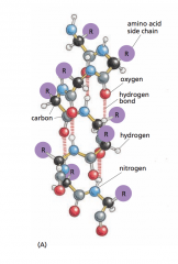
amino acid side chains |
-project from the backbone: are part of the amino acid that is not involved in forming peptide bonds; side chains give each amino acid its unique properties (nonpolar and hydrophobic: "water fear," some –/+ charged, and some chemically reactive) |
|
|
How many different types of amino acids are there in proteins? Chart of polar amino acids and nonpolar amino acids: can you guess polarity or charge? Arginine, Aspartic acid, lysine, glutamic acid, asparagine, histidine, glutamine, serine, threonine, alanine, glycine, valine, tyrosine, leucine, isolucine, proline, phenylalanine, methionine, tryptophan, cysteine |
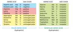
Answer: 20 |
|
|
hydrophobic interaction |
-4th force of attraction -determines shape of a protein -in (aq) solution, hydrophobic molecules + nonpolar side chains of some amino acids tend to be forced together to minimize their disruptive effect on the hydrogen-bonded network of surrounding water molecules -important factor governing folding of protein: distribution of polar and nonpolar amino acids (nonpolar side chains tend to cluster in the interior of the folded protein; polar side chains arrange outside the folded protein and can form hydrogen bonds with water and other polar molecules -if polar molecules are somehow buried inside the folded protein, they are usually hydrogen-bonded to other polar amino acids or to the backbone |
|
|
conformation |
-the final folded structure of the protein, adopted by any polypeptide chain -determined by energetic considerations: • A protein generally folds into the shape in which its free energy (G) is minimized (releases heat and increases disorder of the universe -chaperone proteins assist with protein folding |
|
|
unfolded / denatured protein process |
-by treatment with solvents that disrupt the noncovalent interactions holding the folded chain together -converts the protein into a flexible polypeptide chain that has lost its natural shape -sometimes, when the denaturing solvent is removed, the protein often refolds spontaneously into its original conformation -- called renaturation |
|
|
renaturation |
-when a protein refolds spontaneously into its original conformation once the denaturing solvent is removed -shows that the amino acid sequence contains all the info necessary to specify the 3-D shape of a protein |
|
|
misfolded proteins |
-when proteins fold incorrectly, sometimes they form aggregates that can damage cells and tissues -contributes to neurodegenerative disorders: Alzheimer's, Huntington's, Scrapie, "mad cow" -misfolded proteins = prions...spread from cell to cell -prions infectious b/c can spread via food, blood etc. |
|
|
chaperone proteins |
-proteins that assist protein folding in a cell (efficiency) -bind to partly folded chains and help them to fold along most energetic pathway -can form "isolation chambers" where single polypeptide chains can fold without risk of forming aggregates |
|
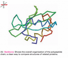
The backbone model shows... |
-the overall organization of the polypeptide chain and provides a straightforward way to compare the structures of related proteins |
|
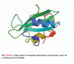
The ribbon model shows... |
-the polypeptide backbone in a way that emphasizes its various folds |
|
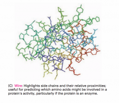
The wire model shows... |
-the positions of all the amino acid side chains -useful for predicting which amino acids might be involved in the protein's activity |
|
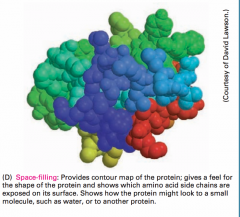
The space-filling model shows... |
-a contour (outline) map of the protein surface, which reveals which amino acids are exposed on the surface and shows how the protein might look to a small molecule such as water or to another macromolecule in the cell |
|
|
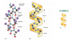
α helix |
-folded structure first found in the protein α-keratin, abundant in skin and derivatives: hair, nails, and horns -these folding patterns result from hydrogen bonds that form between the N-H and C=O groups in the polypeptide backbone -the protein adopts a regular, repeating form -short regions of a-helix are especially abundant in protein in cell membranes (transport proteins and receptors) |
|
|
ß sheet |

-folded structure first found in the protein fibroin; a major constituent of silk -formed when hydrogen bonds form between segments of a polypeptide chain that lie side by side -when the neighboring segments run in the same orientation (N-terminus to C-terminus) the structure is a parallel ß sheet -when the neighboring segments run in opposite directions, the structure is an antiparallel ß sheet -both types of ß sheet produce a very rigid, pleated structure, and they form the core of many proteins -these folding patterns result from hydrogen bonds that form between the N-H and C=O groups in the polypeptide backbone -give silk fibers tensile strength -permit the formation of amyloid fibers: insoluble protein aggregates that include those associated with neurogenerative disorders (Alzheimers, etc.) -the protein adopts a regular, repeating form |
|
|
helix |
-a regular structure that resembles a spiral staircase -generated by placing many similar subunits next to one another -generated when a single polypeptide chain turns around itself to form a structurally rigid cylinder -a hydrogen bond is made between every 4th amino acid, linking the C=O of one peptide bond to the N-H of another --> creates a regular right-handed helix with a complete turn every 3.6 amino acids -can be right-handed or left-handed -handedness is not affected by turning helix upside down, but it is reversed if helix is reflected in a mirror |
|
|
polypeptide backbone |
-is hydrophilic -is hydrogen-bonded to itself in the α helix that is composed of mainly amino acids with nonpolar side chains -is shielded from the hydrophobic lipid environment of the membrane by its protruding nonpolar side chains |
|
|
coiled-coil |
-forms when 2 or 3 α helices wrap around one another to form a particularly stable structure -forms when the α helices have most of their nonpolar (hydrophobic) side chains on one side, so that they can twist around each other with these side chains facing inward (minimizes their contact with the aqueous cytosol) -long, rodlike coiled-coils form the structural framework for many elongated proteins |
|
|
amyloid fibers |
-insoluble protein aggregates that include those associated with neurogenerative disorders (Alzheimers, etc.) -these structures are stabilized by ß sheets and are stacked tightly, with their amino acid side chains interdigitated (like zipper teeth) -we tend to associate amyloid fibers with disease -- but many organisms take advantage of these stable structures (bacteria) |
|
|
primary structure |
-the amino acid sequence is the protein's primary structure |
|
|
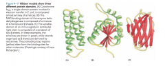
secondary structure |
-the α helices and ß sheets that form certain segments of the polypeptide chain (folds) |
|
|
tertiary structure |
-the full 3-D conformation formed by an entire polypeptide chain (including α helices, ß sheets, random coils, and other loops and folds between N and C termini) |
|
|
quarternary structure |
-if the protein molecule is formed as a complex of more than one polypeptide chain |
|
|
protein domain |
-any segment of a polypeptide chain that can fold independently into a compact, stable structure -contains 40-350 amino acids folded into α helices, ß sheets, and elements of the secondary structure -is the modular unit from which many larger proteins are constructed -different domains of a protein are associated with different functions (ex. catabolite activator protein (CAP) has 2 domains: small domain binds to DNA, large domain binds cyclic AMP |
|
|
intrinsically disordered sequence |
-regions of polypeptide chain lacking any definite structure, which continually bend and flex due to thermal buffeting -short stretches linking domains in otherwise highly ordered proteins -remain undetected for many years -unfolded structure makes them prime targets for the proteolytic enzymes released when cells are fractionated to isolate their molecular components -important functions of unstructured proteins: • able to flex and bend -- can wrap around one or more target proteins like a scarf, binding with both high specificity and low affinity • can help scaffold proteins bring together proteins in an intracellular signaling pathway, facilitating interactions • give some proteins ability to form rubberlike fibers -- allows our tendons and skin to recoil after being stretched • ideal substrates for addition of chemical groups that control the way many proteins behave |
|
|
why there are only a small fraction of potential polypeptide sequences in cells? What are the constraints? |
because many biological functions depend on proteins with stable, well-defined 3-D conformations -- this restricts the list of possible polypeptide sequences because functional proteins must be "well-behaved" and not engage in unwanted associations with other proteins in the cell (ex. forming insoluble protein aggregates) -many of these potential proteins would have been eliminated by natural selection -present-day proteins have evolved to guarantee that the polypeptide will adopt a stable conformation (exact chemical properties to perform a specific function) |
|
|
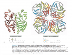
protein families |
-each family member has an amino acid sequence and a 3-D conformation that closely resembles those of the other family members ex) polypeptide chains of digestive enzyme proteins and blood clotting proteases have portions of identical amino acid sequences |
|
|
binding site |
-any region on a protein's surface that interacts with another molecule through sets of noncovalent bonds -if a binding site recognizes the surface of a second protein, the tight binding of two folded polypeptide chains at this site will create a larger protein, whose quarternary structure has a precisely defined geometry • each polypeptide chain in this protein is called a subunit |
|
|
subunit |
-when a binding site recognizes the surface of another protein...at this site, the tight binding of two folded polypeptide chains will create a larger protein, whose quarternary structure has a precisely defined geometry • each polypeptide chain in this protein is called a subunit -when 2 identical, folded polypeptide chains form a symmetrical complex of 2 protein subunits: called a dimer |
|
|
dimer |
-when 2 identical, folded polypeptide chains form a symmetrical complex of 2 protein subunits |
|
|
symmetrical protein complexes are formed from... |
multiple copies of the protein subunit |
|
|
proteins formed from 2 different subunits (multiple subunits) -- give the obvious example |
-hemoglobin: contains 2 copies of α-globin (green) and 2 copies of ß-globin (blue), and each of the 4 polypeptide chains has a heme molecule (red) |
|
|
formation of chains of identical protein molecules |
- when the binding site on one protein molecule is complementary to another region on the surface of another protein molecule of the same type -this is why the molecules will often be arranged in a helix (can be extended in either direction) • this type of arrangement can produce an extended protein filament -identical proteins associate to form tubes -- microtubules of cytoskeleton -identical proteins associate to form shells like the protein coats of viruses |
|
|
formation of viruses and ribosomes |
-built from a mixture of one or more types of protein plus RNA or DNA molecules -structures can be isolated in pure form and dissociated into their constituent macromolecules -- shows that all the info needed for assembly of the structure is contained in the macromolecules themselves -much of the cell is self-organizing -- if req. proteins are in the right amounts, appropriate structures will form |
|
|
globular proteins |
-where the polypeptide chain folds up into a compact shape like a ball with an irregular surface (enzymes, w/ multiple subunits, have a quaternary structure with an overall rounded shape |
|
|
fibrous proteins |
-simple, elongated 3-D structure -span a long distance ex) α-kerratin -- α-helix (keratin filaments are very stable -- hair, horns, nails) • α-keratin is a dimer of 2 identical subunits -- the long α helices of each subunit form a coiled-coil • coiled-coil regions are capped at ends by globular domains containing binding sites that allow them to assemble into ropelike intermediate filaments: component of the cytoskeleton that gives cells mechanical strength |
|
|
intermediate filaments |
-component of the cytoskeleton that gives cells mechanical strength ex) α-kerratin -- α-helix (keratin filaments are very stable -- hair, horns, nails) • α-keratin is a dimer of 2 identical subunits -- the long α helices of each subunit form a coiled-coil • coiled-coil regions are capped at ends by globular domains containing binding sites that allow them to assemble into ropelike intermediate filaments |
|
|
collagen |
-most abundant fibrous extracellular proteins in animal tissues -form gel-like extracellular matrix that helps bind cells together to form tissues -consists of 3 long polypeptide chains, each containing nonpolar amino acid: glycine -generally bind side-by-side / end-to-end -- to create long overlapping arrays called collagen fibrils: very strong and help hold tissues together |
|
|
collagen fibrils |
-long overlapping arrays created to bind molecules to one another -very strong and help hold tissues together |
|
|
elastin |
-formed from loose/unstructured polypeptide chains that are covalently cross-linked into a rubberlike elastic meshwork -enable elastic fibers: skin, arteries, lung, etc. to stretch and recoil without tearing (elasticity is due to ability of protein molecules to uncoil reversibly when they stretch) |
|
|
extracellular proteins are often stabilized by... |
-covalent cross-linkages b/c when protein molecules are attached to the outside of a cell's plasma membrane or secreted as part of the extracellular matrix, they are exposed to extracellular conditions -linkages that can either tie together 2 amino acids in the same polypeptide chains or join together many polypeptide chains in a large protein complex (collagen fibrils and elastic fibers) |
|
|
covalent cross-links |
-linkages that can either tie together 2 amino acids in the same polypeptide chains or join together many polypeptide chains in a large protein complex (collagen fibrils and elastic fibers) -most common covalent cross-links in proteins: sulfur-sulfur bonds / disulfide bonds (S-S bonds) |
|
|
disulfide bonds |
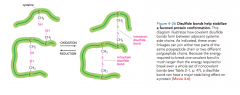
-sulfur-sulfur bonds (S-S bonds) - most common covalent cross-links in proteins -formed before a protein is secreted by an enzyme in ER that links together 2 -SH groups from cysteine side chains adjacent in the folded protein -do not change a protein's conformation, but act as an "atomic staple" to reinforce the protein's most favored conformation -not formed in the cytosol -- proteins don't require this type of structural reinforcement in cytosol |
|
|
translation |
-the conversion of the information in RNA into protein -translation of mRNA into amino acid sequence of protein must follow the genetic code |
|
|
transcription |
-segment of DNA is copied into RNA (mainly mRNA) by enzyme RNA polymerase -mitochondria have their own transciption and protein synthesis |
|
|
start codon |
-AUG -modified methionine |
|
|
genetic code |
-relationship between the sequence of bases in nucleic acid and the order of amino acids in the poypeptide synthesized from it -sequence of 3 nucleic acid bases is a codon for one amino acid |
|
|
codon |
-group of 3 consecutive nucleotides in RNA -specifies one amino acid |
|
|
reading frames |
-way of dividing the sequence of nucleotides in a nucleic acid (DNA or RNA) molecule into a set of consecutive, non-overlapping triplets (codons, during translation) |
|
|
tRNA |
-transfer RNAs -adaptor molecules composed of RNA that can recognize and bind to a codon at one site on their surface and to an amino acid at another side -recognition and attachment of the correct amino acid depend on the enzymes called aminoacyl-tRNA synthetases, which covalently couple each amino acid to its appropriate set of tRNA molecules |
|
|
aminoacyl-tRNA synthetases and how does it work? |
-an enzyme that attaches the appropriate amino acid onto its tRNA -covalently couple each amino acid to its appropriate set of tRNA molecules -there is a different synthetase enzyme for each amino acid -20 synthetases in all amino acids -one attaches glycine to all tRNAs that recognize codons for glycine / another attaches phenylalanine to all tRNAs that recognize codons for phenylalanine -- and so on... -specific nucleotides in both the anticodon and the amino-acid-accepting arm allow the correct tRNA to be recognized by the synthetase enzyme |
|
|
anticodon |
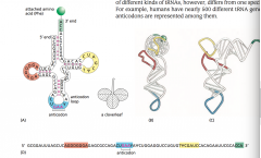
-a set of 3 consecutive nucleotides that bind the complementary codon in an mRNA molecule through base-pairing -forms at one of the 2 regions of unpaired nucleotides of the L-shaped molecule -crucial for tRNA in protein synthesis |
|
|
single-stranded region at the 3' end of the mRNA molecule: |

-site where the amino acid that matches the codon is attached to the tRNA -one of the two regions of unpaired of unpaired nucleotides situated at either end of the L-shaped molecule -- critical to the function of tRNA in protein synthesis |
|
|
genetic code is ____________ this __________ implies that ________________ or that _________________ |
redundant -implies there is more than one tRNA for many of the amino acids or that some tRNA molecules can base-pair with more than one codon -both situations occur |
|
|
Can tRNAs be constructed so that they can tolerate a mismatch (or wobble) at the third position? explain: what does this say about alternative codons? what's the advantage of wobble base-pairing? do all species have the same # of different kinds of tRNA molecules? |
-some tRNAs are constructed so that they require accurate base-pairing only at the first 2 positions of the codon and can tolerate a mismatch / wobble at the 3rd position -this wobble base-pairing explains why so many of the alternative codons for an amino acid differ only in their third nucleotide -wobble base pairing makes it possible to fit the 20 amino acids to their 61 codons with as few as 31 kinds of tRNA molecules (so there aren't so many needed) -No. The exact # of different kinds of tRNAs differs from one species to the next |
|
|
what does it mean when a tRNA molecule becomes charged? |
-it means the tRNA molecule is linked to the one amino acid in 20 that is its right partner |
|
|
True or False: you don't need both synthetases and tRNAS to allow each codon in the mRNA molecule to associated w/ its proper amino acid |
False: both the synthetases and tRNAs are equally important b/c it it is the combined action of them to allow it to occur |
|
|
hydrolysis vs. condensation |
-Condensation is a chemical process by which 2 molecules are joined together to make a larger, more complex, molecule, with the loss of water...synthesis of macromolecules (carbs, proteins, etc.) -Hydrolysis is the opposite to condensation. A large molecule is split into smaller sections by breaking a bond, adding -H to one section and -OH to the other.The products are simpler substances. Since it involves the addition of water, this explains why it is called hydrolysis, meaning splitting by water. |
|
|
the recognition of a codon by the anticodon on a tRNA molecule depends on: (a similar process, which is...) |
-the same type of complementary base-pairing used in DNA replication and transcription |
|
|
ribosome what are the functions of the individual parts? how does the ribosome choreograph all the movements required for translation? / how is an amino acid added to a growing peptide chain? |
-large complex made from >50 different ribosomal proteins and ribosomal RNAs (rRNAs) -required for accurate / rapid translation of mRNA into protein -captures complementary tRNA molecules, holds them in position, and covalently links the amino acids that they carry so as to form a protein chain -small subunit matches tRNAs to codons of mRNA -large sunit catalyzes the formation of the peptide bonds that covalently link the amino acids together into a polypeptide chain how does the ribosome choreograph... -each ribosome contains 1 binding site for a mRNA molecule and 3 binding sites for tRNA molecules (A-site, P-site, E-site) to add an amino acid to a growing peptide chain: 1) the appropriate charged tRNA enters the A-site by base-pairing with the complementary codon on the mRNA molecule 2) the amino acid is then linked to the peptide chain held by the tRNA in the neighboring P-site 3) the ribosome shifts, and the spent tRNA is moved to the E-site before being ejected 4) the cycle of reactions is repeated whenever an amino acid is added to the polypeptide chain (the chain grows from its amino to its carboxyl end 5) once a stop codon is encountered, the addition of amino acids ceases |
|
|
rRNA |
ribosomal RNA -the RNA component of the ribosome -for protein synthesis in living things -folded into highly compact, exact 3-D structures that form the core of the ribosome |
|
|
Process of protein synthesis (mRNA, tRNA, etc.) a.k.a. translation |
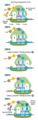
1. the 2 subunits of the ribosome come together on an mRNA molecule (usually at beginning = 5' end) 2. mRNA is pulled through the ribosome (like a long piece of tape) 3. As the mRNA moves through the ribosome, the ribosome translates the nucleotide sequence into an amino acid sequence, one codon at a time, using the tRNAs as adaptors -so each amino acid is added in the correct sequence to the end of the growing polypeptide chain 4. the 2 subunits of the ribosome separate when synthesis of the protein is finished |
|
|
which operates faster, a ribosome of a eukaryotic cell or a ribosome in a bacterial cell? bonus: in 1 second, 1 ribosome in a eukaryotic cell adds ______ amino acids in 1 second, 1 ribosome in a bacterial cell adds _______ amino acids |
-bacterial ribosomes operate faster -eucaryotic cell: 2 amino acids / second -bacterial ribosome: 20 amino acids / second |
|
|
what is responsible for the overall structure of the RNA (and its ability to choreograph protein synthesis)? |
rRNA, which constitutes 2/3 of the ribosome |
|
|
ribosomal proteins function? (btw, not talking about rRNAs) |
-located on the surface of the ribosome -fill the gaps and crevices of the folded RNA -role: to fold and stabilize the RNA core, while permitting the changes in rRNA conformation necessary for RNA to catalyze efficient protein synthesis |
|
|
what primarily forms the 3 tRNA binding sites (sites are: ____, ___, and _____) |
A, P, and E sites -formed primarily by the rRNAs |
|
|
what forms the catalytic site for peptide bond formation? |
-the 23S RNA of the large subunit (ribosome) |
|
|
what is the structure and function of the catalytic site in this RNA-based peptidyl transferase (on the ribosome) |
-highly structured pocket -precisely orients the 2 reactants: the elongating peptide and the charged tRNA -increases the probability of a productive reaction |
|
|
ribozymes |
-RNA molecules that possess catalytic activity (enzymes) |
|
|
describe the beginning of the translation of mRNA: (protein synthesis) -- eukaryotes hint: (selecting a start codon...) |
1. codon AUG, and a special tRNA is required to initiate translation: initiator tRNA, which carries the amino acid methionine (formylmethionine in bacteria) -so that newly made proteins all have methionine as the 1st amino acid at the N-terminal end (synthesized first) -methionine is generally removed later by a protease -the initiator tRNA is not the same as the tRNA that normally carries methionine 2) initiator tRNA is first loaded into a small ribosomal subunit w/ additional proteins called translation initiation factors -only the charged initiator tRNA is capable of binding tightly to the P-site of the small ribosomal subunit b) the loaded ribosomal subunit binds to the 5' end of an mRNA molecule (signaled by the cap on the eukaryotic mRNA) 3) the loaded ribosomal subunit moves forward (5' to 3') along the mRNA -- searching for the first AUG) 4) When the AUG is encountered, several initiation factors dissociate from the small ribosomal subunit to make way for the large ribosomal subunit to assemble and complete the ribosome (protein synthesis is ready to begin with the addition of the next charged tRNA to the A-site b/c the initiator tRNA is bound to the P-site) |
|
|
translation initiation factors |
Initiation factors are proteins that bind to the small subunit of the ribosome during the initiation of translation, a part of protein biosynthesis -the only charged initiator tRNA is capable of binding tightly to the P-site of the small ribosomal subunit |
|
|
mechanism for selecting a start codon in bacteria explain how it is different from that of a eukaryote |
-bacterial (prokaryotic) mRNAs have no 5' caps to tell the ribosome where to begin searching for the start of translation -bacterial mRNAs contain specific ribosome-binding sequences (up to 6 nucleotides long) located a few nucleotides upstream of the AUGs at which translation is to begin -prokaryotic ribosome can bind directly to a start codon in the mRNA as long as a ribosome-binding site precedes it by several nucleotides • why prokaryotic mRNAs are polycistronic: they encode several different proteins and each is translated from the same mRNA • in contrast, a eukaryotic mRNA generally carries the info for a single protein |
|
|
describe the end of the phases of translation (protein synthesis)
|
-same for both prokaryotes and eukaryotes 1. signaled by the presence of a particular codon called a stop codon (UAA, UAG, and UGA) 2. UAA, UAG, and UGA - are not recognized by a tRNA -do not specify an amino acid -signal the ribosome to stop translation 3. proteins called release factors bind to any stop codon that reaches the A-site on the ribosome -this binding alters the activity of the peptidyl transferase in the ribosome 4. causes the ribosome to catalyze the addition of a water molecule instead of an amino acid to the peptidyl-tRNA -this reaction frees the carboxyl end of the polypeptide chain from its attachment to a tRNA molecule -because the carboxyl end is the only attachment that holds the growing polypeptide to the ribosome --> the protein chain is released into the cytosol 5) the ribosome releases the mRNA and dissociates into its 2 separate subunits -these 2 subunits can assemble on another mRNA molecule to begin a new round of protein synthesis -if the mRNA is being translated efficiently, a new ribosome hops onto the 5' end of the mRNA as soon as the preceding ribosome is finished translating enough mRNA to move out of the way --> these mRNAs would be considered: polyribosomes / polysomes: large cytoplasmic assemblies made up of several ribosomes spaced as close as 80 nucleotides apart along a single mRNA molecule |
|
|
describe the newly-created proteins as they are spun out of the ribosome |
-many proteins fold into their 3-D shape spontaneously after being synthesized and released by the ribosome -many proteins require molecular chaperones to help them fold correctly in the cell -chaperones use ATP hydrolysis to bind and release new proteins until they are folded • prevents the creation of aggregates / misfolds |
|
|
polyribosomes / polysomes |
-large cytoplasmic assemblies made up of several ribosomes spaced as close as 80 nucleotides apart along a single mRNA molecule |
|
|
the biological properties of a protein molecule depend on... |
...its physical interaction with other molecules |
|
|
specificity (binding of protein to other molecules) |
-each protein molecule can bind to just one or a few molecules out of the many thousands of different molecules it encounters |
|
|
ligand |
-any substance (ion, small molecule, macromolecule) that is bound by a protein is a ligand for that protein |
|
|
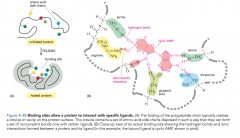
how is a protein able to selectively bind with high affinity to a ligand? |
due to the formation of a set of weak, noncovalent bonds -hydrogen bonds, electrostatic attractions, van der waals attractions, hydrophobic attractions -an effective interaction requires many weak bonds to simultaneously form • only possible if the surface contours of the ligand fit closely with the protein fyi: our bodies produce antibodies that can bind to different molecules |
|
|
what happens when molecules have poorly matching surfaces? |
- few noncovalent bonds are formed -the molecules dissociate as they come together -prevents incorrect / unwanted formations |
|
|
what happens if too many noncovalent bonds are formed between molecules? when would this occur? |
the association can persist for a long time -occurs whenever a biological function requires molecules to remain tightly associated for a long time ex) a group of macromolecules come together to form a ribosome |
|
|
binding site |
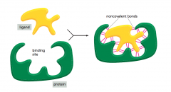
-the region of a protein that associates with a ligand -consists of a cavity in the protein surface formed by a particular arrangement of amino acids -the amino acids can belong to widely separated regions of the polypeptide chain brought together when the protein folds |
|
|
what can a protein do in order to interact with specific ligands? |
|
|
|
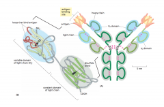
antibodies |
a.k.a. immunoglobins -proteins produced by the immune system in response to foreign molecules -each antibody binds to a particular target molecule -the target the antibody binds to: called antigen (with specificity) -Y-shaped molecules w/ 2 binding sites complementary to parts of the surface of the antigen -formed from loops of polypeptide chain that protrude from the ends of a pair of side-by-side protein domains -diversity of antigen-binding sites just by changing length / amino acid sequence of the loops |