![]()
![]()
![]()
Use LEFT and RIGHT arrow keys to navigate between flashcards;
Use UP and DOWN arrow keys to flip the card;
H to show hint;
A reads text to speech;
56 Cards in this Set
- Front
- Back
|
Reflex |
a rapid, automatic response that occurs to a specific stimulus. Purpose: - Protects the body fromdamage such as from excessive light or heat • Coordinates skeletal musclemovements • Regulates ANS such as heart rate and moving digested substances through the digestivetract |
|
|
Reflex Arch |
thepathway followed by nerve impulses through a reflex (5major parts) Receptor --> Sensory Neuron --> Interneuron --> Motor Neuron --> Effector |
|
|
(1) Receptor |
the structure thatreceives the stimulus; it's a modified dendriteof a neuron |
|
|
(2) Sensory Neuron |
the unipolar neuron that transmits the nerve impulse fromthe receptor to the CNS |
|
|
(3) Interneuron |
the short neuron in thegray matter of thespinal cord that connects the sensory input to the motor output •monosynaptic reflex arc - lacks aninterneuron. It has only one synapsebetween the sensory neuron and the motor neuron • polysynaptic reflex arc - has aninterneuron. |
|
|
(4) Motor Neuron |
the multipolar neuron that transmits the nerve impulse fromthe CNS to the effector |
|
|
Effector |
the muscle or gland at the end of the reflex pathway to which themotor neuron is connected |
|
|
Central Canal |
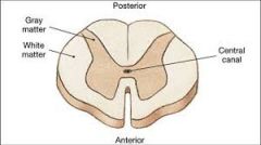
also known as ependymal canal, is the CSF filled space that runs longitudinally through the length of the entire spinal cord. |
|
|
Ependymal Cells |
neuronal cells that form the epithelial lining of the ventricles and the central canal of the spinal cord. Ependymal cells are also the epithelial layer that surrounds the choroid plexus. These cells have tight junctions connecting them together, making it difficult for large cells to pass through (Part of the BBB) |
|
|
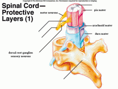
Identify Meninges on a human spinal cord 1) Dura Mater 2) Arachnoid 3) Pia Mater |
|
|
|
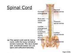
Indentify gross structures on spinal cord (posterior view) 1) cervical enlargement 2) lumbar enlargement 3) conus medullaris 4) cauda equina |
|
|
|
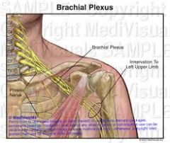
Brachial Plexus |
|
|
|
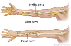
Ulnar nerve Median nerve Radial nerve |
|
|
|
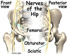
Sciatic Nerve & Femoral Nerve |
|
|
|
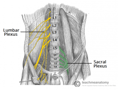
Lumbar & Sacral plexus |
|
|
|
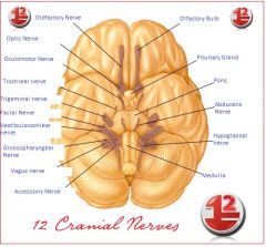
12 Cranial Nerves |
|
|
|
Cranial nerve I |
Olfactory: sense of smell |
|
|
Cranial nerve II |
Optic: provides visual interpretation from the retina |
|
|
Cranial nerve III |
Oculomotor: motor nerve that lifts eyelid up, rotates eyeball up, constricts pupils |
|
|
Cranial Nerve IV |
Trochlear: innervates eye muscles and turns the eye
|
|
|
Cranial Nerve V |
Trigeminal: provides sensory and motor functions for nose, eyes, tongue and teeth
|
|
|
Cranial Nerve VI |
Abducens: motor nerve connected to the pons that turns the eye laterally
|
|
|
Cranial Nerve VII |
Facial: located just over the brain stem; responsible for different facial expressions and senses touch on face and tongue
|
|
|
Cranial Nerve VIII |
Vestibulocochlear (acoustic or auditory):
motor nerve that provides information on balance of head and sense of sound or hearing. |
|
|
Cranial Nerve IX |
Glossopharyngeal: sensory nerve that carries info regarding temperature, pressure and other factors from pharynx and parts of the tongue and palate
- involved with taste buds and salivary glands and some motor functions with swallowing |
|
|
Cranial Nerve X |
Vagus: mixed nerve that carries sensory and motor information involving the pharynx, larynx, esophagus, trachea, bronchi, and parts of the heart and palate.
- Constricts muscles in each of the areas listed above. - Aides in taste |
|
|
Cranial Nerve XI |
Accessory: motor nerve that supplies information about spinal cord, trapezius, and other areas
- provides muscle movement to shoulders and neck |
|
|
Cranial Nerve XII |
Hypoglossal: motor nerve involving the tongue |
|
|
Name a function for each of the regions of the cerebrum: Gustatory Cortex |
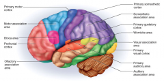
perception of taste |
|
|
Name a function for each of the regions of the cerebrum: Visual Cortex |

visual perception of images |
|
|
Name a function for each of the regions of the cerebrum: Primary Motor Cortex |

controls voluntary muscle movements throughout the body, including those of the hands, feet, arms, legs, face and tongue. |
|
|
Name a function for each of the regions of the cerebrum: Motor Speech (Broca's area) |

located only in the left hemisphere - provides motor movements involving speech production. - damage to this area allows people to understand information without being able to communicate back |
|
|
Name a function for each of the regions of the cerebrum: Frontal eye field |

controls visual attention and eye movements |
|
|
Name a function for each of the regions of the cerebrum: Somatic sensory association area |

Makes sense of sensory input; stores memories of different senses. So if blindfolded this region could interpret what sandpaper feels like and what it feels like to place your hands in a pair of scissors. |
|
|
Name a function for each of the regions of the cerebrum: Sensory speech area (Wernicke's area) |

Only on the left hemisphere; understands speech |
|
|
Name a function for each of the regions of the cerebrum: Auditory association area |
Allows you to recognize a particular sound as speech, music or noise |
|
|
Function of: Cerebrum |
The cerebrum or cortex is the largest part of the human brain, associated with higher brain function such as thought and action. |
|
|
Function of: Cerebellum |
receives information from the sensory systems, the spinal cord, and other parts of the brain and then regulates motor movements. The cerebellum coordinates voluntary movements such as posture, balance, coordination, and speech, resulting in smooth and balanced muscular activity. |
|
|
Functions of: Brain Stem |
regulation of many Autonomic systems involving heart rate, breathing, sleeping, and eating. It includes the medulla oblongata (myelencephalon), pons (part of metencephalon), and midbrain (mesencephalon) |
|
|
function of: Lacrimal gland |
The lacrimal gland produces tears which then flow into canals that lead to the lacrimal sac. |
|
|
function of: ciliary body |
the structure in the eye that releases a transparent liquid (called the aqueous humor) within the eye. The ciliary body also contains the ciliary muscle, which changes the shape of the lens when your eyes focus on something. This process is called accommodation. |
|
|
function of: aqueous humor |
It maintains the pressure needed to inflate the eye and provides nutrition for the central cornea and lens as they do not have their own blood supply. |
|
|
function of: vitreous humor |
The vitreous humour is in contact with the retina and helps to keep it in place by pressing it against the choroid. |
|
|
function of: Choroid coat |
a pigmented, highly vascular membrane of the eye that is continuous with the iris and lies between the sclera and the retina, functioning to nourish the retina and absorb scattered light. |
|
|
function of: Retina |
The purpose of the retina is to receive light that the lens has focused, convert the light into neural signals, and send these signals on to the brain for visual recognition. |
|
|
function of: Fovea centralis |
Provides sharp vision: only cones, no rods |
|
|
function of: optic nerve |
The job of the optic nerve is to transfer visual information from the retina to the vision centers of the brain via electrical impulses. |
|
|
function of: Lens |
The lens, by changing shape, functions to change the focal distance of the eye so that it can focus on objects at various distances, thus allowing a sharp real image of the object of interest to be formed on the retina. |
|
|
Function of: The auricle |
The auricle functions to collect sound and transform it to directional and other information. The auricle collects sound and acting as a funnel amplifies this and directs it to the auditory canal. |
|
|
Function of: tympanic membrane |
It functions by vibrating in response to percussions from compression sound waves in the air. These vibrations are translated into fluid waves by the ossicles in the middle ear. |
|
|
Function of: ear ossicles |
The function of the ossicles is to transmit (and amplify) vibrations of the tympanum across the middle ear to the oval window, which transfers them to the inner ear. |
|
|
Function of: Auditory tube |
to protect, aerate and drain the middle ear (and mastoid). |
|
|
Function of: Cochlea |
The function of the cochlea is to transform the vibrations of the cochlear liquids and associated structures into a neural signal. |
|
|
Function of: Semicircular canals |
provide the brain with information on balance and body positioning |
|
|
Function of: Oval window |
sends amplified sound vibrations from the middle ear to the vestibule of the inner ear. |
|
|
Function of: Round window |
It allows fluid in the cochlea to move, which in turn ensures that hair cells of the basilar membrane will be stimulated and that hearing will occur. |