![]()
![]()
![]()
Use LEFT and RIGHT arrow keys to navigate between flashcards;
Use UP and DOWN arrow keys to flip the card;
H to show hint;
A reads text to speech;
25 Cards in this Set
- Front
- Back
|
The two primary anatomical landmarks for the gallbladder location are: |
-Main lobar fissure -Right portal vein |
|
|
What is the name of the hormone released from the duodenum causing gallbladder contraction? |
-Cholecystokinin |
|
|
The normal distended gallbladder wall should not exceed |
- 3mm |
|
|
A fold in the gallbladder towards the fundus is referred to as: |
-Phrygian cap |
|
|
A shadowing effect may be seen arising from the cystic duct region. What is the source of the shadow? |
-Spiral valves of Heister |
|
|
The common bile duct is formed by the confluence of |
-Cystic duct -Common hepatic duct |
|
|
All of the structures listed are located in the region of the portal triad except: |
-Hepatic vein |
|
|
Differentiation of a bile duct from a blood vessel would require observation of all of the following except: |
-Ducts change in size with changes of patient position |
|
|
Gallstones will produce a shadow when >___mm in size. |
-3.0 |
|
|
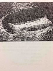
The most likely diagnosis of the gallbladder exam below is: |
-Polyp |
|
|
A female patient presented to the ER with RUQ pain extending to the right shoulder, nausea and vomiting. The ultrasound exam could not detect a distended gallbladder, however a large acoustic shadow was seen in the GB region. What is the most likely diagnosis?
|
-cholelithiasis; "packed" GB |
|
|
Gallbladder wall calcification is referred to as: |
-Porcelain gallbladder |
|
|
Sludge is also known as: |
-Inspissated bile |
|
|
The primary purpose of placing the patient in a LLD position when evaluating the GB is: |
-to determine if suspected stones roll |
|
|
The sonographic criteria for diagnosing cholecystitis is: |
*Fluid filled GB w/o stones *Edematous "halo" surrounding GB *TRV GB diameter >5cm *GB wall thickness >4mm -All of the above |
|
|
A male patient presents with epigastric pain, nausea & vomiting. The U/S exam reveals a markedly dilated GB w/thin walls. The most likely diagnosis would be: |
-Hydrops |
|
|
A patient presents to the ER w/RUQ pain, nausea & vomiting. She also exhibits a positive Murphy's sign. The U/S exam revealed an enlarged, thick walled GB containing echoes that did not layer or shadow. Pericholecystic fluid was also demonstrated. The most likely diagnosis would be: |
-Acalculous cholecystitis |
|
|
A patient presents to the U/S w/loss of appetite, fatty food intolerance, nausea, vomiting and jaundice. The sonogram reveals a GB, which has thick, irregular walls. A gallstone is identified but appears to be surrounded by an echogenic mass. The most likely diagnosis would be: |
-GB carcinoma |
|
|
Small echogenic mass-like areas w/internal cysts are seen in the fundus of the gallbladder that produces a comet-tail artifact. These findings most likely represent: |
-Adenomyomatosis |
|
|
A patient presents w/RUQ pain,fever, chills, & jaundice. The sonogram reveals massively dilated ducts. Stones are visualized in the ducts along w/an enlarged gallbladder. This condition is referred to as: |
-Cholangitis |
|
|
A Klatskin tumor occurs in the: |
-Confluence of the right & left hepatic ducts |
|
|
Anomalous insertion of the CBD into the pancreatic duct allowing reflux of pancreatic juice into the bile duct leading to dilation and cholangitis is referred to as: |
-Choledocolithiasis |
|
|
The normal average size of the common hepatic duct of a patient under the age of 40 would be: |
-6mm |
|
|
Stones impacted in the gallbladder neck will not move with changing patient positions. |
-True |
|
The most likely diagnosis of the gallbladder exam is: |
-Sludge |