![]()
![]()
![]()
Use LEFT and RIGHT arrow keys to navigate between flashcards;
Use UP and DOWN arrow keys to flip the card;
H to show hint;
A reads text to speech;
19 Cards in this Set
- Front
- Back
|
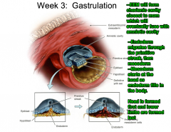
List the tissues that compose the buccopharyngeal and cloacal membranes and identify their locations in the trilaminar embryo. |
|
|
|
List the major derivatives of the ectoderm, endoderm and mesoderm, including paraxial, intermediate and lateral plate mesoderm, as outlined in the resources. |

|
|
|
Describe the formation of the notochord and its function in neurulation. |
|
|
|
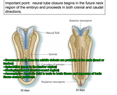
Describe the process by which the neural tube closes. |
|
|
|
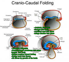
Diagram and describe cranial-caudal folding. What drives caudal folding? What does caudal folding bring into the body? Cranial? |
|
|
|
Diagram lateral folding. What drives this folding? |
|
|
|
Predict the consequences of neurluation defects. What congenital abnormalities are associated with failure of neural tube to close? |

|
|
|
Predict the consequences of cranial-caudal or lateral folding defects |
|
|
|
Predict the consequences of defects in neural crest cell migration. |
|
|
|
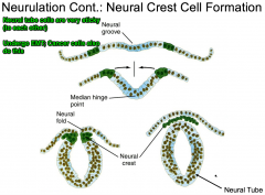
Diagram neural crest cell formation. |
|
|
|
Diagram/describe neural tube closure. |
|
|
|
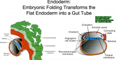
Give an overview of Gastrulation. What the relationship between the amniotic cavity and the chorionic cavity? Though what structure do the mesoderm and endoderm migrate? IN what order? |
|
|
|
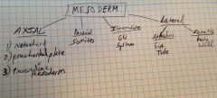
What are the four subdivision of the mesoderm and what do they give rise to? |
|
|
|
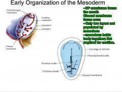
Diagram/Describe early mesoderm organization. Identify the Buccopharyngeal and Cloacal membranes. What do they give rise to? What germ layer tissue is excluded from those structures? |
|
|
|
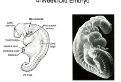
Diagram the 4-week-old embryo |
|
|
|
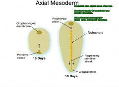
Diagram the axial mesoderm. Identify the pericardial plate and the notochord. What are their respective functions? |
|
|
|
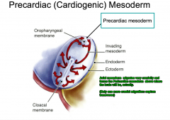
Diagram/ describe the pre-cardiac mesoderm. To ward which structure does it migrate does it migrate? |
|
|
|
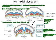
Describe the non-axial mesoderm. Describe the structures and from where the originate. From where are germ cells derived? |
|
|
|
Which layer does not go through the primitive streak. What structure/region signals it to develop? |

|