• Shuffle
    Toggle On
    Toggle Off
  • Alphabetize
    Toggle On
    Toggle Off
  • Front First
    Toggle On
    Toggle Off
  • Both Sides
    Toggle On
    Toggle Off
  • Read
    Toggle On
    Toggle Off
Reading...
Front

Card Range To Study

through

image

Play button

image

Play button

image

Progress

1/111

Click to flip

Use LEFT and RIGHT arrow keys to navigate between flashcards;

Use UP and DOWN arrow keys to flip the card;

H to show hint;

A reads text to speech;

111 Cards in this Set

  • Front
  • Back

Cellular activity

  • Foundation for bodily functions
  • Dysfunction, source of disease
  • Foundation for rest of study of anatomy and physiology

Cytology

Study of cellular structure and function


  • 200 kinds of cells in human body

Components of a cell

  • Plasma membrane (cell membrane)
  • Cytoplasm
  • Cytoskeleton
  • Organelles
  • Inclusions
  • Cytosol

Plasma membrane (cell membrane)

Forms a cell's surface boundary

Define Cytoplasm

Material between plasma membrane and nucleus


  • nucleus largest organelle

Parts of cytoplasm

  • Cytoskeleton: supportive framework of filaments and tubules
  • Organelles: diverse structures performing tasks for the cell
  • Inclusions: accumulated cell products. (lipids, pigments, and bacteria.)
  • Cytosol: clear gel embedding the other components.

Cytosol

Clear gel embedding the other components

ICF

Intracellular fluid

ECF

Extracellular fluid


  • refers to all body fluids on ICF
  • ECF between cells termed tissue fluid
  • also blood plasma, lymph, and cerebrospinal fluid.

micrometer

10-6 meter

size of most cells

10-15 micrometers

Why is cell size limited?

  • If too large it would rupture
  • Diffusion efficiency
  • Lessens impact of single cell death on organism

Activities of cell surface

  • Binding of single molecules
  • Stimulation of cellular activity
  • Attachment of cells to each other
  • Transport of materials into and out of cells

Plasma membrane

  • Membrane at cell surface
  • Defines boundary of cell
  • Governs interactions with other cells
  • Maintains chemical composition differences between ECF and ICF
  • Two layered lipid film with embedded proteins

What lipids are in plasma membrane?


  • Phospholipids
  • Cholesterol
  • Glycolipids

Phospholipids

  • Highly fluid
  • 75% of lipid molecules
  • hydrophilic phosphate head
(faces water on inside and outside of cell)
  • Two hydrophobic fatty acid tails
(forms middle of phospholipid bilayer away from water)

Cholesterol

  • 20% of lipid molecules
  • Affects membrane fluidity
  • If too much, inhibits protein and enzymes in membrane
  • If too little, plasma membrane too fragile

Gycolipids

  • Remaining 5% of lipid molecules
  • Phospholipids with short extracellular carbohydrate chains

Types of membrane proteins

  • Integral proteins
  • Peripheral proteins

Integral proteins

  • Proteins that penetrate cell membrane
  • most glycoproteins (proteins with carbohydrates attached.)

Peripheral proteins

  • Do not protrude into phospholipids
  • usually on intracellular face

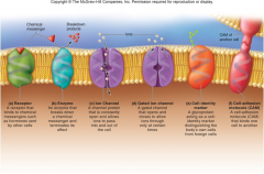
Function of membrane proteins

  • Receptors
  • Enzymes
  • Channel
  • Carriers
  • Cell-identity markers
  • Cell-adhesion molecule (CAMs)

Receptors

  • Membrane protein
  • Receives and binds chemical signals from other cells

Membrane Enzyme function

  • Carry out chemical reactions at membrane surface
  • I.E. degrading signal molecules
  • I.E. breaking down dietary nutrients

Channel proteins

  • Have tunnels through them
  • Allow water and hydrophilic solutes to enter or leave cell
  • some always open
  • other open and close at different times. (gated channel)

Carrier (membrane protein)

  • Bind substance on one side of membrane
  • release it on the other side
  • transport glucose, amino acids, ions, and other substances

Cell-identity markers

  • glycoproteins and glycolipids, cell identification tags
  • unique to an individual
  • enable immune system to distinguish body from anything foreign

Cell-adhesion molecules (CAMs)

  • link cells to each other
  • link cells to extracellular material
  • bind tissue together
  • also needed for sperm-egg binding
  • needed for immune cell binding

Types of Plasma membrane proteins

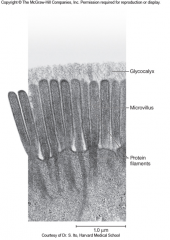
Characteristics of Glycocalyx

  • Fuzzy carbohydrate coat covering cells
  • Short sugar chains of glycolipids and glycoproteins

Functions of Glycocalyx

  • Cell-adhesion molecule
  • Cushions and protects plasma membrane
  • functions in ability to distinguish healthy cells from diseased cells, foreign organisms, and transplanted tissues.
  • Determine human blood types and transfusion compatibility

Microvilli

  • Extension of plasma membrane
  • Serve primarily to increase surface area
  • Better developed in cells specialized for absorption (small intestine)
  • Well developed in taste buds and inner ear

Visual of Microvilli and Glycocalyx (brush border)


Cilia


  • Hairlike extensions
  • Most cells with solitary, nonmotile cilium may serve sensory function
  • Modified cilia in many roles
light absorbing parts of retinal cells in eye

motion and balance in inner ear


monitoring fluid flow in kidneys


sensory cells of nose

Motile cilia

  • Less widespread
  • Abundant in mucous membranes of respiratory tract (to move mucus from lungs up to the throat)
  • Abundant in uterine tubes (move egg or embryo toward uterus)

Flagella

  • Resembles long solitary cilium
  • Has same structure as cilia
  • Stiffened by sheath of microfilaments
  • In tail of sperm (used to crawl up mucous membrane of uterus)

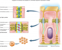
Cell junction function and characteristics

  • Formed by proteins at cell surface
  • Link cells togeather
  • Attach them to extracellular material
  • Enable cells to grow and divide normally, resist stress, and communicate with each other.

Types of Cell junction

  • Tight junctions
  • Desmosomes
  • Gap junctions

Tight junction

  • Completely encircles epithelial cell near upper end. (joins tightly to adjacent cells)
  • Formed by fusion of plasma membrane of adjacent cells
  • Makes it difficult for substances to leak between cells (prevent digestive juices seeping between epithelial cells.)

Desmosome

  • Holds cells together at specific point
  • not continuous
  • Keep cells from pulling apart
  • Enable tissues to resist mechanical stress
  • Common in epidermis and cardiac muscle

Gap Junction


  • Formed by ring of proteins surrounding channel
  • Aids diffusion through channel from cytoplasm of one cell into another. (ions, glucose, amino acids, other solutes.)
  • I.E. in cardiac muscle allows electrical excitation to pass directly from cell to cell.


Cell Junctions (picture)


Types of cell membrane transport


  • Selective permeability
  • Simple diffusion

Selective permeability

  • Allows some substances to pass
  • Holds back others

Simple diffusion

  • Net movement of particles from high concentration to low (down a concentration gradient)
  • How oxygen and steroid hormones enter cell
  • Does not require cell energy
  • Random motion due to heat of molecules provides energy for simple diffusion from.

How do nonpolar and hydrophobic substances pass the plasma membrane?

Diffuse through lipid regions of membrane

How do Hydrophilic substances pass the plasma membrane?

Diffuse through protein channels of membrane

Osmosis

  • Net movement of water from lower solute concentration to higher
  • Water molecules loosely associated with solutes
makes less able to break free

water molecules on low solute side more able to pass through membrane


  • Plays key role in homeostasis (cell volume and capillaries absorbing fluid)

Facilitated diffusion

  • Carrier mediated transport (employ carrier proteins in plasma membrane)
  • Movement of solute down concentration gradient
  • Occurs with aid of carrier (binds particle on side where solute more concentrated and released on lower concentration side.
  • No energy required by cell
  • Transports solutes that can't pass membrane. I.E. sugar absorbtion.

Active transport

  • Carrier-mediated transport
  • Requires energy input (like rolling a wagon uphill)
  • Moves solutes up its concentration gradient (from less concentrated to more)
  • Ceases immediately without ATP energy.

Sodium-potassium (Na-K) pump

  • Important active-transport process (uses ATP and largest source of daily caloric needs)
  • Sodium normally more concentrated in ECF
  • Potassium normally more concentrated in ICF
  • Binds three Na and pumps out of cell
  • Binds two K and pumps into cell

Sodium pump roles

  • Controlling cell volume
  • Generate body heat
  • Providing energy for other transport pumps

Vesicular transport (define and types)

  • Move substances through membrane in vesicles (requires ATP)
  • Endocytosis
  • Exocytosis

Endocytosis

Movement of substances through membrane in vesicles INTO the cell (requires ATP)


Exocytosis

Movement of substances through membrane in vesicles OUT of the cell(requires ATP)

Types of Endocytosis

  • Phagocytosis
  • Pinocytosis
  • Receptor-mediated endocytosis

Phagocytosis


  • Cell eating
  • footlike pseudopods reaching out from cell
  • surrounds particle
  • engulf and enzymatically degrade particle
  • especially in macrophages (white blood cells)

Pinocytosis

  • Cell drinking
  • Occurs in all human cells
  • formation of dimples in plasma membrane
  • cave in and pinch off as vesicles
  • contain droplets of ECF

Receptor-mediated endocytosis

  • more selected membrane transport
  • cell able to take in specific molecules from ECF
  • specific receptor proteins on plasma membrane

  1. cluster together
  2. membrane sinks in here
  3. pit pinches off, forming vesicle
I.E. insulin absorption from blood

Exocytosis

  • Reverse endocytosis
  • Secretory vesicle in cell

  1. migrates to surface
  2. fuses with plasma membrane
  3. releases products from cell
  4. becomes part of plasma membrane
I.E. digestive glands to secrete enzymes and breast cells to secrete milk

Cytoskeleton

  • Network of protein filaments and tubules in cytoplasm
  • Structurally supports cell
  • Determines shape and organizes content
  • Transports substances within cell
  • Contributes to cell movements
  • Connected to proteins in plasma membrane

Microfilaments

  • Thinnest components to cytoskeleton
  • Made of protein, actin
  • Form dense fibrous mesh on internal plasma membrane (terminal web)
  • 6 nm thick

Intermediate filaments

  • Stiff intermediate components of cytoskeleton
  • Contribute to strength of desmosomes
  • Include tough protein keratin
  • fills cells of epidermis and gives strength to the skin
  • 8-10 nm diameter

Microtubules

  • Thickest components of cytoskeleton
  • Hold organelles in place
  • Form bundles that maintain cell shape
  • Like "monorails"
  • Form axonemes of cilia and flagells
  • Form centrioles and mitotic spindle
  • 25 nm diameter

Kinds of inclusions


  • Stored cellular products
I.E. pigments, fat globules, and glycogen granules


  • Foreign bodies
Viruses, bacteria, and dust particles


  • Never enclosed in a membrane
  • Not essential to cell survival

Organelles

  • Play individual roles to survival of cell
  • Compartmentalized components of the cell

Nucleus

  • Largest organelle
  • Most cells with only one nucleus
Mature red blood cells don't have a nucleus

Skeletal and liver cells have multiple nuclei

Nuclear envelope

  • Two parallel membranes surrounding nucleus
  • perforated by nuclear pores
regular traffic into and out of nucleus

bind two membranes togeather

Chromosomes

  • Threadlike bodies of DNA and protein
  • Found in nucleus
  • Most human cells with 46 chromosomes
  • Chromatin, (form in nondividing cells) fine filaments dispersed throughout the nucleus.

Chromatin

Form of chromosomes in non dividing cells


  • fine filaments dispersed throughout nucleus

Nucleoli

  • One or more dense masses in nucleus
  • Where subunits of ribosomes are made

Endoplasmic reticulum

  • System of interconnected channels called cisternae
enclosed by a membrane
  • Synthesizes steroid and other lipids
  • Detoxifies drugs
  • Manufactures cell's membranes
  • Produces phospholipids and plasma membrane proteins
  • Produces proteins for secretion of lysosomes

Smooth endoplasmic reticulum

  • cisternae lack ribosomes
  • continuous with rough ER
  • abundant in cells that synthesize steroid hormones
I.E. the testes, ovaries
  • abundant in cells specializing in detoxification
I.E. liver cells

Rough endoplasmic reticulum

  • cisternae covered with ribosomes
  • continuous with outer membrane of nuclear envelope
  • most abundant in cells making lots of protein

Ribosomes

  • Small granules of protein and ribosomal ribonucleic acid (rRNA)
  • Produced in nucleus
  • "Read" genetic messages from the nucleus
  • Assemble amino acids into proteins specified by code
  • Many attached to nuclear envelope and rough ER
  • Others free in cytoplasm

Golgi Complex


  • Small cluster of cisternae (flattened curved sac)
  • Synthesize carbohydrates
  • Complete protein and glycoprotein synthesis
  • Pinch off membranous sacs, Golgi vesicles
  • Some becoming lysosomes
  • Some becoming part of plasma membrane
  • Others becoming secretory vesicles (stored for later release by exocytosis)

Golgi vesicles

Membranous sacs filled with complex secretory products produced from golgi complex

Lysosomes

  • Packages of enzymes enclosed in a membrane
  • Often round or oval
  • Break down (proteins, nucleic acids, and carbohydrates, phospholipids, and others.)
  • Digest and destroy microbes in white blood cells
  • Digest worn-out organelles
  • Help carry out apoptosis

Apoptosis

Prearranged "cell suicide" in no longer needed cells

Peroxisomes


  • Resemble lysosomes but contain different enzymes
  • Abundant in liver and kidney cells
  • Bread down fatty acids in two carbon molecules (can be used as energy or ATP synthesis)
  • Neutralize free radicals
  • Detoxify drugs
  • Kill bacteria
  • Produce hydrogen peroxide

Mitochondria


  • Specialized for ATP synthesis
  • Surrounded by double membrane

  1. inner membrane with folds (cristae)
  2. Space between cristae (mitochondrial matrix)

  • Contains enzymes, ribosomes, and small DNA molecules call mitochondrial DNA. mDNA


Mitochondria history

  • Were once free-living bacteria
  • Internalized by another primitive cell
  • Evolved to become permanent indispensable residents
  • Mitochondrial DNA
circular loop like modern bacteria

genetically different from DNA in cell nucleus


mutations responsible for some muscle, heart, and eye disease.
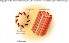
Centrioles


  • Short cylindrical assembly of microtubules
arranged in nine groups of three each


  • Basal bodies of cilia and flagella considered solitary centrioles


Centrosome

  • Patch of cytoplasm near nucleus
  • contains pair of centrioles
  • plays a role in cell division

Visual of Centriole

Steps of protein synthesis

  1. DNA in nucleus with protein codes (genes)
  2. Transcription: Copy made of gene (messenger RNA, mRNA) and mRNA migrates to cytoplasm

  3. Translation: Reading of code by ribosomes (amino acids assembled in correct order)

Transcription


  • First step of protein synthesis
  • Begins at site of particular gene
  • Double helix "unzipped"
  • RNA polymerase (enzyme) reads bases and creates parallel molecule of messenger RNA. Mirror image of gene.
  • mRNA "edited" in nucleus removing noncoding segments and to make mature mRNA molecule
  • Exported through nuclear pore into cytoplasm

DNA-------->mRNA transcription coding

DNA mRNA


A ----------->U


T ----------->A


C----------->G


G----------->C


DNA: A(lways) T(ake) C(are) ATC

around

mRNA: U(nusually) A(gressive) G(eese) UAG

Translation

  • mRNA translated into amino acids
  • Small and large ribosomal subunits assemble on mRNA and begin reading coded message of mRNA

Codons

Three-base segments of mRNA


  • 64 codons in genetic code
  • 20 amino acids composing proteins
  • some redundancy present
  • each codon standing for particular amino acid
I.E. GGU,GGA,GGG glycine, AUG methonine, UAG, UGA, UAA

Transfer RNA (tRNA)

  • Small RNA molecule
  • Bound by ribosome as it reads codon
  • Carries an amino acid
  • Has complementary base series to codon (anticodon)
  • Binds tRNA with anticodon CCA carries glycine added to protein by ribosome
  • Repeats process as ribosome reads next codon
amino acids added one by one

continues until reaches stop codon

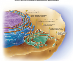
Protein processing and secretion (Nucleus/ribosome/Rough ER)


  1. mRNA "docks" on rough ER
  2. Protein reels into ER cisterna during assembly
  3. Certain amino acid segments cut and spliced
  4. Altered proteins shuffled into transport vesicles and carry protein to nearest cisterna of golgi complex.

Protein processing and secreation (Golgi process)

  1. Sorts proteins
  2. Passes from the one cisterna to the next
  3. Further alters proteins (may add carbohydrates or other components)
  4. budding off of gogli vesicles (become lysosomes or released by exocytosis)

Picture of protein synthesis and secretion


Phases of cell division


  1. Gap 1, G1
  2. Synthesis phase, S
  3. Gap 2, G2
  4. Meitotic phase, M

First Gap phase G1 (cell division)

  • Interval between cell division and DNA replication
  • Time of protein synthesis and growth
  • Completion of normal cell tasks
  • Begin replicating centrioles
  • Accumulation of materials needed for DNA replication

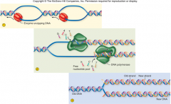
Synthesis phase (cell division)

  • Cell carrying out DNA replication (creates two sets of identical DNA molecules)
  • DNA molecules "unzip" and separate into two strands
  • DNA polymerase (enzyme) reads base sequence of DNA strands and assembles nucleotides to make complementary strand.
  • Two enzymes work in opposite directions

Picture of DNA replication

Second Gap G2 (cell division)

  • Relatively brief
  • Interval between DNA replication and cell division
  • Finishes replication of centrioles
  • Synthesis of enzymes that control cell division

Interphase


  • The time between cell division
  • Consists of G1, S, and G2 phases of cell division.

Phases of Mitotic phase

  • Prophase
  • Metaphase
  • Anaphase
  • Telophase
  • Cytokinesis

Mitotic phase (M) (cell division)

  • period of mitosis
  • Nucleus replicated
  • DNA divided into two identical sets
  • Cell pinching in half and creates two identical daughter cells

Some examples of cell division time of cells

  • Fibroblasts (connective tissue cells) once a day
  • Bone cells: divide slowly
  • Skeletal nerve cells: no dividing at all
  • Cancer: continually cycling

Mitosis


  • Process of cell division in cells (except eggs and sperm which is meiosis)
  • Develops fertilized egg into an individual
  • Continue growth of organs after birth
  • Repairs damaged tissues

Prophase


  • Chromosomes coil into short dense rods
  • Chromosomes have two identical bodies (sister chromatids) joined at center pinched spot (centromere)
  • 46 chromosomes (two chromatics per chromosome (92 DNA molecules in all)
  • Nuclear envelope disintegrates
  • Spindle fibers (elongated microtubules sproute from centrioles pushing them apart)
  • Centrioles attach to centromeres of chromosomes

picture of a centromere/sister chromatid

Metaphase


  • Chromosomes aligned on cell equator
  • Await signal to split in two at centromere

Mitotic spindle

  • Array of spindle fibers
  • Some reaching from centrioles to chromosomes
  • Some anchoring assembly to inside of plasma membrane

Anaphase

  • Centromere cleaved in two by enzyme and forms into sister chromatics NOT chromatids (called daughter chromosomes)
  • Each daughter is genetically identical
  • One daughter chromosome moved to each pole of cell by centromere
  • Almost all body cells genetically identical except egg and sperm cells

Telophase

  • Clustering of chromatics on each side of cell
  • New nuclear envelop developes around each cluster made of rough ER
  • Chromatids beginning to uncoil returning to dispersed form
  • Mitotic spindle breaks up
  • Nuclear division ends

Cytokinesis

  • Division of cytoplasm
  • Overlaps with telophase
  • Motor proteins pulling on membrane skeleton
  • Creates crease, cleave furrow around equator
  • Cell pinches in two
  • Transitions into interphase