![]()
![]()
![]()
Use LEFT and RIGHT arrow keys to navigate between flashcards;
Use UP and DOWN arrow keys to flip the card;
H to show hint;
A reads text to speech;
56 Cards in this Set
- Front
- Back
|
What causes the hyperpigmentation in ephelides (2)? |
1. Sun-induced melanogenesis 2. Transport of increased # of fully melanized melanosomes to keratinocytes |
|
|
What are differences between ephelides and lentigines (3)? |
1. Ephelides are lighter in color 2. Ephelides are confined to sun-exposed skin, while lentigines can appear anywhere 3. Ephelides are responsive to sun exposure, while lentigines do not change |
|
|
Describe the growth of cafe-au-lait macules |
Enlarge proportionately with overall body growth, then remain stable during adulthood |
|
|
Two conditions in which cafe-au-lait macules are part of the clinical presentation? |
1. Neurofibromatosis Type 1 2. McCune-Albright syndrome |
|
|
Which syndrome typically has large cafe-au-lait macules that are generally found midline? |
McCune-Albright syndrome |
|
|
What is a probable cause for the pathogenesis of Becker's melanosis? |
Increase in androgen receptors and heightened sensitivity to androgens (onset during/after puberty, hypertrichosis, dermal thickening, acne, and hypertrophic sebaceous glands are associated with Becker's melanosis) |
|
|
What decade of life do Becker's nevi usually appear? |
2nd or 3rd decade of life |
|
|
Most common distribution of a Becker's nevus? |
Unilateral on an upper quadrant of the anterior or posterior chest + shoulder |
|
|
What genetic disease has solar lentigines as part of its clinical presentation? |
Xeroderma pigmentosum Inability to repair damage from UV radiation leads to lots of solar lentigines, NMSCs, and melanomas |
|
|
Histopathologic differences in solar lentigines vs ephelis? |
Rete ridges are more club-shaped and elongated Solar elastosis is usually present |
|
|
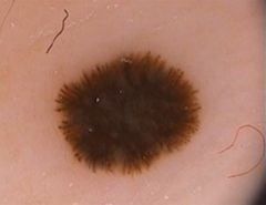
Name for a brown macule with early age of onset and little or no relationship to sun exposure? |
Lentigo simplex |
|
|
Melanosis/lentigines in which location would give you worry for melanoma? |
Conjunctiva; primary acquired melanosis is a precursor lesion of melanoma in this area |
|
|
Clinical presentation of LEOPARD syndrome (acronym)? |
1. Lentigines 2. Electrocardiographic conduction defects 3. Ocular hypertelorism 4. Pulmonary stenosis 5. Abnormalities of genitalia 6. Retardation of growth 7. Deafness |
|
|
Clinical presentation of Carney complex? |
Multiple lentigines + multiple neoplasias (myxomas of skin/heart/breast, schwannomas, blue nevi, pituitary adenoma, sertoli tumors) |
|
|
Clinical presentation of Peutz-Jeghers? |
Mucocutaneous lentigines + intestinal polyposis |
|
|
Primary body location for dermal melanocytosis? |
Lumbosacral region |
|
|
Which ethnicity has the highest prevalence of dermal melanocytosis? |
Asians |
|
|
In Mongolian spots, where are the melanocytes usually found? |
Middle to lower dermis |
|
|
What phenomenon explains the blue color in dermal melanocytosis? |
Tyndall phenomenon I.e. decreased reflectance of light in lower-wavelength region (roygbiv) compared with surrounding skin; blue and violet wavelengths are reflected |
|
|
Unilateral bluish macules with confluence to form an irregularly demarcated and mottled patch on the face? |
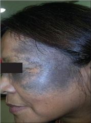
Nevus of Ota Usually V1/V2 distribution, can involve sclera, sometimes bilateral |
|
|
What is the common facial distribution for Nevus of Ota? |
V1 and V2 distribution |
|
|
Most common demographic for Nevus of Ota? |
Asians and blacks, females (80%) |
|
|
What can be said about melanomas that arise from Nevus of Ota? |
Typically present as subcutaneous nodules; do not adhere for the ABCD rules of melanoma Can also have primary melanoma of eye structures (choroid, orbit, iris, meninges) if sclera is involved |
|
|
Most common distribution for Nevus of Ito? |
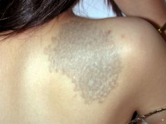
Posterior supraclavicular, along lateral cutaneous brachial nerves (scapular, deltoid, supraclavicular) |
|
|
Which lasers are effective in treating Nevus of Ota/Ito (3)? |
Q-switched ruby Alexandrite Nd:YAG |
|
|
What 3 body locations are the most common for blue nevi? |
1. Scalp 2. Sacral region 3. Dorsal aspects of distal extremities |
|
|
Well-circumscribed, dome-shaped blue-gray papule on the face, hands, or feet? |
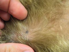
Blue nevus |
|
|
Histologic appearance of blue nevi? |
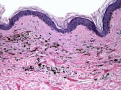
Heavily pigmented, spindle-shaped melanocytes in dense collections within the dermis Long axis of melanocytes is parallel with the epidermis |
|
|
Large (1-3 cm) firm blue plaque on the buttocks, sacral region, or scalp? |
Cellular blue nevi |
|
|
Histologically, what to cellular blue nevi have that common blue nevi don't? |
Plump elongated melanocytes devoid of melanin pigment |
|
|
What are "good" features of benign melanocytic nevi? |
1. Well-circumscribed, regular well-defined borders 2. Round/ovoid 3. Symmetric 4. Size 2-6 mm 5. Contain course and dark hairs |
|
|
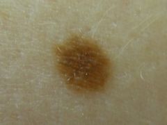
Junctional melanocytic nevus Dark brown macule with uniform pigment network that thins out towards periphery |
|
|
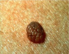
Compound nevus Light to medium brown papule with multiple round/ovoid globules forming a cobblestone pattern |
|
|
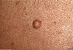
Intradermal nevus Pink to light brown papule with focal globular areas, whitish structureless areas, and fine comma vessels |
|
|
What bullous disorder can have large, asymmetric melanocytic nevi associated with it? |
Epidermolysis bullosa (particularly the AR inherited forms such as simplex) |
|
|
What is the most common dermoscopic pattern in acral melanocytic nevi? |
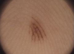
Parallel furrow |
|
|
What mutation is often found in common acquired melanocytic nevi? |
Somatic mutations in BRAF |
|
|
Clinical presentation of Spitz nevi? |
Well-circumscribed dome-shaped papules or nodules that are pink to tan to dark brown; telangiectasias are a frequent finding |
|
|
What type of nevus characteristically has a "starburst" pattern on dermoscopy? |
Reed nevus (likely a variant of Spitz nevus) |
|
|
What are the histologic characteristics of Spitz nevi? |
1. Nests of large spindle and epithelioid cells vertically oriented along the rete ("raining-down pattern", inverted wedge-shape pattern) 2. Sharply defined laterally 3. Orderly infiltration of dermal collagen by these cell nests with "maturation" (gradual diminution of nuclear and cellular sizes) 4. Occasional bizarre cytologic features (necrotic bodies, mitotic figures) |
|
|
Most common location to find Spitz nevi in adults? |
Head (face) and neck, also scalp |
|
|
Why do some dermatologists worry about Spitz nevi? |
- share histologic overlap with melanoma (large spindle and epithelioid cells) - recurrence is sometimes noted |
|
|
Most common location for Reed nevus? |
Extremities; particularly the thigh |
|
|
Characteristic histology of Reed nevus? |
Fascicles of uniform, slender spindle cells that are closely aggregated |
|
|
Most common diagnosis of a recently developed or changing small, well-circumscribed black lesion on the thigh of a young woman? |
Reed nevus |
|
|
Another name for Clark's nevus? |
Atypical (dysplastic) melanocytic nevus |
|
|
For suspected atypical nevi, what margins should you use for the initial biopsy? |
2mm margins; deep shave (i.e. saucerization) is preferred over punch |
|
|
What are the cut-off sizes for small, medium, and large congenital melanocytic nevi? |
Small is < 1.5 cm Medium is b/w 1.5 and 19.9 cm Large is > 20 cm (or > 9cm on scalp for adults, > 6 cm on trunk for newborns) |
|
|
What mutation is commonly seen in congenital melanocytic nevi? |
NRAS mutation |
|
|
What is a complication seen in congenital melanocytic nevi, particularly in cases with multiple satellite-like lesions, a large CMN, or posterior axial location? |
Neurocutaneous melanosis Perform an MRI for evaluation in suspected individuals |
|
|
How does neurocutaneous melanosis typically present? |
Signs and symptoms of increased intracranial pressure (often due to hydrocephalus, mass effect) |
|
|
What is seen on MRI for neurocutaneous melanosis (3)? |
1. Post-gadolinium enhancing mass 2. Post-gadolinium enhancing diffuse thickening of the leptomeninges 3. Focal areas of increased signal on T1 |
|
|
Melanoma risk in congenital melanocytic nevi is related to what? |
Size of the lesion Small and medium-sized CMNs are not associated with significantly increased risk; giant CMNs have substantial melanoma risk |
|
|
Histological difference between congenital melanocytic nevi and common acquired nevi? |
CMNs typically have deeper infiltration (lower reticular dermis, subq fat, surrounding blood vessels, within adnexal structures) |
|
|
Tan macule or patch with darker speckles within? |
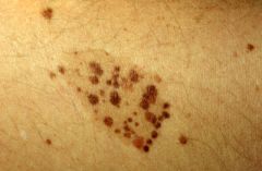
Nevus spilus |
|
|
Nevus spilus can be seen in which conditions (2)? |
1. Phakomatosis pigmentovascularis types III and IV 2. Phakomatosis pigmentokeratotica |