Reading...
![]()
Play button
![]()
Play button
![]()
Use LEFT and RIGHT arrow keys to navigate between flashcards;
Use UP and DOWN arrow keys to flip the card;
H to show hint;
A reads text to speech;
83 Cards in this Set
- Front
- Back
|
How many cranial nerve nuclei are located in the brainstem
|
Nine
Exception: CN 1, 2, (+ XI ?) |
|
|
Alar and Basal Plates: derivatives and location
|
- Motor fibers are derived from the BASAL plate and are located more medially
- Sensory fibers address from the ALAR plates and I located more laterally |
|
|
How many nuclei associated with a cranial nerve
|
Individual cranial nerve carries more than one modality and therefore have more than one nucleus associated with it.
|
|
|
Reticular formation: structure and function
|
– Integration and modulation info going through the brainstem
Structure – Lateral zone processes afferent information – Medial zone processes Eafford information |
|
|
At which level of the spinal cord are all of the following apparent:
spinothalamic tract red nucleus substantia nigra basis pedunculi |
Midbrain
|
|
|
At which level of the spinal cord are all of the following apparent:
Spinal trigeminal nucleus Lateral cuneate nucleus Spinal vestibular nucleus Dorsal Motor nucleus of Vegas Hypoglossal nucleus |
Open Medulla
|
|
|
At which level of the spinal cord are all of the following apparent:
Spinal trigeminal nucleus Nucleus Cuneatus Nucleus Gracialis Hypoglossal nucleus |
Closed medulla
|
|
|
Which landmark can used for the midbrain and micrographs
|
Red nucleus
|
|
|
Functions of the vestibular system
|
– Sensory organ to detect body position and motion
– Equilibrium: balance and postural stability – Motor output: reflexes and controlled motor movements – Vision: control of head and eye motion and stabilization of visual gaze during head/body movement |
|
|
Anatomical structures of the vestibular system
|
– Otic capsule
– Membranous labyrinth – Perilymph – Fine connective tissue |
|
|
Otic capsule
|
In the petrous part of the temporal bone (bony labyrinth?)
|
|
|
Components of the membranous labyrinth
|
1) 5 vestibular sensory organs:
– 3 semicircular canals – 2 otoliths 2) Auditory cochlea 3) Endolymphatic sac |
|
|
What are the three semicircular canals
|
– Superior/anterior
– Posterior – Lateral/horizontal *arranged orthogonally to each other |
|
|
Function of the semicircular canals
|
– Detect angular ACCELERATION = head rotation [ each canal sensitive to specific plane]
– Together, specify the DIRECTION and AMPLITUDE of head rotation |
|
|
Perilymph
|
Fluid between the membranous and bony labyrinth
|
|
|
Function of fine connective tissue in vestibular system
|
To suspend the membranous from the bony labyrinth
|
|
|
What is the bony labyrinth
|
cavity in the petrous part of the temporal bone that contains the membranous labyrinth
|
|
|
What are the otolithic organs?
|
Urticle
Saccule = to saclike organs between the semicircular canals and the cochlea |
|
|
Function of the otolithic organs?
|
Sense body orientation and linear motion
Urticle: horizontal plane Saccule: sagittal plane |
|
|
Composition of Perilymph + drainage
|
– Similar to extracellular fluid and CSF
– Low potassium and high sodium content – Ultrafiltrate of CSF or blood – Drains via venules and middle ear mucosa |
|
|
What is the endolymph
|
Contained within contiguous open lumen of semicircular canals, urticles and saccules
|
|
|
Composition of endolymph + production + absorption
|
– Unique extracellular fluid
– HIGH potassium and LOW sodium content – Produced by the dark cells of the sensory epithelium – Absorbed by the endolymphatic sac |
|
|
What are the five sensory epithelia
|
Cristae of the semicircular canals [3]
Maculae of the utricle and saccule [2] |
|
|
Hair cells function + location
|
– Part of sensory epithelium
– Receptor cells for detecting movement of endolymph |
|
|
Types of hair cells
|
– Stereocilia [60-100/hair cell]
- Kinocilium [1/hair cell] |
|
|
When neurotransmitter is released by the hair cells
|
Glutamate
|
|
|
What are the supporting cells to the hair cells + importance
|
– Microvilli with tight junctions
– Important so that only cilia are exposed to high potassium levels |
|
|
Are there more hair cells in the urticle/saccules or in the ampulla of the semicircular
|
urticle/saccules >> Ampula
35 000 vs 8000 |
|
|
How does endolymph motion transform into neuronal signals (at rest and in motion)
|
– In the stereocilia there are stretch activated potassium channels
• In resting conditions, some channels are open and allows some membrane depolarization to occur causing activation of voltage sensitive calcium channels → glutamate release → VIII nerve excitation • Activation occur when stereocilia are pushed towards kinocilium → opening of more stretch sensitive potassium channels → more Ca enters cell → increased impulse frequency • To inhibits, move stereocilia in opposite direction of kinocilium → closing of strech receptors → hyperpolarization |
|
|
What is the ampula
|
Swelling at the end of semicircular canals
|
|
|
Describe the hair cells in the ampulla
|
All hair cells in the ampulla orient in the SAME direction, with kinocilium closest to utricle
|
|
|
What is the cupula
|
Acellular, gelatinous mass
– Hinged gate spaning the ampulla lumen – Senses motion of fluid to semicircular canals – Hair cells cilia embedded into cupula – Surrounded by endolymph |
|
|
What is the sensory epithelium of the semicircular canals
|
Cristae
|
|
|
What is a sensory epithelium of the otholithic organs
|
Maculae of orticle and saccule
|
|
|
Describe the otholitic membrane
|
acellular gelatinous mass
|
|
|
What is the otoconia
|
Calcium carbonate crystals that sit on top of the oncolytic membrane
– Pressure on the otoconia deflects hair cell cilium – Response to gravitational force – Detects head tilt and acceleration/deceleration |
|
|
During a head turned to the left, what is happening to the firing rates in the left and right ampula
|
– Increase firing rate on the left ampula
– Decreased firing rate on the right ampula |
|
|
Striola
|
Central curvilinear landmark within the otholithic organs
|
|
|
Vestibular afferents: neuron cell type
|
Bipolar
|
|
|
Scarpa's ganglion
|
aka Vestibular ganglion
|
|
|
Path of the vestibularcochlear nerve
|
int. auditory meatus --> cranial cavity --> enters brainstem at junction of pons and medulla (cebellopontine angle) --> vestibular nuclear complex & cerebellum
|
|
|
Location of vestibular nuclear complex
|
Dorsal pons and the medulla beneath the fourth ventricle
|
|
|
What are the four vestibular nuclei
|
1) lateral vestibular nucleus aka Deiter's nucleus
2) medial vestibular nucleus 3) superior & medial vestibular nuclei 4) inferior vestibular nucleus |
|
|
Role of lateral vestibular nucleus
|
Innervates gravity–opposing muscles of LIMBS to maintain POSTURE
|
|
|
Role of medial vestibular nucleus
|
Reflex adjustments of NECK and TRUNK muscles to RESTORE head position after disturbance
|
|
|
Role of superior and medial vestibular nuclei
|
– Eye movements
– This vestibulo-ocular reflux |
|
|
Role of inferior vestibular nucleus
|
Integrates multisensory input, and cerebellum to regulate VOR gain (calibration of system)
|
|
|
Input to lateral vestibular nucleus
|
- urticle
- saccule - semicircular canal |
|
|
Pathway of lateral vestibular nucleus (Deiter's nucleus)
|
Lateral vestibulospinal tract
– Descends entire spinal cord (mostly in ant. cord) – Uncrossed [ipsilateral] |
|
|
Target of lateral vestibular nucleus
|
Ventral horn: at Alpha & Gamma neurons that innervate gravity opposing muscles of limbs
|
|
|
What senses angular acceleration (head rotation)?
|
semicircular canals
|
|
|
What senses linear head motion?
|
urticle and saccules
|
|
|
In someone falling toward their left, how does the lateral vestibular system respond?
|
- innervation of hair cells from left semicirular canals, urticl ena saccule --> vest. ganglion --> CNVIII -- > lat vesticular nucleus
- left lateral vestibular nucleus activated - left lateral vestibular tract activated - uncrossed innervation of spinal cord [IPSILATERAL] - proximal left arm and leg musculature innervated to counter imbalance |
|
|
Function of medial vestibular nucleus
|
- influences neck and axial muscles
- stabalizes head in space |
|
|
Input to medial vestibular nucleus
|
primarily semicircular canals
|
|
|
Medial vestibulospinal tract pathway
|
- medial vestiulospinal nucleus --> tract
- descends in medial longitudinal fasciculus - bilateral projections to cervical and upper thoracic spinal cord (ipsilateral projections more dense) - motor neurons innervate neck musculature - contralateral projection cross at level of medulla |
|
|
In someone falling toward their left, how does the medial vestibular system respond?
|
- innervation of hair cells from left semicirular canals, urticl ena saccule --> vest. ganglion --> CNVIII -- > left MEDIAL vesticular nucleus
- left medial vesticular nucleus activated - activation of medial vestibulocpinal tract (decending MLF bilateral) - innervates cervial motor neurons - neck muscles respond to keep head erect |
|
|
Function of the superior and medial vestibular nuclei
|
Reflex to stabilize visual image in response to head turn
(Vestibulo-ocular reflex VOR) |
|
|
Input to superior and medial vestibular nuclei
|
Semicircular canals
|
|
|
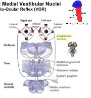
Pathways from superior and medial vestibular nuclei in VOR
|
3 neuron arc
1) bipolar neuron's 2) medial and superior vestibular nuclei 3) Motor neuron's in the abducens nucleus and the oculomotor nuclei that innervate oculomotor muscles |
|
|
Which muscles are innervated by horizontal VOR
|
Lateral recti [L&R]
Medial recti [L&R] |
|
|
Projections to medial rectus originate from
|
Midbrain at oculomotor nucleus
|
|
|
Projections to lateral rectus originate from
|
Pons at abducens nucleus
|
|
|
Describe VOR to medial rectus
|
Semicircular canals --> Scarpa's ganglion --> medial vestiular nucleus (rostra medulla --> synapse --> aducens nucleus (pons) --> synapse --> MLF --> cross over --> oculomotor nucleus (midbrain) --> synapse --> innervate medial rectus
|
|
|
Describe VOR to medial rectus
|
Semicirular canals --> Scarpa's ganglion --> medial vestiular nucleus (rostra medulla --> synapse --> aducens nucleus (pons) --> synapse --> abducens nerve --> innervate lateral rectus
|
|
|
Explain why there are 4 synapses happening in the abducens nucleus
|
At the abducens nuclei, activation of the left nucleus will cause inhibition of right and thus only left lat rectus + right medial rectus will be activated in a given movement
|
|
|
What is in charge of adjusting VOR and control of gain
|
Inferior vestibular nucleus
|
|
|
Function of Inferior vestibular nucleus
|
Eye + Vestibular vestibular system --> cerebellum --> Purkinje cells --> are they them same? --> if not system recalibrate to compensate
- direct connection of where your body is moving, and a visual represeantion of what happening - detects slippage between visual and vestibular input |
|
|
Define : Nystagmus
|
Rhythmic alteration of slow (VOR) and fast (saccades) eye movements during VOR
|
|
|
Pathological vs physiological nystagmus
|
- saccades happen normally during normal eye movement (they are generally not perfectly smooth motions)
|
|
|
Caloric testing for nystagmus
|
- inject warm fluid into ear canal
- induce fluid movement on one side through semicircular - percieved by CNS as head movement ---> cause nystgmus |
|
|
Causes of dizziness and balance disorders
|
– Often difficult to diagnose
– Maybe due to vestibular dysfunction – Non-vestibular causes: fluctuations in blood pressure, Visual system problems, peripheral neuropathies |
|
|
Benign paroxysmal positional vertigo (BPPV)
|
- small part of oticonia breaks off
- can get stuck in ampulla of semicircular canals - sense gravity in linera motion - mismatch between right and left |
|
|
Meniere's disease
|
– A.k.a. endolymphatic hydrops
–problem getting rid of endolymph but you keep making it - increase in pressure of endolymph |
|
|
Vesticular neuritis
|
– Viral infection of the vestibulo-cochlear nerve or facial nerve
|
|
|
Perilymph fistula
|
- hole in the oval or round windows of the cochlea
- lumens are contiguous, so in case of leak you wlil get decrease in pressure of endolymph |
|
|
Ototoxicity
|
- hair cells very metabolically active and sensitive to toxins
- particularly sensitive to aminoglycoside Ab - can damage or kill hair cells |
|
|
Mal de Debarquement
|
- adjust to being on boat
- come back to land - vestibular system cannot undergo necessary plasticity to readjust to life on land - can last for days or can be permanent |
|
|
Aging, dizziness and balance
|
- common deficit of aging are problem with audition, dizziness and stability
- usually problem with hair cells dying - also get problems with visual system |
|
|
Characteristics of bilateral vestibular dysfunction
|
- could be caused by toxicity (aminoglycosides)
- slow onset of loss of vestibular function - instability of eyes with head movements - instability when walking in dark (w/o visual input) |
|
|
Characteristics of unilateral vestibular dysfunction
|
- severe acute symptoms
- extreme diziness, nausea and vomitting - deviation towards side of lesion when walking - abnormal nystagmus - displaced otoconia |
|
|
Characteristics of vestibular compensation
|
- gradual recovery from unilateral lesions
- learning induced changes to central circuits - vestibular inputs ignored in favor of vision and proprioception |
|
|
Some common causes of dizziness due to mismatch bw vestibular and visual inputs
|
- rotation induced dizziness
- false visual motion (optic ilusion) - motion sickness - alcohol |