![]()
![]()
![]()
Use LEFT and RIGHT arrow keys to navigate between flashcards;
Use UP and DOWN arrow keys to flip the card;
H to show hint;
A reads text to speech;
108 Cards in this Set
- Front
- Back
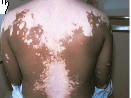
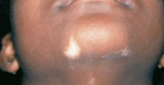
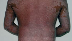
In this condition, depigmented macules appear on the face, hands, feet, extensor surfaces, and other regions and may coalesce into extensive areas that lack melanin. The brown pigment is normal skin color; the pale areas are this condition. In lighter individuals, macules may look reddish or tan instead of pale |
Vitiligo |
|
condition in which a somewhat bluish color is visible in the tonails and toes. This condition, especially when slight, may be hard to distinguish from normal skin color |
Cyanosis |
|
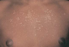
common superficial fungal infection of the skin, causing hypo- or hyper pigmented, slightly scaly macules on the trunk, neck, and upper arms. They are easier to see in darker skin and may be more obvious after tanning. In lighter skin, macules may look reddish or tan instead of pale |
Tinea Versicolor |
|
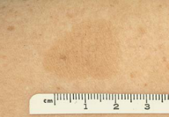
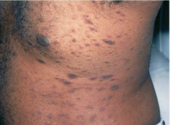
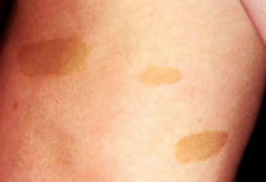
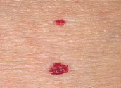
a slightly by uniformly pigmented macule or patch with a somewhat irregular border, usually 0.5 to 1.5 cm in diameter; benign. Six or more such spots, each with a diameter of >1.5 cm, however, suggest neurofibromatosis |
Cafe-Au-Lait Spot |
|
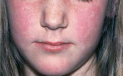
|
Erythema |
|
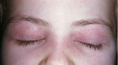
|
Heliotrope |
|
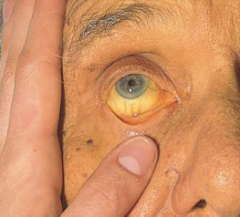
this condition makes the skin diffusely yellow. It is seen most reliably in the sclera. It may also be visible in mucous membranes. Causes include liver disease and hemolysis of red blood cells |
Jaundice |
|
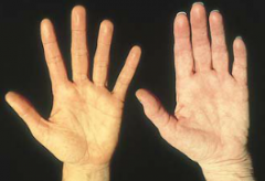
the yellowishness palm of this condition is compared with a normally pink palm. This condition does not affect the sclera, which remains white. The cause is a diet high in carrots and other yellow vegetables or fruits. It is not harmful but indicates need for assessing dietary intake
|
Carotenemia
|
|
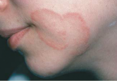
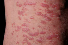
Reddish oval ringworm-like papules or plagues |
Pityriasis Rosea |
|
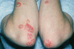
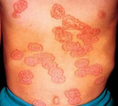
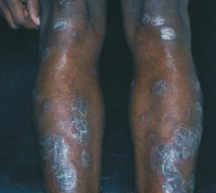
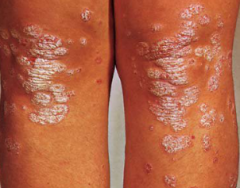
Silvery scaly papules or plagues, mainly on the extensor surfaces |
Psoriasis |
|
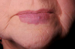
Tan, flat, scaly plaques |
Tinea Versicolor |
|
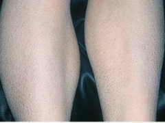
Appears mainly on flexor surfaces (adult form) |
Atopic Eczema |
|
ex. ____ epidermal nevus (patterns & shapes) |
Linear |
|
ex. ______ vesicles of herpes simplex (patterns & shapes) |
Clustered |
|
ex. mycosis fungoides (patterns & shapes) |
Geographic |
|
ex. Tinea corporis (Patterns & Shapes) |
Serpiginous |
|
ex. _______ plaque of tinea faciale (ringworm) (Patterns & Shapes) |
Annular, Arciform |
|
|
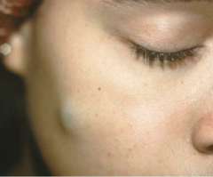
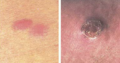
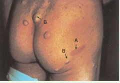
Flat, nonpalpable lesion with changes in skin color, small flat spot, up to 1.0 cm ex. Hemangioma & Vitiligo |
Macule |
|
|
flat nonpalpable lesion with changes in skin color flat spot, 1.0 cm or larger ex. Cafe-Au-Lait Spot |
Patch |
|
(type of macule) |
Hemangioma |
|
(type of patch) |
Cafe-Au-Lait Spot |
|
(type of macule) |
Vitiligo |
|
|
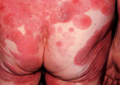
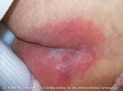
palpable, elevated lesion 1.0 cm or larger, often formed by coalescence of papules |
Plaque |
|
(type of plaque) |
Psoriasis |
|
(type of plaque) |
Psoriasis |
|
|
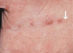
palpable elevated, solid bump up to 1.0 cm |
Papule |
|
(type of papule) |
Psoriasis |
|
(type of nodule) |
Dermatofibroma |
|
|
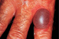
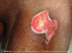
palpable, elevated solid knot-like bump larger that 0.5 cm, deeper and firmer than a papule |
Nodule |
|
|
palpable elevated solid nodule filled with expressible material, either liquid or semisolid |
Cyst |
|
(type of cyst) |
Epidermal Inclusion Cyst |
|
|
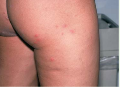
a palpable, elevated, somewhat irregular, relatively transient, superficial area of localized skin edema |
Wheal |
|

(type of wheal) |
Urticaria |
|
|
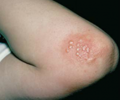
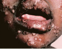
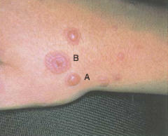
palpable elevation up to 1.0 cm; filled with serous fluid |
Vesicle |
|
(type of vesicle) |
Herpes Simplex |
|
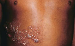
(type of vesicle) |
Herpes Zoster |
|
|
palpable elevation 1.0 cm or larger; filled with serous fluid |
Bulla |
|
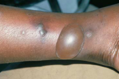
(type of bulla) |
Insect Bite |
|
(type of bulla) |
Insect Bite |
|
|
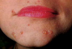
palpable elevation filled with pus (yellow proteinaceous fluid filled with neutrophils) |
Pustule |
|
(type of pustule) |
Acne |
|
(type of pustule) |
Small Pox |
|
|
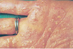
a palpable, elevated, minute, slightly raised tunnel in the epidermis, commonly found on the finger webs and on the sides of the fingers. It looks like a short (5-15 mm), linear ir curved gray line and may end in a tiny vesicle. Skin lesions include small papules, pustules, lichenified areas, and excoriations. With a magnifying lens, look for the _____ of the mite that causes scabies. |
Burrow (scabies) |
|
(type of burrow) |
Scabies |
|
|
a thin flake of dead exfoliated epidermis |
Scale |
|
(type of scale) |
Icthyosis Vulgaris |
|
(type of scale) |
Dry Skin |
|
|
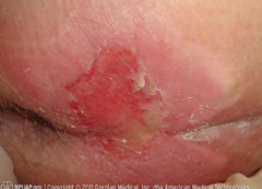
the dried residue of skin exudates such as serum, pus, or blood |
Crust |
|
(type of crust) |
Impetigo |
|
|
visible and palpable thickening of the epidermis and roughening of the skin with increased visibility of the normal skin furrows (often from chronic rubbing) |
Lichenification |
|
(type of Lichenification) |
Neurodermatitis |
|
|
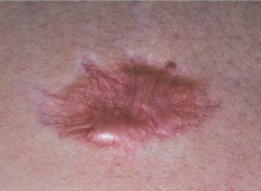
increased connective tissue that arises from injury or disease |
Scars |
|
(type of scar) |
Hypertrophic Scar from Steroid Injections |
|
|
hypertrophic scarring that extends beyond the borders of the initiating injury |
Keloids |
|
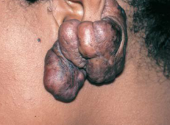
(type of ____) |
Keloid on the ear lobe |
|
|
nonscarring loss of the superficial epidermis; surface is moist but does not bleed |
Erosion |
|
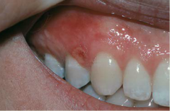
(example of erosion) |
Aphthous stomatitis |
|
|
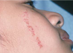
linear or punctate erosions caused by scratching |
Excoriation |
|
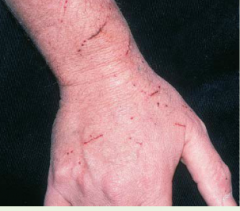
(example of excoriation) |
Cat Scratches |
|
|
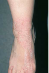
A linear crack in the skin, often resulting from excessive dryness |
Fissure |
|
(example of a fissure) |
Athlete's Foot |
|
|
a deeper loss of epidermis and dermis; may bleed and scar |
Ulcer |
|
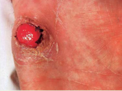
(type of ulcer) |
Syphilitic chancre |
|
|
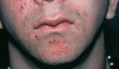
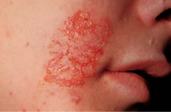
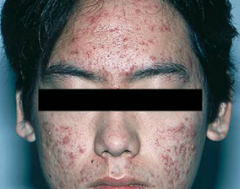
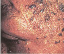
disorder that affects 85% of adolescents in the U.S. Disorder of the pilosebacceous unit that involves proliferation of the keratinocytes at the opening of the follicle; increased production of sebum, stimulated by androgens, which combines with keratinocytes to plug the follicular opening. Lesions appear in areas with the greatest number of sebacceous glands, namely the face, neck, chest, upper back, and upper arms. |
Acne vulgaris |
|
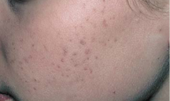
(type of primary lesion) open and closed comedones, occasional papules |
Mild Acne |
|
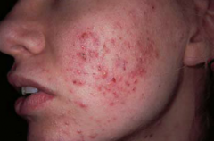
(type of primary lesion) comedones, papules, pustules |
Moderate Acne |
|
(type of primary lesions) |
Severe Cystic Acne |
|
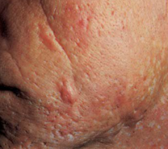
(type of seconary lesions) |
Acne with Pitting and Scars |
|
Color & Size: Fiery red. From very small to 2 cm Shape: Central body, sometimes raised, surrounded by erythema and radiating legs Pulsating & Effect of Pressure: Often seen in center of the spider, when pressure with a glass slide is applied. Pressure on the body causes blanching of the spider Distribution: Face, neck, arms, and upper trunk; almost never below the neck Significance: Liver disease, pregnancy, vitamin B deficiency; also occurs normally in some people |
Spider Angioma |
|
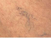
Color & Size: Bluish. size variable, from very small to several inches Shape: Variable. May resemble a spider or be linear, irregular, cascading Pulsatility and Effect of Pressure: Absent. Pressure over the center doesn't cause blanching, but diffuse pressure blanches the veins Distribution: Most often on the legs, near veins; also on the anterior chest |
Spider Vein |
|
Color & Size: bright or ruby red; may become purplish with age. 1-3 mm Shape: Round, flat or sometimes raised, may be surrounded by a pale halo Pulsatility and Effect of Pressure: Absent. May show partial blanching, especially if pressure applied with edge of pinpoint Distribution: Trunk; also extremeties Significance: None; increases in size and numbers with aging |
Cherry Angioma |
|
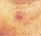
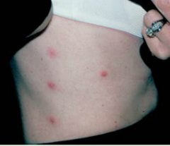
Color & Size: Deep red or reddish purple, fading away over time. Petechia, 1-3 mm; purpura are larger Shape: Rounded, sometimes irregular; flat Pulsatility and Effect of Pressure: Absent Distribution: Variable Significance: Blood outside the vessels; may suggest a bleeding disorder or, if petechiae, emboli to skin; palpable purpura in vasculitis |
Petechia/ Purpura |
|
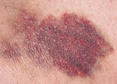
Color & Size: Purple or purplish blue, fading to green, yellow, and brown with time. Variable in size, larger than petechiae, >3 mm Shape: Rounded, oval, or irregular; may have a central subcutaneous flat nodule (a hematoma) Pulsatility and Effect of Pressure: Variable Significance: Blood outside vessels; often secondary to bruising or trauma; also seen in bleeding disorders |
Ecchymosis |
|
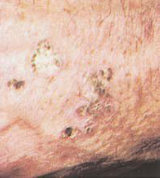
superficial, hyperkeratotic papules. Often multiple; can be round or irregular; pink, tan, or grayish. Appear on sun-exposed skin of older, fair-skinned people. Considered to be dysplastic or precancerous; one of every 1,000 per year develop into squamous cell carcinoma. Usually found on the face and hands |
Actinic Keratosis |
|
common, benign, whitish-yellowish to brown raised papules or plaques that feel slightly greasy and velvety or warty and have a "stuck on" appearance. Typically multiple and symmetrically distributed on the trunk of older people, but may also appear on the face and elsewhere. In black people, often in younger women, may appear as small, deeply pigmented papules on the cheeks and temples |
Seborrheic Keratosis |
|
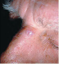
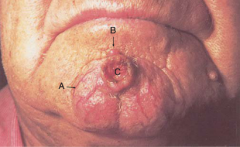
Though malignant, grows slowly and almost never metastasizes. It is common in fair-skinned adults 40 years or older, and usually appears on the face. An initial red macule or papule may develop a depressed center and a firm, elevated border. Telangiectatic vessels are often visible |
Basal Cell Carcinoma |
|
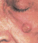
Usually appears on sun-exposed skin of fair-skinned adults older than 60 years. May develop in an actinic keratosis. Usually grows more quickly than a basal cell carcinoma, is firmer, and looks redder. The face and the dorsum of the hand are often affected, as shown here |
Squamous Cell Carcinoma |
|
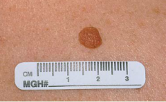
common mole. usually appears in the first few decades. Several nevi may arise at the same time, but their appearance usually remains unchanged. Typical Features: -round or oval shape -sharply defined borders -uniform color, especially skin-colored, tan or brown -diameter <6 mm but >10 mm if congenital -flat or raised surface |
Benign Nevus |
|
|
Atypical moles that are varied in color but often dark and larger than 6 mm, with irregular borders that fade into the surrounding skin. Found primarily on the trunk. May number more than 50 to 100. |
Atypical (dysplastic) Nevi |
|
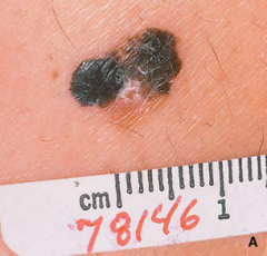
asymmetric, with irregular borders, variations in color, diameter >6 mm, evolution in size, symptom or morphology |
Malignant Melanoma |
|
|
ABCDE's of Melanoma |
Asymmetry, Borders, Color, Diameter (>6mm), Evolution |
|
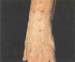
macules on the dorsum of the hand, wrist, and forearm |
Solar lentigines |
|
papules and pustules |
folliculitis from Pseudomonas |
|
pustules on the palm |
pustular psoriasis |
|
vesicles |
Chickenpox |
|
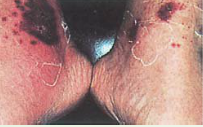
A) Bulla B) target lesion |
erythema multiforme |
|
A) Telangiectasia B) Nodule C) Ulcer |
Squamous Cell Carcinoma |
|
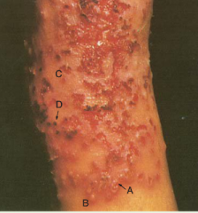
A) Vesicle B) Pustule C)Erosions D) crust |
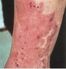
Infected Atopic Dermatitis |
|
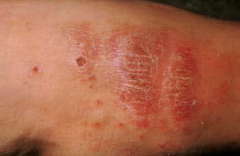
A) Excoriation B) Lichenification on the leg |
Atopic Dermatitis |
|
|
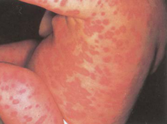
Wheals (urticaria) |
|
|
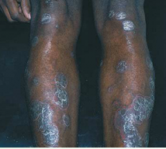
Psoriasis |
|
malignant tumor may appear in many forms: macules,papules, plaques, or nodules almost anywhere on the body. Lesions are often mulitiple and may involve internal structures. |
Kaposi's sarcoma in AIDS |
|
combination of A) Patch B) Nodules |
Neurofibromatosis |
|
presence of reddened area that fails to blanche with pressure, and changes in temperature (warmth or coolness), consistency (firm or boggy), sensation (pain or itching), or color (red, blue, or purple on darker skin; red on lighter skin) |
Stage 1 Pressure Ulcer |
|
The skin forms a blister or sore. Partial thickness skin loss or ulceration involving the epidermis, dermis, or both |
Stage 2 Pressure Ulcer |
|
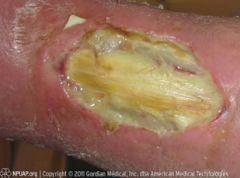
a crater appears in the skin, with full-thickness skin loss and damage to or necrosis of subcutaneous tissue that may extend to, but not through, underlying muscle |
Stage 3 Pressure Ulcer |
|
Full-thickness skin loss, with destruction, tissue necrosis, or damage to underlying muscle, bone, and sometimes tendons and joints |
Stage 4 Pressure Ulcer |
|
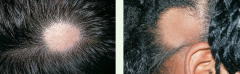
clearly demarcated round or oval patches of hair loss, usually affecting young adults and children. no visible scaling or inflammation |
Alopecia Areata |
|
Hair loss from pulling, plucking, or twisting hair. Hair shafts are broken and of varying lengths. More common in children, often in settings of family or psychosocial stress |
Trichotillomania (Trichotillosis) |
|

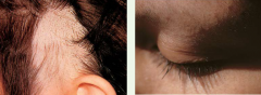
Round scaling patches of alopecia. Hairs are broken off close to the surface of the scalp. Usually caused by fungal infection from Trichyophyton tonsurans fromm humans, less commonly from microsporum canis from dogs or cats. |
Tinea Capitis ("Ringworm") |
|
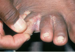
superficial infection of the proximal and lateral nail folds adjacent to the nail plate. The nail folds are often red, swollen, and tender. Represents the most common infection of the hand, usually from S. aureus or Strep species. Arises from local trauma due to nail biting, manicuring, or frequent hand immersion in water |
Paronychia |
|
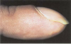
bulbous swelling of the soft tissue at the nail base. Seen in congenital heart disease, interstitial lung disease and lung cancer, inflammatory bowel diseasesm and malignancies |
Clubbing of the Fingers |
|
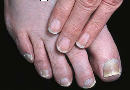
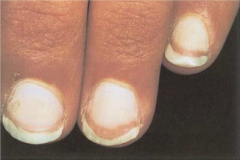
painless separation of the whitened opaque nail plate from the pinker translucent nail bed. Local causes include excess manicuring, psoriasis, fungal infection, and allergic reactions to nail cosmetics. Systemic causes include diabetes, anemia, photosensitive drug reactions, hyperthyroidism, peripheral ischemia, bronchiectasis, and syphilis |
Onycholysis |
|
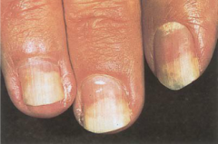
nail plate turns white with a ground-glass appearance, a distal band of reddish brown, and obliteration of the lunula. Commonly affects all fingers, although may appear in only on finger. Seen in liver disease, usually cirrhosis, heart failure, and diabetes. May arise from decreased vascularity and increased connective tissue in nail bed |
Terry's Nails |
|
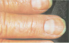
trauma to the nails is commonly followed by nonuniform white spots that grow slowly out with the nail. Spots in the pattern illustrated are typical of overly vigorous and repeated manicuring |
Leukonychia (white spots) |
|
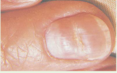
curving, transverse white bands that cross the nail parallel to the lunula. Arising from the disrupted matrix of the proximal nail, they vary in width and move distally as the nail grows out. Seen in arsenic poisoning, heart failure, Hodgkin's disease, chemotherapy, carbon monoxide poisoning, and leprosy |
Mees' Lines (Transverse White Bands) |
|
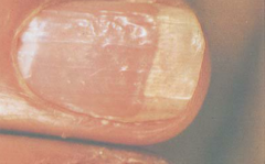
transverse depressions of the nail plates, usually bilateral, resulting from temporary disruption of proximal nail growth from systemic illness. Seen in severe illness, trauma, and cold exposure if Raynaud's disease is present |
Beau's Lines (Transverse Linear Depressions) |
|
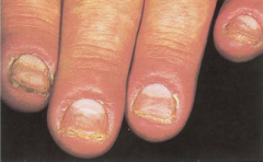
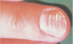
Punctate depressions of the nail plate caused by defective layering of the superficial nail plate by the proximal nail matrix. Usually associated with psoriasis but also seen in Reiter's syndrome, sarcoidosis, alopecia areata, and localized atopic or chemical dermatitis |
Pitting |