![]()
![]()
![]()
Use LEFT and RIGHT arrow keys to navigate between flashcards;
Use UP and DOWN arrow keys to flip the card;
H to show hint;
A reads text to speech;
239 Cards in this Set
- Front
- Back
|
Name the parts of the dogs skeleton |
Back (Definition) |
|
|
What are the four functions of bones? |
-Support and protect soft tissues and organs -Assist in movement -Storage of mineral salts especially calcium and phosphorus - manufacture blood cells in red bone marrow |
|
|
What are the two types of bone tissue? |
Compact bone Spongy bone |
|
|
What is compact bone? |
A hard whitish substance. It appears to be solid but under the microscope a series of canals called the haversian canals run the length of the bone. The canals carry blood vessels, nerves and lymphatics |
|
|
What are the canals surrounded by bone tissue? |
Haversian canal Lamellae Lacunae Canaliculi |
|
|
What is the haversian canal |
Contains blood vessels, lymphatics and nerves |
|
|
What is lamellae |
Plates of bone arranged around the haversian canal |
|
|
What is lacunae? |
Spaces between the lamellae (contains osteocytes and lymph) |
|
|
What is canaliculi? |
Fine channels between the lacunae and haversian canals. They carry lymph |
|
|
What is canaliculi? |
Fine channels between the lacunae and haversian canals. They carry lymph |
|
|
What does compact bone look like under a microscope? |

Back (Definition) |
|
|
What does bone tissue consist of? |
Calcium Collagen fibres (prevents brittle bone) Nucopolysaccharides - ground substance Ostecites (mature blood cells) - within the laccuni |
|
|
What does bone tissue consist of? |
Calcium Collagen fibres (prevents brittle bone) Nucopolysaccharides - ground substance Ostecites (mature blood cells) - within the laccuni |
|
|
Where is compact bone found? |
In the outer layer if all bones known as the Cortex |
|
|
What is spongy bone? |
The haverseous system are spread widely and the spaces between are filled with red bone marrow (fat and blood cells) it is hard with a spongy appearance. All bones are covered by periosteum which is a tough, fibrous membrane. It helps to provide blood to the bome and it is also very sensitive to pain |
|
|
Where is spongy bone found? |
At the end of the long bones and the core of short and flat bones |
|
|
What is bone formation known as? |
Ossification or osteogenisis |
|
|
How do bones develop? |
By endochondrial or interchomdrial ossification. In the growing animal some bone begins as cartilage. When its time for bone to be produced the cartilage becomes calcified and acts as a kind of scaffold. Bone is then laid down upon the scaffolding and bone gradually replaces it. |
|
|
What develops by endochondrial ossification? |
Limbs bones develop |
|
|
What is intramembranous ossification? |
When bones develop without the aid of scaffolding. The bone is formed between two layers of membrane (dense connective tissue) for example the skull |
|
|
Can bone have up to two centres of ossification? |
Yes, the tissue between continues to reproduce itself until maturity. This is an important factor in the growth of long bones and some irregular bones |
|
|
When growth is complete the centres of ossification are said to be what? |
Fused |
|
|
Name the centres of ossification in the long bone |

Back (Definition) |
|
|
Name the centres of ossification in the long bone |

Back (Definition) |
|
|
What is the area of bone development within the shaft is? |
The diaphysis |
|
|
The areas of bone development at either end is? |
The epiphysis |
|
|
What is between the diaphysis and epiphysis? |
The epiphyseal plate. It appears as a much darker area on X-rays compared the bony epiphysis or diaphysis. Longitudeal growth continues until the bone is taken over and the growth plate closes. |
|
|
What is between the diaphysis and epiphysis? |
The epiphyseal plate. It appears as a much darker area on X-rays compared the bony epiphysis or diaphysis. Longitudeal growth continues until the bone is taken over and the growth plate closes. |
|
|
How does the medullary cavity develop? |
By the activity of osteoclasts |
|
|
What do osteoclasts do? |
Change insoluble calcium phosphate inti soluble calcium salts which are carried away in the blood, therefore break down bone |
|
|
What is bone like in young animals? |
Relatively smooth |
|
|
What is bone like in older animals? |
It has more roughened areas |
|
|
What do osteoblasts do? |
Build up the exterior of bone |
|
|
How do osteoblasts and osteoclasts work? |

Back (Definition) |
|
|
What do long bones usually have? |
There is at least one small opening the shafts cortex called the nutrient formane |
|
|
What does the medullary cavity contain in young animals? |
Red marrow to produce white and red blood cells but is replaced by yellow marrow (fat) in ageing |
|
|
Where is the nutrient Forman on a diagram? |

Back (Definition) |
|
|
What are the four bone groups? |
Long bone Flat bone Irregular bone Short bone |
|
|
What are the four bone groups? |
Long bone Flat bone Irregular bone Short bone |
|
|
What is long bone and where is it found? |
They have a shaft (diaphysis) and have an epiphysis at two ends. There is an outer layer of compact bone. At the extremities there is spongy bone which gives strength without adding much weight. It is found in limbs such as the femur and humerus |
|
|
What is flat bone and where is it found? |
Two layers of compact bone with a layer of spongy bone between. Found in the skull, pelvis and scapula |
|
|
What is short bone and where is it found? |
Sometimes classed as irregular bones for example the carpal and tarsal |
|
|
What is short bone and where is it found? |
Sometimes classed as irregular bones for example the carpal and tarsal |
|
|
What are sesamoid bones and where are they found? |
These develop within the tendon ( occasionally ligaments especially when tendons operate over a ridge of bone) for example the patella |
|
|
What are pneumatic bones and where are they found? |
filled cavities called sinuses. These enable the bone to maintain its strength while remaining light weight. For example the frontal and maxillary sinuses |
|
|
Why do bones have surface irregularities? |
These areas are where muscles attach to bones and where bones articulate |
|
|
What are articular irregularities? |
Articulating surfaces with enter into the formation of joints and are smooth |
|
|
What are articular irregularities? |
Articulating surfaces with enter into the formation of joints and are smooth |
|
|
Give examples of Articular irregularities? |
The femoral head (femur fits into acetabulum) Trochlea of the humerus with the trochlear notch of the ulna Capitulum if the humerus with the head of the radius |
|
|
What are non articular irregularities? |
These are the areas which give attachment to the muscles/ligaments and are rough projections. |
|
|
What are non articular irregularities? |
These are the areas which give attachment to the muscles/ligaments and are rough projections. |
|
|
What are examples of non articular irregularities? |
Process - rough projection for muscle attachment e.g. Anconeal process of the ulna Spine - pointed rough projection e.g. Spine of scapula Tuberosity - broad rough projection e.g. The tibial tuberosity. Trochanter - broad rough projection e.g. Greater trochanter of the femur Tubercle - small tuberosity e.g. Greater tubercle of the humerus Crest - long rough narrow projecting surface e.g. Iliac crest |
|
|
How does muscle strength effect projections? |
The stronger the muscle the larger and rougher the projection. |
|
|
What are depressions? |
Fossa - a notch in the bone e.g. Trochanteric fossa (femur) Groove - a long narrow depression e.g. Trochlear groove (femur) |
|
|
What is a foramen? |
A long narrow depression e.g. Nutrient foramen in a long bone |
|
|
What is a sinus? |
A hollow cavity e.g. Frontal sinus |
|
|
What are the three skeleton types? |
Axial Skull, Vertebral column, Ribs and Sternum Appendicular Limbs and limb girdles Splanchnic Os penis |
|
|
What is the skill composed of? |
Cranium - encloses the brain Maxilla - upper jaw with nasal chambers Mandible - Lower jaw |
|
|
What does the base of the brain box consist of? |
Occipital and sphenoid bones |
|
|
What does the base of the brain box consist of? |
Occipital and sphenoid bones |
|
|
What is the foramen magnum? |
The hole where the spinal cord passes from the spine into the head and brain |
|
|
What does the base of the brain box consist of? |
Occipital and sphenoid bones |
|
|
What is the foramen magnum? |
The hole where the spinal cord passes from the spine into the head and brain |
|
|
What are the occipital chondyles? |
Where the first cervical vertebrae articulates |
|
|
What does the temporal bone contain and protect? |
The tympanic bulla, it consists of thick, bony tissue |
|
|
What is the tympanic bulla? |
A small bony chamber containing the middle ear. The exit from the bulla to the external ear is the external auditory meatus |
|
|
What does the lateral view of the skull look like? |

|
|
|
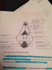
What does the ventral view of the skull look like? |
Back (Definition) |
|
|
where do the frontal sinus' lie? |
over each orbit connecting the nasal cavity. they are lined with a mucous membrane which also lines the respiratory tract. |
|
|
what is the ethmoid bone? |
a sieve like bone which separates the cavity of the nose from the cranium. the olfactory nerves pass through its perforations. These nerves determine our sense of smell. |
|
|
What does the her palate consist of? |
the palatine, the maxilla and the incisive bone |
|
|
what are the nasal chambers? |
the nasal chamber is made up of bone and cartilage, it separates the nasal cavities. It is attached to the middle of the ethmoid bone. |
|
|
What are the walls of each nasal cavity formed by? |
The incisive the maxilla the palatine The roof of the nasal cavity of the nasal bone. ALL the bones are flat bones |
|
|
what are the bony scrolls attached to the nasal cavity? |
turbinate bones. They are covered with mucus membrane |
|
|
where is the lacrimal bone? |
it carries lacrimal drainage and is near the nasal chambers and the cranium. It lies between the frontal palatine and the maxilla bones |
|
|
What does the zygomatic bone form? |
The zygomatic arch or the cheek bone. it protects the eye on its lateral side. The eyes lie in the orbits. |
|
|
What is the mandibular sympahsis? |
there are right and left mandibles which join at the centre. the centre is the mandibular symphasis which is a point of weakness. In RTAs this point commonly seperates as its its only a weak joint. Then the lower jaw becomes unstable. |
|
|
What is the horizontal part of the mandible called? |
The horizontal ramus |
|
|
what is the vertical part of the mandible called? |
the vertical ramus |
|
|
where do teeth fit into? |
the horizontal ramus |
|
|
where do the jaw muscles attach? |
they attach to one end of the skull and at the other to a process |
|
|
What are the vertical ramus's 2 strong processes? |
Coronoid process - forms the point of attachment for some jaw Chondyloid process - articulates with the temporal bone |
|
|
what is the hyoid apparatus? |
small bones and cartilages hanging down from the tympanic bone of the skull. The larynx is suspended below the pharynx and it swings as the animal swallows. |
|
|
where are the upper teeth embedded? |
the maxilla |
|
|
where are the lower teeth embedded? |
the horizontal ramus of the mandible |
|
|
what are the teeth sockets called? |
alveoli |
|
|
what is the part of the tooth projecting from the jaw called? |
the crown. The root is buried in the jaw |
|
|
what are the layers of teeth? |
the pulp cavity is the innermost, containing blood vessels and nerves. this is surrounded by a hard layer called dentine which makes up the bulk of the tooth. the room has an extra layer covering the dentine called cement fixing the tooth into the socket. the tooth outermost layer is enamel. |
|
|
What is the masticatory surface? |
the surface which comes into contact with the tooth in the opposite jaw |
|
|
What are the four types of teeth found in dogs and cats? |
Incisors - for incising canines - sharp, catch and hold prey pre-molars Molars |
|
|
What are molars? |
cheek teeth. They have a masticatory surface containing 2-4 rounded cones called tubercles or cusps. The last upper premolar and the 1st lower molar are very large for cutting. they are called carnassial . |
|
|
how many sets of teeth do they have? |
deciduous teeth (baby teeth) permanent adult teeth |
|
|
when do animals lose their deciduous teeth? |
4-5 month old starting at the incisors and working caudally. the process is usually complete by 6-7 months old. |
|
|
deciduous dentition of the dog? |
I - 3/3 c 1/1 m 3/3 = 28 Total teeth |
|
|
permanent dentition of the dog? |
I - 3/3 C 1/1 PM - 4/4 m 2/3 = 42 total teeth |
|
|
deciduous dentition of the cat? |
I 3/3 C 1/1 M3/2 = 26 total teeth |
|
|
permanent dentition of the cat? |
I3/3 C 1/1 PM 3/2 m1/1 = 30 total teeth |
|
|
what is doliocephalic? |
dogs with long, thin head and muzzle. E.g. Greyhound, whippet |
|
|
what is mesocephalic? |
dogs with medium, length head and muzzle. E.g. labrador, collie, springer spaniel |
|
|
what is brachycephalic? |
dogs with short head and muzzle. E.g. pugs, bulldog, chihuahua |
|
|
what is the vertebral column? |
irregular bones, that are firmly connected with some degree of movement. running from the skull to the tip of the tail. |
|
|
what is the function of the vertebral column? |
supports the trunk and contributes to the support of the appendicular skeleton protection of the spinal cord which it surrounds. |
|
|
what are the vertebral column regions? |
cervical - neck - 7 bones C1 - C7 - the first 2 cervical vertebrae are called Atlas and Axis Thoracic- chest - thorax - 13 bones - T1-T13 Lumbar - lions - lower back - 7 bones L1-L7 Sacral - top of pelvis - sacrum - 3 bones fused together -S1-S3 Coccygeal - tail - the number varies - can be up to 20 or more (Co) |
|
|
What is the cervical vertebrae? |
total of 7 bones with intervertebral foramina. The first two have individual names. Atlas and Axis. |
|
|
What is the the thoracic vertebrae? |
Total of 13 bones. tall spinous processions and depressions. These make a synovial joint the ribs. |
|
|
What are lumbar vertebra? |
A total number of 7 with a large flat, laterally projecting transfers processes. |
|
|
what are sacral vertebrae? |
3 fused vertebrae that support the pelvis. The sacroiliac ligament runs from the sacrum to the sciatic tuberosity on the pelvic bone. |
|
|
What are coccygeal vertebrae? |
Gradually decrease in size so the last few are like rods of bone. The number varies depending on the breed. |
|
|
What are the ribs? |
there are 13 pairs of ribs (26 in total) T1-T13. Ribs are partly bone and partly cartilage (Chostochondral) they are long arched bones. |
|
|
What do ribs function to do? |
protect the thoracic viscera - heart and lungs. aid respiration. |
|
|
What is the sternum? |
composed of 8 bones called sternebrae. the most cranial sternebrae called the manbrium. The most caudal is called the xiphoid notch. They function to act as an anchor for the rib cage and to be the ventral boundary for the thoracic cavity. |
|
|
What are two girdles? |
Pectoral and pelvic. |
|
|
What is the pectoral girdle? |
Consists of 4 bones. Two scapulae and two clavicles. Fused together. |
|
|
What is the pelvic girdle? |
It supports the hind limbs and is composed of 4 bones. Ilium, Ischium, pubis, acetabulum, |
|
|
What is the acetabulum? |
It articulated with the head of the femur to form the hip. The iliac bones have long processes which articulate with the sacrum called the iliac sacral joint. |
|
|
What is the obturator foramen? |
Lateral to the pubic symphysis on each side is a hole.To allow blood vessels to pass into the hind limbs. |
|
|
What is the scapula? |
flat, approximately triangular with an obvious spine. The articular surface is the glenoid cavity. |
|
|
What us an arthrosis? |
A joint at which 2 or more bones meet. There are 3 types |
|
|
What are the three types? |
Fixed Cartilagenous Synovial |
|
|
What is the splanchnic skeleton? |
Skeleton has developed within tissue and is unattached to other parts of the skeleton. E.g. OS penis |
|
|
what are the bones of the forelimb? |
spine of scapula, scapula, (glenoid cavity, clavicle = shoulder joint) humerus, radius, olecranon, ulna, carpal joint, accessory carpal, metacarpal joints, sesamoid, phalanges |
|
|
what are the bones of the hindlimb? |
ilium, ischium, acetabulum, obturator foramen, head of femur, femur, fabella, patella, tibial tuberosity, tibia, femoral chondyl, fibula, calcareous, tibia tarsal bone, metatarsals, phalanges. |
|
|
what is an arthrosis? |
A joint at which the point of 2 or more bones meet, there are three types: fixed, cartilaginous and synovial. |
|
|
what is a fixed joint? |
this is where two bones meet and are separated by only a thin bond of fibrous tissue. These joints are generally immovable. E.g the skull. |
|
|
What is a cartilaginous joint? |
Where 2 bony surfaces are covered with hyaline cartilage and are connected by a pad of fibril cartilage and ligaments. These do not form a capsule around the joint. There is a limited amount of movement possible. e.g. between the bodies of the vertebrae. |
|
|
what is a synarthrosis? |
cartilaginous joints allowing little or no movement e.g. pelvic symphysis between the sterna brae. |
|
|
What is a amphiarthrosis? |
cartilaginous joints allowing a reasonable degree of movement e.g. intervertebral joints. |
|
|
what is a synovial joint? |
consists of 2 or more bones where the of the bones are covered with hyaline cartilage. there is a joint cavity which contains synovial fluid. the synovial provides fluid and nutrition to the articulating cartilage and lubricate the joint. the fluid is quite viscous. the fluid is surrounded by a fibrous capsule which is lined with synovial membrane. the bones are also connected by a number of ligament. some movement is always possible, some may be divided into an articular disc or menisci e.g. stifle joint. |
|
|
what are the 5 types of synovial joint? |
1 hinge joint - allow movement in one direction only to a straight line e.g. elbow joint 2 pivot joint - allow only rotation 3 condylar joint - allows flexion and extension to a straight line and then over-extension 4 ball and socket - fit into a cup shaped socket, great range of movement, 5 plane joints - gliding movements are restricted by ligaments or by bony prominences such as the carpal and tarsal joints |
|
|
What are the 3 classifications of joint movement? |
Gliding Rotary/Circular Angular |
|
|
What is gliding movement? |
No angular or rotary movements |
|
|
What is rotary movement? |
Rotation - turn on axis e.g. head/neck Circumduction - extremity of a part moves in a circle so that the whole part draws a cone. e.g. rigid hind limb circumducting the hip joint |
|
|
What is angular movement? |
flexion - decreasing the angle between two bones Extension - increasing the angle between two bones e.g. straightening the stifle abduction - moving a part away from the midline e.g. dog cocks its leg adduction - moving a part towards the midline |
|
|
What is the type of muscles tissue found in the muscles attached to the limb bones? |
voluntary |
|
|
what is the type of muscles found in the bowel, walls of blood vessels, walls of the bladder, uterus and respiratory tract? |
involuntary |
|
|
Where is cardiac muscles found? |
The heart, it contains specialised cells that are able to generate spontaneous electrical impulses. |
|
|
where is epithelial tissue found? |
Covering the organs, in the skin, lining the body cavities and in blood vessels, for example, dilated columnar epithelium in the trachea, stratified squamous epithelium in the skin, simple cuboidal epithelium in the kidney, transitional in the bladder. |
|
|
Where is muscular tissue found? |
smooth muscle in the bladder, cardiac muscle in the heart and skeletal muscle around the skeleton. |
|
|
Where is connective tissue found? |
cartilage (joint end of humerus), bone (scapula), blood, lungs and trachea (loose connective tissues, tendons (dense connective tissue) |
|
|
Where is nervous tissue found? |
brain, spinal cord and peripheral nerves. |
|
|
what are the four tissue types of the body? |
muscle epithelial connective nervous |
|
|
what are muscle cells like? |
long, thin and are often called muscle fibres |
|
|
What are the 3 muscle tissues? |
voluntary involuntary cardiac |
|
|
what is voluntary muscle like? |
under the voluntary control of animals. they are attached to the bones of the body. they consist of long parallel, multi-nucleated cells, which may be a few millimetre in small muscles and up to 30cm in long muscles. they are made up of fibres called myofibrils. |
|
|
What is voluntary muscle made up of? |
myofibrils, these are striped across in alternate light and dark bands. Each fibril is enveloped in a sheath of connective tissue called sacrolemma. A bunch of fibrils are bound together by endomysium. A number of these bunches are enclose in perimysium and larger muscles are surrounded by epimysium. |
|
|
what are the advantage/disadvantage of voluntary muscle? |
contract strongly when stimulated by a nerve fibre but they tire quickly. therefore they must have high blood supply |
|
|
What are the functions of muscles? |
cause movement of bones at a joint move complete limbs support of structures of abdominal walls opening and closing of sphincter opening and closing of eyes ridgetity of limb muscles if neccesary |
|
|
what is involuntary muscle? |
forms the walls of internal organs (stomach, bowel etc) the muscle consists of spindle shaped cells containing a nucleus. The cells do not have stripes and have not sheath. They are bound together by connective tissue. They act without any conscious knowledge or effort, therefore under the control of the autonomic system. They do not tire easily. |
|
|
What is cardiac muscle? |
Involuntary muscle which is irregularly striped. It is only found in the heart. It consists of short, cylindrical, branched fibres with centrally placed nuclei. It contracts automatically in a rhythmic manner. |
|
|
How does muscle attach to bone? |
Daigram, pg 4 |
|
|
What is the origin? |
point of attachment which moves the least the the muscle contracts. It is usually the most cranial or proximal point of attachment. |
|
|
What is the insertion? |
It is the point of attachment which moves the most |
|
|
What is the belly? |
It is the swollen part of muscle |
|
|
what is the head? |
the tapered origin of muscle |
|
|
what are thick fibrils made up of? |
Myosin proteins |
|
|
what are thin fibrils made up of? |
actin proteins |
|
|
what is isometric contrition? |
Increases the muscle tone without shortening the muscle |
|
|
What is isotonic contraction? |
No change in tension and muscle shortens |
|
|
what is hypertrophy? |
Increased in strength and bulk |
|
|
What is atrophy? |
Muscle wastage |
|
|
what is the muscle contraction like? |
the myosin fibrils have side arms which attach to the actin at various points. When a muscle contracts the attachments breakdown and reform in a different position along the fibrils as they slide over each other. |
|
|
What is the muscle contraction mechanism? |
muscle contraction is controlled by motor neurone nerves, via the CNS. The nerves fibres enter the belly, they then split and a nerve fibre will supply bundles of muscle fibres. the more delicate a movement the smaller the motor unit. |
|
|
What is a motor unit? |
All the muscle fibres activated by one nerve fibre. |
|
|
What is muscle tone? |
Muscles in a slight state of tension. |
|
|
how are muscle contractions monitored? |
Stretch receptors within muscles Stretch receptors in tendons stretch receptors in joint capsules |
|
|
what are intrinsic muscles? |
Originate within the head region and change the position of a part of the head. They do not change the position of the head relative to the body |
|
|
What are extrinsic muscles? |
Where the muscle originates externally and it changes the position of the whole head in relation to the body. |
|
|
what is the temporal muscle? |
Origin - temporal foss on cranium Insertion- medial vertical ramus of mandible Action- closing the jaw |
|
|
what is the master muscle? |
Origin- zygomatic arch Insertion- lateral vertical ramus of mandible Action- closing the jaw |
|
|
what is the digastricus? |
Origin- jugular process of occipital bone Insertion- lateral vertical ramus of mandible Action- opening the jaw |
|
|
what is the epaxial vertebral? |
origin and Insertion- run dorsal to transverse processes. Attach to pelvis, sacrum, vertebrae, ribs. Some long and some short Action- to support the spine and extend the vertebral column |
|
|
what is the hay-axial vertebral? |
origin and Insertion- ventral to transverse processes less bulky than epaxial Action- flexing the head, neck and lumbar spine. Bends neck downwards. |
|
|
what is the intercostal muscles? |
origin and Insertion: External - these muscles run from an origin on one rib to a termination of next. Internal - Lie more deeply within the intercostal space, they run from one rib to the next. Action- involved in respiration |
|
|
what is the Diaphragm |
origin and Insertion- ventral vertebrae (crura) to sternum. Action-divides body cities and is involved with respiration. |
|
|
What is the external abdominal oblique? |
origin- last ribs and ventral thoracolumbar . Insertion- linea alba, punic tendon action- ventral and lateral abdominal wall |
|
|
What is the internal abdominal oblique? |
origin- Thoracolumbar area Insertion- costal arch, rectus abdominus, linea alba action-ventral and lateral abdominal wall |
|
|
What is the rectus abdominus? |
origin-sternum Insertion- pubis action- ventral abdominal wall Deepest of the lateral abdominal muscles, runs ether side of the line alba |
|
|
What is the transversalis? |
origin- medial surface of ribs 8-13 transverse processes of lumber vertebrae Insertion- linea alba action- ventral abdominal wall |
|
|
what is an inguinal canal? |
A slit between the abdominal muscles, especially in the aponeurosis of the external abdominal oblique and the transversalis. |
|
|
what is an inguinal hernia? |
the inguinal ring enlarges and some of the abdominal contents may slip out to lie under the skin. |
|
|
what is the trapezius? |
origin- Dorsal midline cervical (C3-T9) Insertion- spine of scapula action-abductor of forelimb |
|
|
What is the brachiocephalic? |
origin-clavicle, dorsal medical neck, occipital bone, temporal bone. Insertion- distal cranial humerus and axilla (armpit) action-Advances the forelimb |
|
|
what is the latissimus dorsi? |
origin-thoracolumbar spinous processes T6-L7 and last 2-3 ribs. Insertion-Cranial medial humerus action- retraction of the limb and flexion of the shoulder |
|
|
What is the pectorals? |
Origin- sternum Insertion-medial cranial humerus Action-adducts, protracts and retracts the forelimb |
|
|
what is the supraspinatus? |
Origin-supraspinuus fossa of scapula (cranial to spine of scapula) Insertion- proximal cranial humerus Action- maintains shoulder extension |
|
|
What is the infraspinatus? |
Origin- Infraspinus fossa of scapula, caudal to spine of scapula) Insertion- proximal lateral humerus Action- stabilises and abducts the shoulder |
|
|
What is the triceps brachii? |
Origin- caudal scapular lateral, medial and caudal proximal humerus. Insertion- Olecranon of ulna Action-Extension of the elbow joint |
|
|
What is the biceps brachii? |
Origin- Cranial, distal scapula Insertion- proximal medial radius and ulna Action- flexion of the elbow |
|
|
What is the brachialis? |
Origin- proximal third of lateral humerus Insertion- proximal medial radius and ulna Action- flexes the elbow |
|
|
What is the carpal flexors? |
Origin- in the group of muscles that run behind the carpus Insertion- carpal bones Action-flex the carpus |
|
|
What is the carpal extensors? |
Origin- humerus (muscle group that runs on the front of the carpus) Insertion- carpal bones Action- extend the carpus |
|
|
What is the digital flexors? |
Origin- In the group of muscles that run behind the paw Insertion- digits Action- flex the digits |
|
|
What is the digital extensors? |
Origin- humerus Insertion- 3rd phalanx Action- extend the digits |
|
|
where the muscles of the forelimb found? |
Pg 19 |
|
|
what is the quadriceps femoris? |
Origin- ilium and proximal femur Insertion- consists of four parts all inserting into the tibial tuberosity (vastus lateralis, vests intermedius, vastus medialis and rectus femoris.) Action- extends the stifle |
|
|
What is the semimembranosus? |
Origin- pelvic ischium Insertion- medial, distal femur, proximal medial tibia Action- retracts the hip and flexes the stifle |
|
|
What is the biceps femoris? |
Origin- ischium Insertion- patella, patellar ligament, cranial tibia (tibia and calcaneous) Action- extend the hock |
|
|
what is the pectineus? |
Origin- pubic ligament, iliopubic area Insertion- distal medial femur Action- abducts the hip |
|
|
What is the gastrocnemius? |
Origin- Caudal femur Insertion- calcaneus Action- flexes the stifle and extends the hock |
|
|
What is the cranial tibialis? |
Origin- lateral, cranial tibia Insertion- proximal, plantar metatarsus I & II Action- flax the hock |
|
|
what is the tendon composed of? |
gastrocnemius biceps femoris semitendinosis gracialis digital flexors |
|
|
where does the sciatic nerve run through? |
the hamstring |
|
|
where are the muscles on the hindlimb? |
Pg 22 |