![]()
![]()
![]()
Use LEFT and RIGHT arrow keys to navigate between flashcards;
Use UP and DOWN arrow keys to flip the card;
H to show hint;
A reads text to speech;
21 Cards in this Set
- Front
- Back
|
What are the main different types of bacterial cell shape? |
1. Spherical - coccus 2. Rod - bacillus 3. Spiral - Spirillum / spirochete |
|
|
What are the numerical varieties of bacterial cells? |
1. Pairs - diplo 2. Clusters - staphylo 3. Chains - stepto |
|
|
What is the shape of a bacteria determined by? |
Structural factors: i) cell wall ii) Bacterial cytoskeleton |
|
|
What does the cell wall of a bacteria dictate? |
1. Size of bacteria 2. Shape of bacteria 3. Prevents osmotic lysis |
|
|
What is the role of peptidoglycan in bacterial cell walls? |
Confers rigidity to the cell walls in bacteria |
|
|
Describe the chemical structure of peptidoglycan. |
A backbone of alternating sugars 1. N - acetylglucosamine (NAG) 2. N - acetylmuramic acid (NAM) |
|
|
NAM is different compared to NAG. What is different about it? |
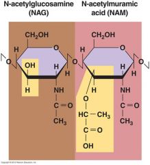
Has amino acid side chains: 1. D amino acids 2. Diaminopimelic acid (DAP) |
|
|
What kinds of crosslinks between backbones are there? |
1. Direct bonds - the back bones attach via the amino acid side chains in the NAM units A Lysine -> Alanine attachment 2. Pentapeptide side chain - bacteria linking via a pentapeptide modification (5 x glycine) of the NAM amino acid side chain |
|
|
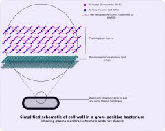
Describe the key features of a gram positive cell wall. |
Thick peptidoglycan (60-90%) 1. Interwoven teichoic acids (acidic polysaccharides) 2. Surface studded with proteins 3. No lipids in the cell wall |
|
|
How does the flow of molecules differ between the gram positive cell wall and the gram negative cell wall? |
Gram positive Molecules pass freely through the peptidoglycan so it does not trap molecules unless they are tethered to cell membranes or walls Gram negative Contains a gelatinous periplasm (which blocks molecule flow) between the outer membrane, peptidoglycan and cytoplasmic membrane. The periplasm contains proteins required for transport of material to and from cells. |
|
|
Describe the key features of a gram negative cell wall. |
1. Thin layer of peptidoglycan (10-20%) 2. Outer membrane - permeability barrier 3. Inner cytoplasmic membrane |
|
|
Describe the key features of the gram negative outer membrane. |
1. Phospholipid bilayer 2. Lipopolysaccharide (LPS) embedded in the membrane via the lipid A portion with the polysaccharide area extending outwards from the surface. 3. Surface is studded with proteins 4. Semi permeable via porins (protein like channels) |
|
|
What is LPS & how is it attached to the outer membrane? |
Lipopolysaccharides are molecules made up from: 1. Lipid A 2. Polysachharide The lipid A portion is embedded in the outer membrane and the main portion - the polysaccharide points outwards of the cell |
|
|
How are the cell walls of Mycobacterium and nocardia different to other cell walls? How can we detect these types? |
Gram +ve cell wall with mycolic acid which makes it highly resistant to chemicals. The basis for the acid-fast stain. |
|
|
What is unique about Mycoplasma? What advantage does this give? |
Lack a cell wall. Instead they are stabilised by sterols. They are pleomorphic and can assume many shapes. Due to their lack of cell wall they are resistant to many antibiotics which target the cell wall. |
|
|
What is different about the cell walls of some Archaea? |
Lack peptidoglycan and instead have pseudo peptidoglycan. |
|
|
Where is pseudo peptidoglycan found? What differentiates it from regular peptidoglycan? |
1. Found in the Archaea cell wall 2. Have N-Acetyltalosaminuronic acid instead of NAM |
|
|
Describe the overall process of how the cell wall grows. |
1. NAM and NAG are synthesised in the cytoplasm 2. Linked together via peptide crosslinks 3. Some links broken by autolysis 4. New peptidoglycan monomers insterted 5. Peptide crosslinks resealed |
|
|
How are NAM-NAG dimers attached to the membrane? |
Attached to a membrane carrier molecule called bactoprenol. |
|
|
How are NAM-NAG dimers linked to existing peptidoglycan polymers? |
Linked via the action of Transglycosidase enzymes |
|
|
What enzyme reseals the peptide crosslinks? |
Transpeptidase enzymes |