![]()
![]()
![]()
Use LEFT and RIGHT arrow keys to navigate between flashcards;
Use UP and DOWN arrow keys to flip the card;
H to show hint;
A reads text to speech;
8 Cards in this Set
- Front
- Back
|
Toreview the anatomy of a growth cone |
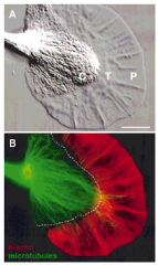
Periphery (drives movement):
Lamella (podium)
Filopodium
Centre: Microtubules
|
|
|
To understand the different localisation and roles of actin and tubulin in the growth cone |
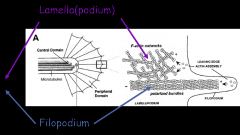
F-actin: Periphery Lamella: actin bundles cross-linked into a net Filopodia: actin bundles polarised to form larger bundles treadmills in resting growth cone, from periphery to central
Tubulin: centre Microtubules dragged sporadically into filopodia -much more dramatically when growth cone comes into contact with attractive cue. |
|
|
To understand that axons do not actually turn but theyreorganise •attraction
|
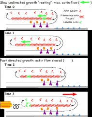
Resting: slow undirected growth
Attractive cue F-actin treadmilling is attenuated (molecular clutch engaged) Stabilises filopodium, resulting in forward movement Drags MTs into back of filopodium Establishing new growth direction |
|
|
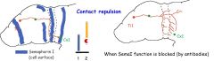
To understand that axons do not actually turn but they reorganise To show that repulsion results in... |
...localised disassembly of the actin cytoskeleton. Growth cones repulsed by each others axons. Lead to growth cone collapse destabilises F-actin [F-actin] drops repulsive signal Localised collapse of filopodia contributing to growth cone reorganisation. eg Semaphorins membrane-bound=retinal axons secreted eg Sema3A |
|
|
Tolearn about axon guidance tactics – How permissive substrates can be used to “channel” axons |
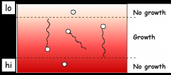
Permissive substrates = contact attractants
eg Lamin - a growth promoting ECM protein localised in optic nerve
Permissive for growth in specific conc range (not instructive-doesn't dictate direction)
Need for growth but non-permissive channel= need balance |
|
|
Tolearn about axon guidance tactics – How non-permissivesubstrates can be used to “channel” axons |
Non-permissive substrates = contact repellants eg Semaphorins Can channel axon growth but need permissive factors for growth so need balance |
|
|
Tolearn about axon guidance tactics – How long-distance diffusiblesubstances - chemoattractants and chemorepellants - can tell growth cones whichway to grow |
Long distance guiding molecules secreted by key patterning organisers. Floorplate secretes chemoattractant Netrin (&Shh) turns commissural axons Roofplate secretes chemorepellant BMP7 commissural axons repelled Work together to guide C axons towards floorplate |
|
|
BMP and Shh |
Molecules used early on to pattern embryo are reused to guide axons Shh BMP |