![]()
![]()
![]()
Use LEFT and RIGHT arrow keys to navigate between flashcards;
Use UP and DOWN arrow keys to flip the card;
H to show hint;
A reads text to speech;
63 Cards in this Set
- Front
- Back
|
axial skeleton (trunk to head) |
consists of the bones that lie around the longitudinal axis of the human body:
|
|
|
appendicular skeleton ( outside of the trunk) |
Made up of
|
|
|
Long bones |
greater in length than in width and are often slightly curved for the purpose of weight bearing. Examples:
|
|
|
Short bones |

|
|
|
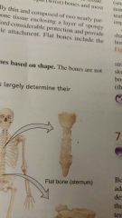
Flat bones |
|
|
|
Irregular bone |
complex shapes like the vertebrae and some facial bones |
|
|
Sesamoid bones |
Example:
Seamoid bones can develop fractures. |
|
|
Another name for Sutural bones?
|
|
|
|
The two major types of surface markings |
Allow the passage of blood vessels and nerves. Form joints
Projections out growths that form joints . Serve as attachment points for ligaments and tendons. |
|
|
spinous process ( vertebra) (Processes) |
slender projection from a vertebrae. The tail of the vertabrae
|
|
|
foramen (Vertebra) (depressions and Openings) |
|
|
|
condyle (Processes that forms joints) |
|
|
|
meatus ( depressions and opening) |
Example the external auditory (In the cranial temple, is like a whole ) |
|
|
What is the purpose of the cranial? |
small cavities of the skull:
Other functions
|
|
|
What are the skull's two grouped categories? |
|
|
|
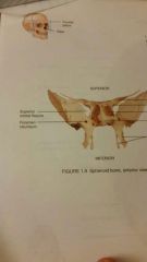
suture (“seam”) |
The three different parts:
|
|
|
Fontanels (“little fountains”) |
|
|
|
Paranasal sinuses features |
paranasal sinuses are prominent features of the
|
|
|
ethmoid sinuses ( paired) |
|
|
|
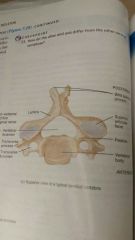
vertebrae |
|
|
|
intervertebral disc |
|
|
|
vertebral foramen
|
vertebra has a large central hole which allow the spinal cord to travel down the vertebrates.
|
|
|
intervertebral foremen |
|
|
|
Vertebral column regions |
|
|
|
How many curves as a babe to an adult? |
Adults have 3 more curves than babes.
|
|
|
cervical vertebrae |
Holds up the head ( Titan of Greek mythology supported the world) provides a pivot, allowing the head to turn on the neck
|
|
|
thoracic vertebrae |
|
|
|
lumbar vertebrae
|
|
|
|
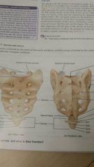
sacrum
|
|
|
|
coccyx
|
|
|
|
thoracic cage
|
formed from:The sternum, the ribs and costal cartilages |
|
|
enclose and protect |
thoracic and abdominal cavities:
|
|
|
Frontal Bone (1) (cranial bone) |
|
|
|
Parietal Bone (2) (Cranial bone) |
Sagittal suture (connects the two parietal bone) |
|
|
Temporal Bone (2) (Cranial Bone) |
Squamous (Patieral and temporal bone)
|
|
|
Occipital Bone (1) |
|
|
|
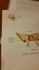
Spheniod bone (1)
(Cranial Bone) |
|
|
|
Epthmoid bone (1) (Cranial Bone) |
|
|
|
Nasal Bone (1) (Facial Bone) |
|
|
|
Maxillae Bone (2) (Facial Bone) |
Depressions and opening
|
|
|
Zygomatic Bone (2) cheek bone
(Facial Bone) |
|
|
|
Lacrimal Bone (2) (Facial Bone) |
Lacrimal Fossa ( tear duct) |
|
|
Palatin Bone (2) (Facial Bone) |
|
|
|
Inferior Nasal Conchae (2) |
|
|
|
Vomer (1) (Facial Bone) |
|
|
|
Inferior Nasal Conchae (2) |
|
|
|
What are the 4 curves of the vertebrae? |
Cervical (1-7) Thoracic (1-12) Lumbar (1-5) Sacrum and Coccyx |
|
|
Cervical vertebrae |
(feature) |
|
|
Thoracic vertebrae |
|
|
|
Sturnum (In Thoracic vertebrae) |
Made of three parts:
|
|
|
Lumber vertebrae |
|
|
|
Sacrum 1 |
|
|
|
Coccyx |
"Tailbone" made of 4 fused coccygeal vertebrae |
|
|
What type of disorders are their for the vertebrae? |
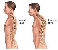
The thoracic is build extremely backed out of its normal alignment
The lumber bone is to inward
The thoracic and lumber become more of to the side. |
|
|
Hyoid Bone |
|
|
|
Cribriform Plate (foramina) |
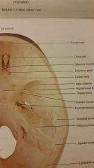
Cribriform Plate- CN I, Olfactory |
|
|
Optic foramen |
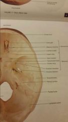
CN II, Optic |
|
|
Superior Orbital fissure |
CN III, IV, V1, VI |
|
|
Foramen Rotundum |
CN V2, maxillary |
|
|
Foramen Ovale |
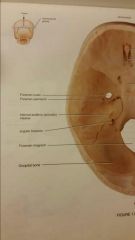
CN V3, mandibular |
|
|
Internal Auditory Meatus |
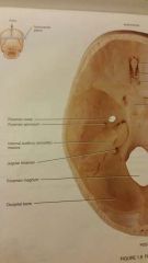
CN VII,VIII |
|
|
Jugular foramen |
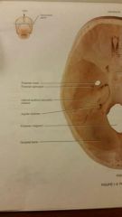
CN IX, X, XI, internal jugular vein |
|
|
Hypoglossal canal |
|