Reading...
![]()
Play button
![]()
Play button
![]()
Use LEFT and RIGHT arrow keys to navigate between flashcards;
Use UP and DOWN arrow keys to flip the card;
H to show hint;
A reads text to speech;
81 Cards in this Set
- Front
- Back
|
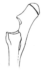
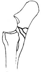
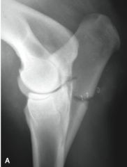
type 1a olecranon fracture (through apophysis)
|
|
|
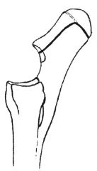
type 1b olecranon fracture (starts through apophysis and break into metaphysics to joint)
|
|
|
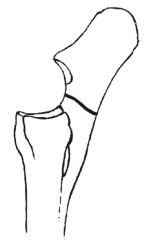
type 2 olecranon fracture (simple fracture involves joint)
|
|
|
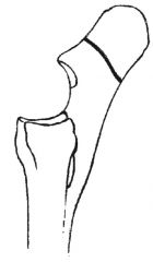
type 3 olecranon fracture (simple does not involve joint)
|
|
|
type 4 olecranon fracture (comminuted, involves joint)
|
|
|
type 5 olecranon fracture (distal body, minimal articular involvement)
|
|
|
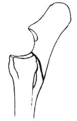
displaced type 1b olecranon fracture
|
|
|
type 1b olecranon fracture
|
|
|
type 1b olecranon fracture
|
|
|
type 2 olecranon fracture
|
|
|
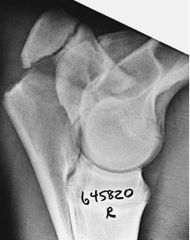
Most common ulnar fracture in horses less than 1 year:
|
type 1 b
|
|
|
Most common ulnar fracture on horses older than 1 year:
|
type 5
|
|
|
Type 1a ulnar fracture:
|
along apophyseal physis
|
|
|
Type 1b ulnar fracture:
|
starts along apophyseal physis and breaks into metaphysis to exit in the joint
|
|
|
Type 2 ulnar fracture:
|
simple fracture through body with articular involvement
|
|
|
Type 3 ulnar fracture:
|
simple fracture through body without articular involvement
|
|
|
Type 4 ulnar fracture:
|
comminuted fracture with or without articular involvement
|
|
|
Type 5 ulnar fracture:
|
distal oblique fracture with marginal articular involvement
|
|
|
What does marked displacement of ulnar fracture indicate?
|
Disruption of the aponeurosis of the ulnar head of the DDFT (lateral) and ulnar head of the flexor carpi ulnaris (medial)
|
|
|
Differential for dropped elbow stance:
|
ulnar fracture, humeral fracture, radial neuropathy
|
|
|
First aid for olecranon fracture:
|
wound care, caudal splint from ground to proximal antebrachium
|
|
|
Indications for non-surgical management of ulnar fracture:
|
non-displaced, non-articular type 5
|
|
|
Non-surgical management of ulnar fracture:
|
full limb splint, prolonged stall rest
|
|
|
Disadvantages of non-surgical management of type 5 fractures:
|
communication with joint results in inflammation in joint and development of OA, synovial fluid enters fracture line and disrupts healing and leads to development of pseudarthrosis (non-union)
|
|
|
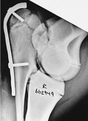
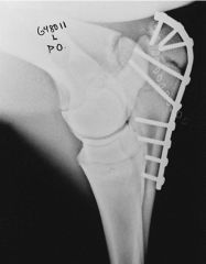
Surgical options for ulnar fractures:
|
tension-band fixation with plate & screws or tension band wire or cable
|
|
|
Incisional approach to ulnar for fracture repair:
|
curved skin incision proximally to avoid point of elbow, dissection between ulnaris lateralis and ulnar head of DDFT
|
|
|
Access to ulnar for fracture repair (after skin incision):
|
sharp dissection of connective tissue attachments of aponeurosis of ulnaris lateralis and ulnar head of DDFT, sagittal division of insertion of triceps over olecranon tuberocity to accommodate width of plate
|
|
|
How many screws are placed during ulnar fracture repair?
|
At least 5 cortices on either side of the fracture
|
|
|
Plate selection for ulnar fractures:
|
narrow LCDCP, DCP, LCP in most cases, in large horses or comminuted fractures, a broad plate may be chosen
|
|
|
When is double plate fixation of the ulnar indicated?
|
Adult horses with comminuted fractures plates are applied caudally and laterally
|
|
|
What occurs with caudolateral plate placement with ulnar fracture:
|
screws can exit medial aspect of olecranon tuberosity proximally or penetrate radial cortex distally
|
|
|
How are the first 2 screws placed during ulnar repair?
|
First screw in proximal fragment with neutral guide in compression hole without penetration of medial cortex and not tightened completely, plate is pulled distally and 2nd screw is placed in distal fragment with load drill guide, both screws tightened alternately to compress fragments
|
|
|
What results from overcompression of comminuted ulnar fractures?
|
Displacement of articular comminution fragments
|
|
|
How is overcompression of comminuted ulnar fractures prevented?
|
Screws are not applied in compression only in neutral
|
|
|
Precautions in screw placement in ulnar repair:
|
proximal fragment screws need to avoid concave medial surface penetration, screws at the level of the trochlear notch should not penetrate the joint, distal screws should not engage the caudal cortex of the radius
|
|
|
What happens if the caudal radius to fixed to the body of the ulna in foals (< 1 year)?
|
Growth of the proximal radial epiphysis forces the anconeal process into the humeral condylar notch causing luxation of the elbow joint and elbow dysplasia
|
|
|
What can be done if the subluxation occurs secondary to fixing the caudal radius to ulnar body in foals?
|
Osteotomy of the body of the ulna distal to the joint and either leaving the gap or cycling the proximal fragment through flexion and extension then bridging the osteotomy with a narrow plate
|
|
|
How is the plate fixed in a type 1b ulnar fracture?
|
Contoured to the dorsal aspect of the apophysis and engages the apophysis with 3 short screws, 4th and 5th screws are placed in lag across the metaphyseal portion of the fracture with engagement of the cranial cortex of the olecranon
|
|
|
Indications for tension wire/ cable of ulnar fracture:
|
weight < 250kg
|
|
|
What can enhance repair with tension wire/ cable of ulnar fractures?
|
Placement of pins or screws cranial to apophysis directed distally into ulnar body
|
|
|
Number of wires & wire size for ulnar repair in foals and weanlings?
|
2-3 wires 1.2mm diameter
|
|
|
Number of wires & wire size for ulnar repair in older horses:
|
4-6 wires 1.5mm diameter
|
|
|
Advantages of tension band wire/ cable repair for ulnar fracture:
|
less expensive than plates, less risk of screws entering joint or engaging caudal radius, less risk of apophyseal fracture, less tissue dissection
|
|
|
Complications of ulnar fracture repair:
|
infection, fixation failure, tendon contracture, support limb varus deformity, suspensory apparatus fatigue, support limb laminitis
|
|
|
What minimizes risks of apophyseal fracture as a complication to ulnar fracture repair?
|
Placement of place proximal to physeal scar of olecranon
|
|
|
Where are locking head screws not indicated in ulnar repair?
|
Distal aspect of the plate because perpendicular placement may weaken bone and cause radial fracture, especially with 5.0 screw
|
|
|
Best prognosis for radial fracture repair:
|
weight < 250 kg
|
|
|
Tension and compression surfaces of radius:
|
caudal cortex under compression, proximal and distal physis under compression, cranial and craniolateral aspect under tension
|
|
|
Etiology of mid-diaphyseal radial fractures:
|
cranial trauma
|
|
|
Result of lateral trauma to radius:
|
oblique fracture of proximal metaphysis
|
|
|
What occurs with SH type 1 & 2 fractures of the proximal radial physis?
|
Fracture of the ulna
|
|
|
What occurs with cranial displacement of the radial metaphysis?
|
Radial nerve dysfunction
|
|
|
First aid for radial fracture:
|
wound management, placement of caudal splint to level of elbow and lateral splint to level of mid-scapula
|
|
|
Indications for non-surgical management of radial fracture:
|
incomplete, complete non-displaced fractures
|
|
|
non-surgical management of radial fracture:
|
stall rest on tie line for 3-4 months, limited use of NSAID to provide some pain relief but not allow overuse of limb
|
|
|
Surgical management of radial fracture in adult:
|
cranially applied 4.5 LCDCP or DCP or 5.5 LCP and laterally applied DCS plate with plates offset to prevent interference of screws and prevent plates for ending at same level in transverse plan
|
|
|
Surgical management of diaphyseal or comminuted radial fracture in foals:
|
broad DCP, LCDCP, or LCP cranially with 5.5 cortex and 5.0 locking head screws and narrow or broad DCP, LCDCP, or LCP laterally with 4.5 cortex screws and 5.5 cortex or 5.0 locking head screws nearest the fracture and at ends of plate
|
|
|
Surgical management of transverse mid-diaphyseal radial fractures in foals:
|
single broad DCP, LCDCP, or LCP applied cranially with 5.5 cortex screws throughout or with LCP 5.0 locking head screws on either side of fracture and plate ends
|
|
|
Surgical management of type 3 proximal radial physeal fractures:
|
if non-displaced, conservative management
|
|
|
Surgical management of type 1 and 2 proximal radial physeal fractures:
|
plate applied to ulnar fracture engaging both cortices of radius where possible, laterally placed narrow plate with proximal screws in radial epiphysis or transphyseal screw & wire in small foals
|
|
|
Surgical management of distal radial physeal fracture:
|
transphyseal bridging with screw and wire or T plate
|
|
|
Prognosis for radial fracture repair:
|
poor in adults; excellent for physeal and transverse fractures in foals
|
|
|
Complications of radial fracture repair:
|
infection, instability, fixation failure, support limb laminitis, ALD, or suspensory apparatus fatigue
|
|
|
Fixation options for ruminant or radial or ulnar fracture:
|
Thomas splint +/- cast, ESF with TPC (closed fracture) or EF (open fracture), internal fixation
|
|
|
Type of internal fixation for radial fractures in ruminants:
|
calves < 200kg single 4.5 DCP cranial, > 200 kg double plating (either craniolateral and craniomedial or cranial and lateral), DCS for distal radial SH type 2 fracture, lag or cancellous screws with cast up to elbow in small calves
|
|
|
Why are casts not used with internal fixation of the radius?
|
Changes tension surface from cranial cortex to the caudal cortex
|
|
|
How does ulnar fracture repair in ruminant differ from horse?
|
Ulna is thin in ruminants, needs to be chiseled to create a flat surface to receive bone plate, must engage caudal cortex of radius
|
|
|
How does the ulna of ruminants differ from horses?
|
Complete, articulates with AC joint
|
|
|
Use of TPC for radial fracture in ruminants:
|
if <150 kg and pins can be placed proximal and distal to fragment, TPC spans antebrachium only, if >150 kg then full limb cast
|
|
|
indications for elbow arthroscopy:
|
septic arthritis, OC chip fracture, assisted reduction of epiphyseal fractures, DOD, staging of OA
|
|
|
important neurovascular structures surrounding the elbow:
|
cranial joint pouch is deep to radial nerve, median artery and nerves course over medial and caudomedial radius
|
|
|
arthroscopic approaches to elbow:
|
craniolateral, caudomedial, proximocaudal
|
|
|
craniolateral approach to elbow joint:
|
2cm cranial to cranial lateral humeral condyle with arthroscope inserted along cranial aspect of humeral condyles caudal to lateral digital extensor, instrument portals made through extensor muscles
|
|
|
what is visualized with craniolateral elbow approach?
|
articular surface of medial and lateral humeral condyles, proximal perimeter of radius, synovial fossa between humeral condyles is not an OCD or cartilage lesion
|
|
|
caudomedial approach to elbow:
|
3 cm distal to humeroradial articulation with scope directed from distal to proximal to penetrate medial elbow along caudal radius, instrument portals slightly caudal and proximal to scope usually through flexor carpi ulnaris
|
|
|
disadvantage of caudomedial approach to elbow
|
greatest risk of inadvertent injury to neuromuscular structures, instrument portals that are made too proximally at level of joint can injure ulnar nerve
|
|
|
what is visualized with caudomedial elbow approach?
|
humeral condyles, ulnar notch, humeral head of DDFT
|
|
|
caudoproximal approach to elbow:
|
slight flexion, between lateral humeral epicondyle and cranial border of olecranon at the level of the proximal olecranon, angled distal and cranial
|
|
|
what is visualized with caudoproximal elbow approach?
|
anconeal process, humeral condyles with flexion & extension, humeral trochlea
|
|
|
origin of DDFT
|
3 heads, humeral (medial humeral epicondyle), ulnar (medial olecranon), radial (caudal middle radius)
|
|
|
origin of SDFT:
|
2 heads, humeral (medial humeral epicondyle), radial (caudal mid-distal radius)
|