Reading...
![]()
Play button
![]()
Play button
![]()
Use LEFT and RIGHT arrow keys to navigate between flashcards;
Use UP and DOWN arrow keys to flip the card;
H to show hint;
A reads text to speech;
74 Cards in this Set
- Front
- Back
|
Size and shape of ovary:
|
The ovaries are bean or kidney shaped and approximately 7-8 cm by 3-4 cm.
|
|
|
Where is the ovulation fossa located?
|
ventral free border of the ovary
|
|
|
What is the mesovarium?
|
cranial aspect of the broad ligament
|
|
|
Where does the mesovarium attach?
|
dorsal border of the ovary
|
|
|
What is the blood supply to the ovary?
|
ovarian branch of the ovarian artery
|
|
|
Where is the oviduct?
|
extending from the cranial tip of the horn of the uterus to a loose attachment in the region of the ovulation fossa
|
|
|
Structure of the oviduct:
|
cranial or ovarian end of the oviduct is wider than the narrow, uterine end
|
|
|
Entry of the oviduct to the uterus:
|
tubal papilla
|
|
|
What is the mesosalphinx?
|
fold of the broad ligament containing oviduct
|
|
|
What portions of the uterus are peritoneal/ retroperitoneal?
|
The uterine horns and the majority of the uterine body are within the peritoneal cavity. The caudal aspect of the uterine body and the cervix are retroperitoneal.
|
|
|
What is the broad ligament composed of and where attached?
|
folds of the peritoneum and attached dorsally and laterally to the abdominal and pelvic cavity wall
|
|
|
Broad ligament of the uterus AKA:
|
mesometrium
|
|
|
Major arterial supply to the uterus:
|
uterine artery, the uterine branch of the vaginal artery, and the uterine branch of the ovarian artery
|
|
|
Conditions of the ovary requiring surgery:
|
neoplastic and non-neoplastic causing enlargement of the ovary with infertility
|
|
|
Most common neoplastic condition:
|
granulosa cell tumors, which is a sex-cord-stromal tissue tumor affecting granulosa and thecal cells
|
|
|
Clinical signs of GCT:
|
anestrus, intermittent or continuous estrous, or stallion like behavior.
|
|
|
Diagnostic findings of GCT:
|
ovarian enlargement on rectal palpation or ultrasound, a multicystic appearance on ultrasound, and elevated levels of testosterone and inhibin
|
|
|
Non-neoplastic ovarian enlargement:
|
hematomas, cysts, or abscesses.
|
|
|
Removal of ovaries can be by:
|
colpotomy, flank or ventral laparotomy, or flank laparoscopy
|
|
|
Describe Colopotomy:
|
standing position, sedation and analgesia, 4 to 5 cm incision into the vaginal wall at either the 2-4 o’clock position (right handed) or 8-10 o’clock position (left), ovary grasped with lidocaine soaked gauze, placement of the ecraseur, ecraseur is tightened until the ovary is freed, +/- losse apposition of vaginal incision
|
|
|
Why are incision made at 2-4 or 8-10 position during colpotomy?
|
avoid inadvertent laceration of bladder, rectum, or uterine branch of the urogenital artery
|
|
|
Advantages of laparotomy ovariectomy:
|
exteriorization of the ovary and direct control of hemorrhage
|
|
|
Approaches to laparotomy ovariectomy:
|
flank, ventral midline, ventral paramedian, or diagonal paramedian approach
|
|
|
When is flank laparotomy chosen?
|
recommended for an ovary less than 10 cm in diameter
|
|
|
Flank approach generally chosen for ovariectomy:
|
modified grid
|
|
|
Describe modified grid:
|
vertical incision midway between the last rib and the tuber coxae with the proximal aspect of the incision just dorsal to the dorsal margin of the internal AO muscle. Incision through the skin, SQ tissue and the external AO muscle. internal AO muscle and the transverse abdominal muscle bluntly separated
|
|
|
Why ventral midline approach for ovariectomy chosen?
|
very large tumors
|
|
|
Describe diagonal paramedian approach:
|
similar to parainguinal, bit closer to inguinal region than midline, over ovary
|
|
|
How is ovary removed for all laparotomy approaches?
|
Exteriorized, stay sutures placed in ovarian pedicle, TA-90 discharged, removed after incising between the 2 rows of staples
|
|
|
Complications of laparotomy ovariectomy:
|
hemorrhage, abdominal pain, incisional infections, and abdominal adhesions
|
|
|
Advantages of laparoscopic ovariectomy:
|
visualization of the surgical site, minimal tension on ovarian pedicle, direct hemostasis, avoidance of GA
|
|
|
Methods of laparoscopic ovary removal:
|
ligating loop sutures, stapling instruments, bipolar vessel sealing device
|
|
|
Complications of laparoscopic ovariectomy:
|
retroperitoneal insufflation, abdominal organ penetration, hemorrhage, loss of the ovary in the abdomen, post-operative colic or emphysema, incisional infection
|
|
|
Conditions of the uterus potentially requiring surgical intervention:
|
uterine cyts, pendulous uterus, neoplasia, pyometra, torsion, tears, or dystocia.
|
|
|
Methods to remove uterine cysts:
|
manual curettage or rupture, lavage with hypertonic solutions, electrocoagulation, laser ablation
|
|
|
Most successful and accepted technique of uterine cyst management:
|
Laser ablation
|
|
|
Descibe surgical correction of pendulous uterus reported by Brink et al in 2010 EVJ:
|
standing, laparoscopy, 3 incisions (midway between the last rib & tuber coxae at the level of the dorsal aspect of the IAO, 2 cm dorsal to this incision in the 17th ICS, and 6 cm distal and 2 cm caudal to the central incision) mesometrium infiltrated with lidocaine, polyglactin 910 number 6 continuous pattern to imbricate the uterus, starting caudally at the cranial body of the uterus and extending cranially to the horn, needle was passed through the seromuscular layer of the uterus then through the mesometrium 3-12 cm dorsal to its attachment to the uterus
|
|
|
Describe total ovariohysterectomy:
|
caudal ventral midline incision ovarian pedicles ligated and transected, broad ligament transection with vessel ligation, uterine body is transected as far caudal as possible, stump closed with a double inverting pattern
|
|
|
Chaney et al 2007 direction of uterine torsion:
|
59% of the torsion were clockwise
|
|
|
Chaney et al 2007 foal survival:
|
Overall 54%, 72% GE < 320 days, 32% GE > 320 days
|
|
|
Direction to roll mare with uterine torsion:
|
in the direction of the torsion, plank is placed on caudal abdomen to keep uterus in place
|
|
|
Complication of rolling:
|
uterine rupture
|
|
|
Surgical management of uterine torsion:
|
flank in a standing mare, grasp ventral gravid horn rock until returns to its normal position, or ventral midline celiotomy
|
|
|
When is VM celiotomy chosen for uterine torsion:
|
uterine rupture, tears or devitalization suspected, foal known to be dead, standing attempts unsuccessful
|
|
|
How is uterine prolapse corrected?
|
standing mare, replacement and eversion after lavage, if unsuccessful, mare anesthesized, hoisted to replace the uterus
|
|
|
Association of uterine tears to pathogenesis:
|
near tip of horn thought to be related to thrust of foals feet, in the body from force of limb or muzzle during dystocia
|
|
|
Javsicas 2010 Vet Surg paper location of tears:
|
horn > body, in horns, right > left
|
|
|
Javsicas 2010 Vet Surg palpation of tears:
|
All body tears palpable per rectum, 24% of horn tears
|
|
|
Javsicas 2010 Vet Surg differences between medically and surgically managed uterine tears:
|
none, other than medically managed mares had a higher RR at admission
|
|
|
Javsicas 2010 Vet Surg overall survival:
|
76%
|
|
|
Javsicas 2010 Vet Surg parameters associated with survival:
|
heart rate, total CO2 on admission
|
|
|
Size and shape of ovary:
|
The ovaries are bean or kidney shaped and approximately 7-8 cm by 3-4 cm.
|
|
|
Ueno et al 2010 EVJ article most common site of periparturient hemorrhage:
|
uterine artery in 24/31
|
|
|
Ueno et al 2010 EVJ article other affected arteries involved in PPH:
|
internal pudendal, caudal mesenteric, internal iliac
|
|
|
Ueno et al 2010 EVJ article ruptures location:
|
bifurcations, curves, or flexures within the vessels
|
|
|
Ueno et al 2010 EVJ article histopathology affected vessels with PPH:
|
smooth muscle atrophy with fibrosis of tunica media, disruption or calcification of tunica intima
|
|
|
Arnold et al 2008 JAVMA PPH variables associated with outcome:
|
tachycardia at presentation or during hospitalization, hemorrhage localized to vagina
|
|
|
Treatment of PPH:
|
avoiding excitement, restoring blood volume, controlling pain, facilitating hemostasis, preventing infection.
|
|
|
Describe C section:
|
caudal ventral midline incision, gravid horn exteriorized, stay sutures placed, uterotomy from hocks to feet, foal extracted, several cm of placenta peeled into lumen of uterus, closed in 2 layers, outer layer inverting
|
|
|
Dystocia resolution:
|
assisted delivery, controlled vaginal delivery, fetotomy, or cesarean section
|
|
|
Assisted delivery:
|
mare standing, assisted
|
|
|
Controlled vaginal delivery:
|
mare anesthetized, assisted delivery
|
|
|
Where is the proper ligament of the ovary?
|
connects the caudal end of the ovary to the uterus
|
|
|
Complications of colpotomy:
|
hemorrhage from the ovarian stump, eventration through the vaginal incision, abscessation or hematoma formation at the vaginal incision, and abdominal adhesions
|
|
|
Chaney et al 2007 mare survival:
|
84%, survival associated with gestational age and HR at admission, 97% with GE < 320 days, 65% > 320
|
|
|
Options of ruminant c-section:
|
standing paralumbar fossa celiotomy, ventral midline celiotomy, paramedian celiotomy, ventrolateral celiotomy, left oblique celiotomy
|
|
|
Disadvantages of VMC approach ruminant c-section:
|
dorsal recumbency is labor intensive and compromises CV status of cow, not good for daily cattle with abundant ventral vasculature
|
|
|
Advantages of VMC approach ruminant c-section:
|
better exteriorization of uterus for less risk of abdominal contamination, better for fractious beef cattle
|
|
|
Disadvantages of paramedian ruminant c-section:
|
dorsal recumbency is labor intensive & compromises CV status of cow, more hemorrhage because of multiple layers of tissue, poor holding power of closure
|
|
|
Disadvantages of ventrolateral ruminant c-section:
|
long incision in a vascularized region increases surgery time, surgeon is kneeling, increased post-operative incisional infections
|
|
|
Advantage of ventrolateral ruminant c-section:
|
good exteriorization of the uterus, especially with emphysematous fetus
|
|

|
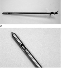
chain ecraseur
|
|
|
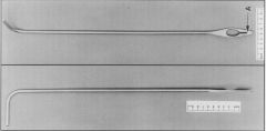
kimberly rupp spay
|
|
|
willis-drop spay
|