Reading...
![]()
Play button
![]()
Play button
![]()
Use LEFT and RIGHT arrow keys to navigate between flashcards;
Use UP and DOWN arrow keys to flip the card;
H to show hint;
A reads text to speech;
15 Cards in this Set
- Front
- Back
- 3rd side (hint)
|
What happens to the walls of the aortic arch and the carotid sinus as the left ventricle contracts?
|

As the left ventricle contracts, the sudden ejection of a bolus of blood into the aorta, common carotid artery and carotid sinus stretches the walls of these flexible vessels, resulting in a rise in intraluminal pressure. Stretch receptors located in these walls have an increased rate of fire as the vessel stretching accelerates, and the associated afferents of CNs IX and X project the information (accelerated firing rate) to the nucleus of the solitary tract in the open medulla that the blood pressure is soaring in these great vessels.
|
|
|
|
After the nucleus of the solitary tract in open medulla gets afferent info from CN IX and CN X, what does it do?
|

This nucleus in turn relays this information to the nearby nucleus ambiguus, whose cell bodies are the preganglionic parasympathetics which comprise the vagus nerve.
|
|
|
|
How do axons from the vagus nerve decrease heart rate?
|

These vagal axons inhibit the postganglionic parasympathetic neurons located close by the S-A and A-V nodes that they connect to, resulting in a decline in heart rate and an increased A-V conduction time.
|
|
|
|
As the axons of the vagus nerve are decreasing heart rate, what is the simulataneous role of the nucleus of the solitary tract?
|

Simultaneously, the nucleus of the solitary tract projects to other cells in the nucleus ambiguus that, when activated, projects caudally down the brainstem and spinal cord and inhibits tonic firing rate of the cells of the intermediolateral cell column, T1-T-12. These latter are preganglionic sympathetic cells. A decrease in their firing rate results in a decreased firing rate of the cells they project to-the postganglionic neurons of the paravertebral ganglia. The axons of these latter end on the smooth muscle of the terminal arterioles of the skin and cause constriction of the vessel so less blood flows there. Sympathetic inhibition causes the opposite effect- the smooth muscle of he arterioles relaxes passively so more blood flows from the central vessels into the large potential space of the terminal arterioles of the skin.
|
|
|
|
Describe baroreceptor reflex and what are the net paraysympathetic and sympathetric effects?
|
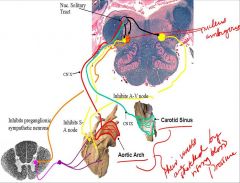
Rise in pressure in the aorta (purple dashes in previous slides) stretches the aorta thus sending some signals. We are talking about the ANS– ventricle contracts stretching walls of carotid sinus and the aortic arch. There are stretch receptors in those walls and when stretched they activate stretch receptors and CN X and CN IX.
They ascend in the caudal tract of the nucleus of the solitary tract in the open merdulla. This reflex pathway is called the baro-receptor reflex. It is active when you suddenly rise from a prone postion to upright. This is because blood “falls” due to gravity and less blood is getting to the brain—may lose consciousness. This relfex controls how blood gets to the brain and it happens in the cardiac cycle where pressure in aorta and carotid sinus are accelerating (is a very short time). There are many other factors affecting BP and this is only one of them Like all reflexes this has sense afferents, interneurons and efferents Black are interneurons from solitary tract nucleus to nucleus ambiguous. Nucleus of solitary tract are the efferent limb. Left one inhibits the sino-atrial node (pre-gang paraS). Post-gang is next to the SA node. Right side inhibits the AV node. SA node determines heart rate (pacemaker) and AV node regulates conduction time from AV node to the ventricular walls. If you inhibit heart rate through the AV node = lengthening of AV conduction time. If heart rate goes down there is less cardiac ouput and it takes longer for ventricles to contract. Together they decrease cardiac output for the moment (100ths of a second). Other neurons (orange) inhibit the pre-gang S axons. They go out by way of sympathetic chain. The system is inhibited and it usually causes constriction of the blood vessels so now they would be free to open up. The net parasympathetic and sympathetic effects is momentary decline in cardiac output and central blood pressure. |
|
|
|
Describe the descending path of the nucleus ambiguous, stretch receptors, and location of taste receptors, and differentiate between the taste and visceral pathway?
|
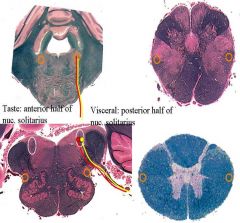
Ambiguos goes down thru medulla and spinal cord, laterally (intermedial lateral cell column)
In thoracic cord they act on the afore-mentioned cell column Aortic arch and carotid sinus stretch receptors go to nucleus of solitary tract via CN IX and X (chemoreceptor reflex follows same pathway from aortic and carotid bodies which sense O2 levels in the blood– if too low it causes change in respiratory rate) Taste is anterior nucleus of solitary tract, they ascend as shown in the red |
|
|
|
Describe what happens to the nucleus ambiguos after it gets information from the nucleus of solitary tract and what is unique about stretch receptors and visceral sensations?
|
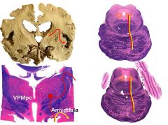
It goes up through the midbrain from the pons, projects taste input to the amygdala, and then the taste receptors terminate in the parvo cellularis of thalamus VPM. From the thalamus, the axons go via the internal capsule to the insula. The anterior insula is the primary sensory cortex for taste in the anterior third. The stretch receptor axons do not go into thalamus but to the amygdala (visceral sensation). There is NO primary cortex for visceral sensation but there are areas that influence heart rate.
|
|
|
|
Give an example of a neuron system linking the cerebral cortex, the limbic system, and the autonomic nervous system?
|
You see or hear something frightening and it influences your heart and respiration rate so sensory brain influences structures and activity.
|
|
|
|
Describe the corticocortical auditory connections?
|
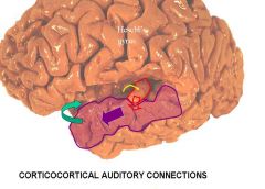
Medial geniculate projected to hescl’s gyrus which went to planum temporale and it also goes to superior temporal gyrus (auditory input) and it relays it to the amygdala.
|
|
|
|
Describe the corticortical visual connections?
|
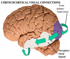
Primary visual cortex is medial parts of calcarine fissure and it continues to medial temporal gyri and then to the amygdala.
|
|
|
|
Describe the sensory - limbic pathways for conditioning?
|
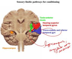
Insula gets input from VPMpc and projects to amygdala.
|
|
|
|
-Describe the role of the hypothalamus in the descending pathway?
-What is the role of edinger westphal? |
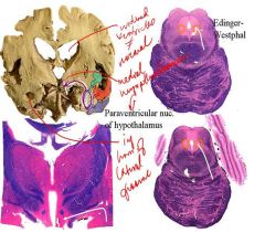
Sensory events happening in the world has an effect on internal organs by way of hypothalamic control.
There is a sensory order of relays from cortex to limbic system Edinger Westphal is a ganglion for pupillary reflex in the midbrain. Axons travel where ascending afferents travel in lateral position and go to superior and inferior salivatory nuclei. |
|
|
|
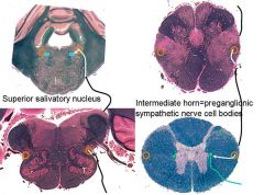
1. pre-ganglionic parasympathetic innervate what?
2. Inferior salivatory nucleus activates what? |

1. pre-ganglionic parasympathetic innervate sublingual and submandibular glands causing salivation.
2. Inferior salivatory nucleus activates the parotid gland |
|
|
|
What happens if you sever descending control of parasympathetic pre-ganglion?
|
function of pre-gangs to blood v is to cause contraction of the arterioles. If they can no longer have control the axons no longer cause firing of the interior muscle, allowing blood to flow to periphery and fill anterior arterioles making skin warm and dry = horner’s syndrome
Pupils also are constrcited because the SNS is disconnected from the brain so they no longer contract Horner’s syndrome can be a lesion from hypothalamus origination to sympathetic chain ganglion (lesion may thus be central or peripheral) |
|
|
|
Describe horner's syndrome and its 3 characteristics?
|
A disconnection of the descending autonomic pathway from the preganglionic sympathetic nerve cell bodies of the intermediolateral column of the spinal cord, T1-L4, after which the following signs appear. Its three characteristics are:
1. Miosis, a constricted pupil due to the unopposed action of the pupillary muscle. 2. Pseudo-ptosis, a drooping eyelid due to paralysis of the superior tarsal muscle. 3. Anhydrosis, an inability to activate the sweat glands. NOTES: Is the way external world affects the viscera Due to disconnection of descending control of brain over lower centers for the ANS. |
Horny PAM
|