![]()
![]()
![]()
Use LEFT and RIGHT arrow keys to navigate between flashcards;
Use UP and DOWN arrow keys to flip the card;
H to show hint;
A reads text to speech;
12 Cards in this Set
- Front
- Back
|
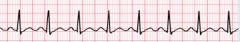
Atrial Fibrillation: Features: Irregular QRS No P Waves Variable rate Cause: SAN overwhelmed by impulses created by atria. |
|
|
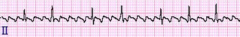
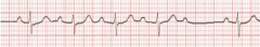
Atrial Flutter: Features: Saw tooth pattern (lots of p waves) Cause: Re-entry in atria |
|
|
Ventricular tachycardia: Features: High Rate (>120 bpm) Regular, broad QRS P waves usually absent Cause: Improper electrical activity in the ventricles |
|
|
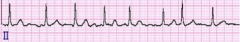
Sinus Tachycardia: Features: Normal, elevated rate Everything else normal Cause: Exercise |
|
|
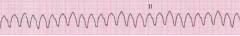
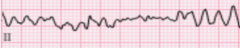
Ventricular Fibrillation: Features: Completely messed up Very high rate Loss of CO Cause: Uncoordinated contraction of ventricular muscle, leading them to quiver instead of contract |
|
|
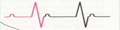
AV nodal reentrant tachycardia (AVNRT) Features: Long PR segment P wave found immediately after QRS Cause: Reentrant loop in AVN Atria and ventricles contract at the same time |
|
|
Junctional Bradycardia: Features: Low rate (<60 bpm) No P wave Cause: SAN doesn't work, reliant on AVN intrinsic heart rate |
|
|
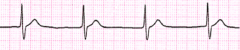
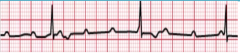
Sinus Bradycardia Features: Everything normal Lower rate Cause: Naturally low HR |
|
|
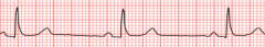
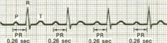
1st Degree AV block Features: Abnormally long PR interval Cause: Slowing of conduction through AVN |
|
|
2nd Degree AV block: Mobitz I type Features: Low rate PR gets larger until after 3-4 beats one is dropped Cause: |
|
|
2nd Degree AV block: Mobitz II type Features: Normal QRS Sporadic dropping of a beat |
|
|
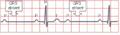
3rd Degree AV block: complete block Features: P waves unassociated with QRS P:QRS is not 1:1 Low rate Cause: No SAN impulses reach the ventricles Ventricles still contract thanks to escape rhythm |