• Shuffle
    Toggle On
    Toggle Off
  • Alphabetize
    Toggle On
    Toggle Off
  • Front First
    Toggle On
    Toggle Off
  • Both Sides
    Toggle On
    Toggle Off
  • Read
    Toggle On
    Toggle Off
Reading...
Front

Card Range To Study

through

image

Play button

image

Play button

image

Progress

1/37

Click to flip

Use LEFT and RIGHT arrow keys to navigate between flashcards;

Use UP and DOWN arrow keys to flip the card;

H to show hint;

A reads text to speech;

37 Cards in this Set

  • Front
  • Back

SA Node blood supply

RCA (55%)


Left circumflex (45%)

Normal Heart rates of different intrinsic pacemakers

SA Node: 60-90 bpm


AV Node: 45-60 bpm


His-Prkinje/BB Fibers: 30-45bpm

AV Nodal Blood supply

RCA (90%)


Left circumflex (10%)

Blood supply of Bundle Branches

RBB: LAD


LAF: LAD


LPF: RCA or Left circumflex

Mechanisms of DYSRHYTHMIA

(1) Increased automaticity


(2) Triggered activity


- Early Afterpolarizations


- Late Afterpolarizations


(3) Reentry circuits


- Two paths, one with a relatively slower refractory period

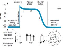
Action of myocardial cell and relation to ion flow

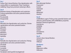
Overview of Anti-Arrhythmics

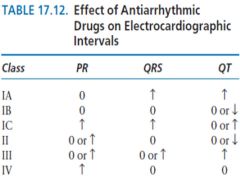
Effect of of Anti-Arrhythmics on ECG intervals

Adverse effects of Amiodarone

Acute Effects
Hypotension
Slowing of heart rate
Decreased contractility


Long-Term Effects


Common
Corneal deposits
Photosensitivity
Gastrointestinal intolerance


Less Common
Hyperthyroidism
Heart failure
Pulmonary toxicity, fibrosis
Hypothyroidism
Bradycardia
Prodysrhythmic effect


Drug Interactions
Increase Levels
- Phenytoin
- Procainamide
- Warfarin
- Digoxin
- Flecainide

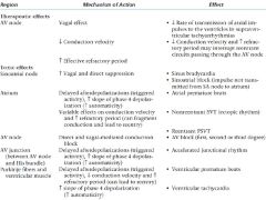
Electrophysiological effects of digoxin

Causes of sinus bradycardia


Pharmacological


  • Beta-blockers
  • Calcium-channel blockers
  • Digoxin
  • Central alpha-2 agonists
  • Amiodarone
  • Opiates
  • GABA-ergic agents
  • Organophosphate poisoning


Non-pharmacological


  • Normal during sleep
  • Increased vagal tone (e.g. athletes)
  • Vagal stimulation (e.g. pain)
  • Inferior myocardial infarction
  • Sinus node disease
  • Hypothyroidism
  • Hypothermia
  • Anorexia nervosa
  • Electrolyte abnormalities – hyperkalaemia, hypermagnesaemia
  • Brainstem herniation (the Cushing reflex)
  • Myocarditis

Differential Diagnosis of causes of bradycardia

  • Cardiac disease
    • Myocardial ischaemia / infarction
    • Myocarditis
    • Congenital disorders
    • Cardiomyopathies
    • His-Purkinje fibre degeneration
  • Hypoxia
  • Electrolyte disturbance (e.g. hyperkalemia)
  • Medications, poisonings and toxic exposures (e.g. digoxin, beta blockers, calcium channel blockers, amiodarone, clonidine, organophosphates)
  • Hypothermia
  • Hypothyroidism
  • Raised intracranial pressure (Cushing response)
  • Infections (e.g. Lyme disease, diphtheria, typhoid fever)
  • Autoimmune (e.g. systemic lupus erythematosis)
  • Infiltrative disorders (e.g. sarcoidosis, amyloidosis)
  • Physiological causes (e.g. athletes, vagal stimuli)
Differential of regular vs irregular bradycardia
Regular
Sinus bradycardia
Junctional bradycardia/Idioventricular
Complete AV block (junctional escape)
Atrial flutter with high degree block

Irregular
Sinus arrhythmia, pause or arrest
Sinoatrial exit block (second degree)
Atrial fibrillation with slow ventricular response
Atrial flutter with variable block
Second degree AV block, type I
Second degree AV block, type II

Sick Sinus Syndrome

group of dysrhythmias from disease
of the sinus node and its surrounding tissues,including sinus bradycardia, sinus arrest, and SA exit block

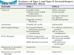
Type I vs Type II AV BLOCK

Bigeminy

Extrasystole after every native beat

Ashman phenomenon

wide complex QRS complexes that follow a short R-R interval preceded by a long R-R interval


This wide QRS complex represents an aberrantly conducted complex that originates above the AV node


occurs because the duration of the refractory period of the myocardium is proportional to the R-R interval of the preceding cycle
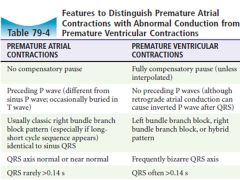
PAC vs PVC

Cause of PVCs and Vtach

Acute or previous myocardial infarction or ischemia
Hypokalemia
Hypoxemia
Ischemic heart disease
Valvular disease
Catecholamine excess*
Other drug intoxications (especially cyclic antidepressants)
Idiopathic causes†
Digitalis toxicity
Hypomagnesemia
Hypercapnia
Class I antidysrhythmic agents
Ethanol
Myocardial contusion
Cardiomyopathy
Acidosis
Alkalosis
Methylxanthine toxicity

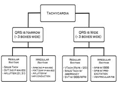
Tachycardia Algorithim

Causes of sinus tachycardia

Drugs/Meds/Iatrogenic


Anticholinergic


Catecholamines


Alcohol


Caffeine


Tobacco


Cocaine


β-blocker withdrawal


Supraventricular tachycardia ablation


Medical


Anemia


Dehydration


Exercise


Anxiety


Pain


Pulmonary embolus


Fever


Infection


Pericarditis


Aortic or mitral regurgitation


Myocardial infarction


Pneumothorax


Hyperthyroidism


Hypoglycemia

Potential Causes of A Fib

Cardiac causes
Hypertension
Heart failure
Coronary artery disease/ prior MI
LV Dysfunction
Cardiomyopathies
Valvular heart disease
Congenital heart disease
Pericardial disease
Postsurgical (particularly cardiac surgery)
Sick sinus syndrome
AF as a result of ventricular pacing
Supraventricular tachycardia (including WPW, atrial tachycardia, a flutter, ect)
Genetic/familial


Noncardiac causes
OSA
Obesity
Excessive alcohol ingestion
Hyperthyroidism
Pulmonary disease (pneumonia, COPD, PE, Pulm HTN)
Lone (idiopathic) AF

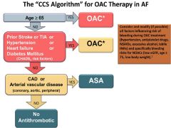
2014 CCS Algorithim for who needs anticoagulation with a fib

Relative risk reduction for stroke in patients with afib:


- ASA


- Wargarin

ASA: 22% (NNT = 34)


Warfarin: 64% (NNT = 14)

CVA risk at different CHADS2 scores

0: 1.9


1: 2.8


2: 4.0


3: 5.9


4: 8.5


5: 12.5


6: 18.2

2014 CCS reccomendations regarding OACs

(1) for OAC with nonvalvular AF, dabigatran, rivaroxaban or apixaban in preference to warfarin


(2) for OAC patients with a mechanical prosthetic valve, rheumatic mitral stenosis or eGFR of 15-30 ml warfarin is prefered over NOACs


(3) Consider lower dose age >75 and CRF

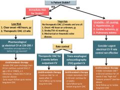
CCS ED algorithim for management of A Fib

AVRT with orthodromic conduction (AV node 1st)

  • Rate usually 200 – 300 bpm
  • P waves may be buried in QRS complex or retrograde
  • QRS Complex usually <120 ms unless pre-existing bundle branch block, or rate-related aberrant conduction
  • QRS Alternans – phasic variation in QRS amplitude associated with AVNRT and AVRT, distinguished from electrical alternans by a normal QRS amplitude
  • T wave inversion common
  • ST segment depression

AVRT with antidromic conduction (bypass 1st)

  • Rate usually 200 – 300 bpm.
  • Wide QRS complexes due to abnormal ventricular depolarisation via accessory pathway

Diseases associated with WPW

Idiopathic*
Cardiomyopathy (especially hypertrophic)
Transposition of great vessels
Endocardial fibroelastosis
Mitral valve prolapse
Tricuspid atresia
Ebstein’s disease

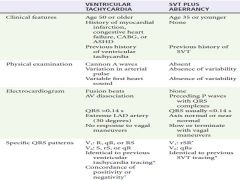
SVT with aberrancy versus VT

Criteria for Torsade de Pointes

1. Ventricular rate greater than 200 beats/min
2. Undulating QRS axis, with the polarity of the complexes
appearing to shift about the baseline
3. Paroxysms of less than 90 seconds

Classification of Conditions that can lead to to Torsade de Pointes

Pause Dependent (Acquired)
Drug induced: Class IA and IC antidysrhythmics; many phenothiazines and butyrophenones (notably haloperidol and
droperidol), cyclic antidepressants, antibiotics (especially macrolides), organophosphates, antihistamines, antifungals,
antiseizure and antiemetic agents
Electrolyte abnormalities: hypokalemia, hypomagnesemia, hypocalcemia (rarely)
Diet related: starvation, low protein
Severe bradycardia or atrioventricular block
Hypothyroidism
Contrast injection
Cerebrovascular accident (especially intraparenchymal)
Myocardial ischemia


Adrenergic Dependent (Tachycardia Prompted)
Congenital
Jervell and Lange-Nielsen syndrome (deafness, autosomal recessive)
Romano-Ward syndrome (normal hearing, autosomal dominant)
Sporadic (normal hearing, no familial tendency)
Mitral valve prolapse
Acquired (Rare)
Cerebrovascular disease (especially subarachnoid hemorrhage)
Autonomic surgery: radical neck dissection, carotid endarterectomy, truncal vagotomy

Rx for Torsade de Pointes (acquired)

Empiric magnesium


D/C any QT pronlonging agents


Correct Electrolyte abnormalities


Increased HR to shorten QT
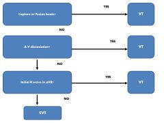
Ainsworth SVT vs VT

ECG Findings to search for in syncope

(1) Acute coronary syndrome
(2) Tachycardia/Bradycardia and AV blocks
(3) Intervals
- Short PR suggesting WPW/Pre-excitation
- Long or Short QT interval
(4) Hypertrophic cardiomyopathy
(5) Brugada syndrome
(6) Arrhythmogenic right ventricular dysplasia

Critical diagnoses to consider in patient with syncope

Life threatining dysrhythmias


Myocardial infarction


Outflow obstruction


Critical aortic stenosis


PE


Pericardial tamponade


Hypertrophic cardiomyopathy


Vascular catastrophes


SAH


Thoracic aortic dissection


Abdominal aortic aneurysm


Ectopic pregnancy


Hypovolemia/Hemorrhage/Anemia


Toxic/Metabolic derangements


Stroke