Reading...
![]()
Play button
![]()
Play button
![]()
Use LEFT and RIGHT arrow keys to navigate between flashcards;
Use UP and DOWN arrow keys to flip the card;
H to show hint;
A reads text to speech;
70 Cards in this Set
- Front
- Back
|
Contractility |
Muscle can contract (shorten) forcefully.
|
|
|
Excitability
|
This means it can be stimulated by nerves and hormones. This allows the nervous system and the endocrine system to control the muscle system.
|
|
|
Extensibility
|
Under normal conditions, it can be stretched out.
|
|
|
Elasticity
|
This means that when the muscles are extended beyond their normal length, they can recoil back to their normal length.
|
|
|
three kinds of muscle tissue?
|
skeletal muscle, smooth muscle, and cardiac muscle
|
|
|
6 Skeletal Muscle Characteristics
|
1. striated (striped)
2. multiple nuclei 3. arranged as many, long, cylindrical cells bundled together 4. several bundles enclosed in tough connective tissue sheath to form a muscle 5. they are responsible for voluntary movement 6. generally connected to bones via tendons. |
|
|
Belly of the muscle
|
is composed of the actual muscle cells.
|
|
|
Fascicles
|
whole muscle is divided into bundles of cells
|
|
|
Muscle fiber (cell)
|
Very long compared with most other cells, up to several cm long, 10-100 micrometers in diameter
|
|
|
Endomysium
|
a thin collagen layer that wraps each cell.
|
|
|
Perimysium
|
the collagen layer that wraps each fascicle.
|
|
|
Epimysium
|
another collagen layer that wraps the perimysium fascicles and differentiates one muscle from the other.
|
|
|
Fascia
|
a sheet of tissue that lies underneath the skin.
|
|
|
Sarcolemma
|
which is the plasma membrane of the muscle fiber. The prefix “sarco” means literally “flesh” or “meat.”
|
|
|
Sarcoplasmic reticulum
|
the endoplasmic reticulum of a muscle fiber. the sarcoplasmic reticulum surrounds regions which are packed with myofibrils.
|
|
|
T-tubules
|
form pits in the sarcolemma. allows a muscle signal to get down to the sarcoplasmic reticulum and make it possible for the message to get to where it needs to be in order to stimulate muscle movement.
|
|
|
Myofibrils
|
thread-like structures which are only a few micrometers in diameter. They extend from one end of the cell to the other. They are structured in repeating units that causes striations.
|
|
|
Myofilaments
|
two filaments of the myofibril which are further separated into two types:actin myofilaments and myosin myofilaments .
|
|
|
How do you know if a sample is complete muscle?
|
If an epimysium is present, it is a complete muscle.
|
|
|
Sarcoplasm
|
is the cytoplasm of a muscle fiber
|
|
|
In the contraction of a sarcomere, the following things happen:
|
1. Ca2+ binds to troponin, exposing the active sites on the actin, allowing the myosin heads to grab onto the actin.
2. The power stroke. 3. ATP binds to the myosin heads, making them release the active sites on the actin. 4. The return stroke. |
|
|
actin
|
thin filament proteins
|
|
|
myosin
|
thick filament proteins
|
|
|
2 other proteins associated with thin filaments
|
tropomyosin and troponin
|
|
|
Z-line
|
= disc-like structures to which actin filaments attach on both sides; composed of alpha-actinin and a dense amorphous matrix
|
|
|
A-band
|
"anisotropic" band: thick dark stripes are indicative of where the myosin is.
|
|
|
I-band
|
"isotropic" band: lighter areas where the actin is.
|
|
|
H-zone
|
pale central region in A-band; due to absence of thin filaments; is the area between the ends of the actin myofilaments which point to one another from opposite ends of the sarcomere.
|
|
|
M-line
|
thick filaments interconnected by cross-linking fine radial filaments, acts to maintain regular spacing and arrangement of thick filaments
|
|
|
cross bridge
|
the combination of a myosin head with the active site of an actin myofilament.
|
|
|
ADP
|
Adenosine Diphosphate
|
|
|
P molecules
|
Phosphate molecules
|
|
|
Motor neurons
|
cells specifically designed to carry signals from the brain and spinal cord to the skeletal muscles at a high velocity.
|
|
|
Neuron
|
The functional unit of the nervous system, a nerve cell
|
|
|
Neuromuscular junction
|
When the axon of a motor neuron reaches the perimysium surrounding several muscle fibers, it branches several times, each branch connecting with one muscle fiber.
|
|
|
Explain the 1st step of a sarcomere contraction
|
Before contraction, the active sites on the actin on the myofilament are not exposed. The myosin myofilament heads are ready, but there isn't anything to do yet.
|
|
|
Explain the 2nd step of a sarcomere contraction
|
start contraction - Ca2+ ions bind to the tropinin, causing the proteins in the actin myofilament to re-arrange. This exposes active sites where the heads can bind. Phosphate ejects when the head binds.
|
|
|
Explain the 3rd step of a sarcomere contraction
|
Power Stroke - the heads bend pulling the actin myofilament. ADP ejects.
|
|
|
Explain the 4th step of a sarcomere contraction
|
ATP molecules bind to the heads, releasing the active sites, the actin myofilament moves.
|
|
|
Explain the 5th step of a sarcomere contraction
|
Return Stroke - ATP molecules break down into ADP and P still bound to the myosin head. Energy releases, heads spring back - repeats.
|
|
|
Synaptic cleft
|
a gap between the end of the axon and the muscle fiber.
|
|
|
Synapse
|
The interface between a nerve cell and another cell
|
|
|
Presynaptic terminal
|
the very end of the nerve
|
|
|
Postsynaptic membrane
|
the membrane of the muscle fiber in the synaptic cleft region.
|
|
|
Synaptic vesicles
|
mitochondria which produce energy and stimulate muscle with acetylcholine Ach – a neurotransmitter.
|
|
|
what are the 7 steps of muscle contraction?
|
1. An action potential travels down the axon of a motor neuron.
2. ACh is released from the presynaptic terminal. 3. ACh travels across the synaptic cleft. 4. ACh interacts with the muscle fiber membrane to create a muscle action potential. 5. The action potential travels down a T-tubule. 6. Calcium ions are released from the sarcoplasmic reticulum. 7. Calcium ions bind to the troponin in an actin myofilament. |
|
|
what are the 6 steps of muscle relaxation?
|
1. an enzyme called acetylcholinesterase inactivates ACh.
2. the muscle action potential is gone, the diffusion of calcium ions out of the sarcoplasmic reticulum stops. 3. Sarcoplasmic reticulum goes back to actively transport calcium ions back inside itself. 4. The active transport of calcium ions into the sarcoplasmic reticulum removes those ions from the troponin that is on the actin myofilaments. 5. troponin and tropomyosin rearrange again to hide the active actin sites. 6. the myosin heads have nothing to grab onto, and no cross bridges can form. |
|
|
ATP molecules are bound to the myosin heads in a sarcomere and are breaking down. Is this the return stroke or the power stroke?
|
return stroke
|
|
|
Suppose you were looking at the Ca2+ concentration in the sarcoplasmic reticulum of a muscle fiber. If the Ca2+ concentration in the sarcoplasmic reticulum suddenly increased dramatically, is the muscle starting its contraction or has it finished contracting?
|
finished contracting
|
|
|
When the sarcoplasmic reticulum releases Ca2+ ions in response to the action potential, do the ions move through the membrane of the sarcoplasmic reticulum via osmosis or diffusion? Is this active or passive transport?
|
this is diffusion, and it is passive transport
|
|
|
Suppose someone is working out and is pushing himself well beyond the limit. In fact, he gets so fatigued that he runs out of energy, and his muscle fibers can no longer produce ATP. What will happen in his sarcomeres?
|
the muscle would temporarily cramp or stiffen, much like rigor mortis.
|
|
|
When a muscle fiber contracts, what happens to the distance between the Z disks?
|
The distance between the Z disks decreases, because the contraction pulls the Z disks
together. |
|
|
When a muscle fiber contracts, what happens to the length of the A band?
|
The length of the A band remains the same. Remember, the length of the myosin
myofilament does not change. Thus, the A band, which is defined by the length of the myosin myofilament, stays the same. |
|
|
When a muscle fiber contracts, what happens to the length of the I band?
|
The length of the I band decreases. The actin myofilaments are pulled towards the center of
the sarcomere. This increases the amount of overlap between the actin and myosin myofilaments. The I band is defined as the region with actin myofilament only. Since more actin overlaps with myosin, the length of the actin myofilament only region goes down. |
|
|
When a muscle fiber contracts, what happens to the length of the H zone?
|
The length of the H zone decreases. The actin myofilaments from each side of the sarcomere
are pulled closer. This reduces the length of the region in which there is only myosin, which is the definition of the H zone. |
|
|
When a muscle fiber contracts, what happens to the length of the myosin myofilament?
|
The length of the myosin myofilament remains the same. The myofilaments never change in length. The amount that they overlap changes.
|
|
|
When a muscle fiber contracts, what happens to the length of the actin myofilament?
|
The length of the actin myofilament remains the same. The myofilaments never change in length. The amount that they overlap changes.
|
|
|
The concentration of calcium ions in the sarcoplasmic reticulum is decreasing. Is the muscle fiber starting to contract or has it finished contracting?
|
If the calcium ion concentration is decreasing, that means calcium ions are heading into the
sarcomere. This means the muscle is starting to contract, because calcium ions bind to the troponin on the actin myofilaments to begin contraction. |
|
|
The myosin heads of a sarcomere have just received a boost of energy. Is the power stroke or the return stroke about to happen?
|
If the myosin heads have just received a boost of energy, ATP must have bound to them and
then broken down into ADP and P. This means the return stroke is about to start. Remember, the myosin heads get “primed” with energy during the return stroke. They then use that energy on the power stroke. |
|
|
A myosin head has ADP attached to it but not an individual phosphate. Which is going to happen next: the return stroke or the power stroke?
|
If ADP but no P is attached, that means the myosin head has bound to the active site (that’s what bumps the P off). Thus, the power stroke is about to happen. That will kick off the ADP.
|
|
|
If you could look at several muscle fibers while they are in action, how could you determine which cells are a part of the same motor unit?
|
All of the cells in the same motor unit will contract identically at the same time. If you could see several muscle cells always contracting in unison, that is a motor unit.
|
|
|
What is the function of acetylcholinesterase?
|
Acetylcholinesterase inactivates ACh after it has interacted with the postsynaptic membrane.
|
|
|
If it were not for acetylcholinesterase, what
would happen to a muscle? |
the muscle cell could never relax once it started contracting!
|
|
|
There are two major roles that ATP plays in muscle contraction and relaxation. The first involves the sarcoplasmic reticulum, while the second involves the myosin head. What are those
two roles? |
ATP provides the sarcoplasmic reticulum with energy for the active transport of calcium
ions into itself. This is critical for muscle relaxation. ATP attaches to the myosin heads, making them release the active sites and giving them energy for the return stroke. Without this step, the myosin heads would never let go of the active sites, and the muscle could neither relax nor contract. |
|
|
A muscle is stiff. It can neither contract nor relax. What is wrong in the sarcomere? What causes this?
|
The myosin heads must be gripping the active sites and not letting go. This must be due to a lack of ATP in the sarcomere.
|
|
|
When a muscle fiber relaxes, does it automatically stretch back to its resting size?
|
No. When a muscle cell relaxes, the myosin heads let go of the active sites. This makes it very easy for the muscle to stretch out, but that does not automatically happen. Something else (gravity or another muscle’s contraction) must cause the relaxed muscle to extend.
|
|
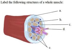
Label the following structures of a whole muscle:
|
a. epimysium b. perimysium c. endomysium d. muscle cell (fiber) e. fascicle
|
|
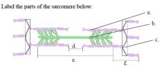
Label the parts of the sarcomere below:
|
a. actin myofilament b. myosin myofilament c. Z disk d. H zone e. A band f. I band
|
|
|
In terms of their nuclei, skeletal muscle fibers are different than most cells in two ways.
What are those ways? |
Skeletal muscle cells have many nuclei rather than just one. Also, the nuclei are at the edge of the cell rather than near the center.
|
|
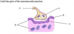
Label the parts of the neuromuscular junction:
|
a. presynaptic terminal b. mitochondria c. synaptic vesicle d. synaptic cleft
e. postsynaptic membrane |