• Shuffle
    Toggle On
    Toggle Off
  • Alphabetize
    Toggle On
    Toggle Off
  • Front First
    Toggle On
    Toggle Off
  • Both Sides
    Toggle On
    Toggle Off
  • Read
    Toggle On
    Toggle Off
Reading...
Front

Card Range To Study

through

image

Play button

image

Play button

image

Progress

1/13

Click to flip

Use LEFT and RIGHT arrow keys to navigate between flashcards;

Use UP and DOWN arrow keys to flip the card;

H to show hint;

A reads text to speech;

13 Cards in this Set

  • Front
  • Back

Small Intestine Histology


  • Mucosa - what epithelium?
  • What are gastric pits? What do they communicate with?
  • Three specialized gastric cells?

Small intestine mucosa: simple columnar epithelium


Gastric pits are in the stomach, communicate with gastric glands lined with mucous cells and specialized gastric cells:


  • Parietal cells
  • Chief cells
  • Enteroendocrine cells

How long is the small intestine?


Diameter?


Duodenum length?


Jejunum length?


Ileum length?


Intestine is 20ft


1 inch in diameter


Duodenum 10 inches


Jejunum 8 feet


Ileum 12 feet

4 Layers of the stomach? Innermost to outermost

Mucosa: simple columnar


Submucosa: nerve plexus


Muscularis: oblique, circle, longitudinal


Serosa

Liver


  • How much does it weigh?
  • Where is it located?
  • Composed of what?
  • Supported by what?
  • Which lobe is larger?
  • Where is the gall bladder located?
  • Weighs 3 pounds

  • Inferior to diaphragm

  • Composed of four lobes

  • Supported by two ligaments (falciform ligament and round ligament)

  • Right lobe is larger

  • Gall bladder on right lobe

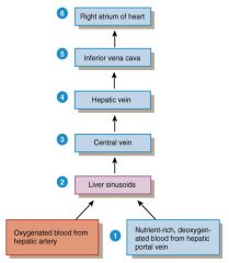
Blood supply to liver?


6 Steps

Urinary System


  • 4 Structures
  • 6 Functions

4 Structures:


  • Kidney
  • Ureters
  • Urinary bladder
  • Urethra


Functions:


  • Regulate plasma concentrations of ions
  • Regulate BP and blood volume
  • Stabilize blood pH
  • Conserve nutrients
  • Eliminate organic waste
  • Assist liver in detoxifying blood

Kidney: External Anatomy


  • Location
  • At which levels of spine?
  • Protected by which ribs?
  • Located between the peritoneum and the posterior abdominal wall
  • Retroperitoneal along with adrenal glands and ureters
  • At level of T12 to L3
  • Protected by 11th and 12th ribs

Kidney: 3 Layers of Tissues?

Outermost: renal fascia - thin layer of tissue that holds against posterior body wall


Middle: Adipose capsule - protect from trauma


Innermost: renal capsule (fibrous capsule) - dense connective tissue

Parenchyma of Kidney


Made up of two parts? Renal_________


1. Renal cortex = superficial layer of kidney


2. Renal medulla


Inner portion consisting of the pyramids


Bases facing renal cortex, apex at papilla


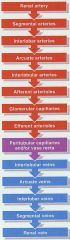
Blood into kidneys, out as urine

Nephron - Two parts


-Renal_________

1. Renal corpuscle: glomerulus and Bowman's capsule


2. Renal tubules: proximal convoluted tubules, loop of Henle, distal convoluted tubules, collecting duct

Renal Corpuscle


  • Glomerular capillaries arise from _______________ and form a ball before being emptied into the _________________ - glomerulus
  • Bowman's capsule - 2 layers?
  • Glomerular capillaries arise from afferent arteriole and form a ball before being emptied into the efferent arteriole - glomerulus

  • Bowman's Capsule:

  • Visceral (inner) layer - podocytes, modified simple squamous epithelial cells

  • Parietal (outer) layer - simple squamous cells

hi

bye