Reading...
![]()
Play button
![]()
Play button
![]()
Use LEFT and RIGHT arrow keys to navigate between flashcards;
Use UP and DOWN arrow keys to flip the card;
H to show hint;
A reads text to speech;
344 Cards in this Set
- Front
- Back
|
Plasma membrane
|
-Forms a cell's flexible outer surface
-Separates the cell's internal environment form its external environment -Regulates the flow of materials into and our of a cell -Communication among cells and between cells and their external environment. |
|
|
Cytoplasm
|
Consists of all the cellular contents between the plasma membrane and the nucleus
|
|
|
Cytosol
|
The liquid portion of cytoplasm that consists mostly of water plus dissolved solutes and suspended particles.
|
|
|
Organelles
|
Several different types in the cytosol. Each has a characteristic structure and specific functions.
|
|
|
Nucleus
|
The largest organelle. Acts as the control center for a cell because it contains the genes, which control cellular structure and most cellular activities.
|
|
|
Lipid bilayer
|
Basic framework of the plasma membrane, two back-to-back layers made up of 3 types of lipid molecules.
|
|
|
Phospholipids
|
Lipids that contain phosphorus
|
|
|
Amphipathic
|
Having both a polar (charged) and nonpolar (uncharged) aspect.
|
|
|
Glycolipids
|
Lipids attached to carbs
|
|
|
Integral proteins
|
Proteins that extend into or through the lipid bilayer
|
|
|
Transmembrane proteins
|
Proteins that span the entire lipid bilayer and protrude into both the cytosol and extracellular fluid.
|
|
|
Peripheral proteins
|
Proteins that are loosely attached to the exterior or interior surface of the membrane.
|
|
|
Functions of membrane proteins
|
Ion channels
Transporters Receptors Enzymes Cell-Identity Markers Linkers (anchor the proteins of two neighboring cells together) |
|
|
Glycoproteins
|
Peripheral proteins that are attached to carbs
|
|
|
Selective permeability
|
The property of membranes that allows some substances to move into and out of the cell but restricts the passage of other substances.
|
|
|
Ion channels
|
Channels formed by integral proteins through which specific ions can move into and out of cells.
|
|
|
Carriers (transporters)
|
Membrane proteins which change shape as they move a substance from one side of the membrane to the other.
|
|
|
Receptors
|
Integral proteins that recognize and bind a specific molecule that governs some cellular function.
|
|
|
Enzymes
|
Integral proteins that speed up specific chemical reactions.
|
|
|
Cell identity markers
|
Membrane glycoproteins and glycolipids that enable a cell to recognize other cells of its own kind during tissue formation, or to recognize and respond to potentially dangerous foreign cells.
|
|
|
Concentration gradient
|
The difference in the concentration of a chemical between two different places (in the case of a cell, the difference is between the intracellular and extracellular fluid).
|
|
|
Electrical gradient
|
The difference in the electric charge between two places, aka the membrane potential.
|
|
|
Electrochemical gradient
|
The combination of the concentration and electrical gradients. Maintaining these gradients is important to the life of the cell.
|
|
|
Passive processes
|
Processes driven by gradients and do not require ATP input.
|
|
|
Types of passive processes
|
Simple diffusion
Facilitated diffusion Osmosis Bulk flow |
|
|
Active processes
|
Processes that go against the concentration gradient and require energy, typically in the form of ATP.
|
|
|
Types of active processes
|
Active transport
Vesicular transport |
|
|
Diffusion
|
The random movement of particles that occurs as a result of multiple collisions between particles.
|
|
|
Diffusion rates
|
Influenced by the steepness of the concentration gradient, temperature, size of the diffusing substance, diffusion distance, and surface area of the membrane.
|
|
|
Simple diffusion
|
Nonpolar, hydrophobic molecules diffuse across the lipid bilayer without the help of transport proteins.
|
|
|
Facilitated diffusion
|
An integral protein assists the transport of a solute across the otherwise impermeable lipid bilayer.
|
|
|
Channel-mediated diffusion
|
The transmembrane protein provides a route or channel through which the ion can pass.
|
|
|
Ion channels
|
Selective and specific membrane proteins and some have gates that open and close.
|
|
|
Carrier-mediated diffusion
|
A solute binds to a specific transporter on one side of the plasma membrane an dis release on the other after the transporter undergoes a conformational change.
|
|
|
Glucose carriers
|
Membrane proteins that facilitate the transport of glucose across the plasma membrane.
|
|
|
Osmosis
|
The net movement of a solvent through a selectively permeable membrane or in living systems, the movement of water across the membrane.
|
|
|
Osmotic pressure
|
A pressure exerted on the cell membrane by a solution containing solute particles that cannot pass through a membrane.
|
|
|
Aquaporins
|
Transmembrane proteins that act as water channels.
|
|
|
Tonicity
|
Relates to the pull that the solutes exert on the water molecules.
|
|
|
Isotonic solution
|
A solution in which the concentration of solutes are the same on both sides, resulting in the cells maintaining their normal shape and volume.
|
|
|
Hypotonic solution
|
A solution that has a lower concentration of solutes.
|
|
|
Hemolysis
|
The rupture of red blood cells.
|
|
|
Hypertonic solution
|
A solution that has a higher concentration of solutes.
|
|
|
Crenation
|
Shrinkage of red blood cells.
|
|
|
Active transport
|
Energy derived from ATP is used to pump a substance across a plasma membrane against its concentration gradient.
|
|
|
Sodium-potassium pump
|
The most important active transport pump. Expels sodium ions from cells and brings in potassium ions. Also acts as an enzyme to split ATP.
|
|
|
Vesicular transport
|
Vesicles help transport substances into or out of a cell.
|
|
|
Endocytosis
|
Material is moved into a cell via a vesicle formed when the plasma membrane invaginates and pinches off. The material located in the invaginated portion ends up inside the cell.
|
|
|
Types of endocytosis
|
Phagocytosis
Pincytosis |
|
|
Phagocytosis
|
The ingestion of solid particles.
|
|
|
Pinocytosis (aka bulk-phase)
|
The ingestion of extracellular fluid.
|
|
|
Exocytosis
|
Membrane-enclosed structures that form inside the cell fuse with the plasma membrane and release their contents into the extracellular fluid. The vesicle's membrane "merges" with the cell's membrane and becomes incorporated into it.
|
|
|
Vesicle
|
A small round sac formed by budding of from an existing membrane that transports substances from one structure to another.
|
|
|
Nucleus
|
The organelle that houses the chromosomes.
|
|
|
Cytosol
|
Intracellular fluid and the gel-like portion of cytoplasm in which the organelles are suspended.
|
|
|
Organelles
|
Specialized structures that have characteristic shapes and perform specific functions that are necessary for the cell's growth, maintenance, and reproduction.
|
|
|
Cytoskeleton
|
A network of several kinds of protein filaments that extend throughout the cytosol, providing a structural framework for the cell and aiding in cellular movement.
|
|
|
Microfilaments
|
The smallest of the three types of protein filaments similar to two intertwined chains. Example: actin filaments in muscle cells.
|
|
|
Intermediate filaments
|
Slightly larger in diameter that microfilaments. Example: Mysosin filaments in muscle cells.
|
|
|
Microtubules
|
Largest of the protein filaments. Include centrioles, cilia and flagella.
|
|
|
Centrioles
|
Organelles that are paired cylinders arranged at right angles to one another. Made of microtubules and play a role in spindle fiber formation during cellular division.
|
|
|
Cilia
|
Numerous, short, hair-like projections that extend from the surface of a cell and function to help move material along the surface of a cell. Help liquid and particles move over the surface of the cell, not into the cell.
|
|
|
Flagella
|
Move an entire cell (cellular locomotion). Example: The tail of the mature sperm.
|
|
|
Ribosomes
|
Tiny spheres, consisting of a large and small subunit. Occur free-floating in the cytosol or are associated with the rough ER. The sites of protein synthesis within a cell.
|
|
|
Endoplasmic reticulum (ER)
|
A network of membranes that form flattened sacs or tubules. Transports substances, synthesizes molecules, detoxifies chemicals, and releases Ca2+ involved in muscle contraction.
|
|
|
Rough ER
|
ER that is continuous with the nuclear membrane. Its outer membranous surface is studded with ribosomes.
|
|
|
Smooth ER
|
ER that extends from the rough ER to form a network of membranous tubules, but is not associated with ribosomes.
|
|
|
Golgi body
|
Consists of four to six membranous sacs that are flattened and stacked. Modifies, sorts, and packages proteins into vesicles for transport within the cell, or to the extracellular space.
|
|
|
Lysosomes
|
Vesicles that form from the Golgi body and contain powerful digestive enzymes. Function in intracellular and extracellular digestion.
|
|
|
Mitochondria
|
Bound by a double membrane. The outer membrane is relatively smooth and straight, while the inner membrane is arranged in numerous folds. The intracellular sites of ATP production.
|
|
|
Nuclear envelope
|
A double membrane surrounding the nucleus.
|
|
|
Nuclear pores
|
Channels that perforate the nuclear membrane.
|
|
|
Nucleolus
|
A specific region within the nucleus where ribosomal subunits are synthesized.
|
|
|
DNA
|
The hereditary material within a cell. It is used as the template for the synthesis of all proteins made within a cell.
|
|
|
Histones
|
"Protein spools"
|
|
|
Chromosome
|
Consists of one DNA molecule and all of its associated histones. There are 46 found within a human cell.
|
|
|
Somatic cells
|
Body cells
|
|
|
Gametes
|
Eggs and sperm
|
|
|
Genes
|
Regions of DNA which code for specific proteins.
|
|
|
Cell division
|
The process by which cells reproduce themselves.
|
|
|
Nuclear division
|
Cell division including mitosis or meiosis.
|
|
|
Cytokinesis
|
The division of a cell's cytoplasm.
|
|
|
Somatic cell division
|
Cell division that results in an increase in the number of body cells and involves mitosis plus cytokinesis.
|
|
|
Reproductive cell division
|
Cell division that results in the production of gametes and consists of meiosis plus cytokinesis.
|
|
|
Aging
|
A normal process accompanied by a progressive alteration of the body's homeostatic adaptive responses.
|
|
|
Gerontology
|
The scientific study of the process and problems associated with aging.
|
|
|
Geriatrics
|
The specialized branch of medicine that deals with the medical problems and care of the elderly.
|
|
|
Physiological signs of aging
|
1. A gradual deterioration in function
2. A gradual decrease in the capacity to respond to environmental stresses |
|
|
Apoptosis
|
An orderly, genetically programmed cell death in which "cell suicide" genes become activated either by an intracellular or extracellular trigger.
|
|
|
Necrosis
|
A pathological cell death due to injury.
|
|
|
Tumor
|
AKA neoplasm. The excess tissue developed when cells in a part of the body divide without control.
|
|
|
Malignant
|
Cancerous
|
|
|
Non-malignant
|
Benign (not cancerous)
|
|
|
Metastasizing
|
Spreading cancerous cells to other parts of the body.
|
|
|
Angiogenesis
|
The growth of new networks of blood vessels.
|
|
|
Carcinogen
|
A chemical agent or radiation that produces cancer. Induces mutations in DNA.
|
|
|
Mutations
|
Permanent changes in the DNA base sequence of a gene.
|
|
|
Viruses
|
Tiny packages of nucleic acids, that can reproduce only while inside the cells they infect.
|
|
|
Oncogenes
|
Cancer causing genes.
|
|
|
Types of tissues
|
Epithelial
Connective Muscular Nervous |
|
|
Types of epithelium
|
Simple epithelium
Stratified epithelium |
|
|
Types of simple epithelium
|
Simple squamous
Simple cuboidal Simple columnar Pseudostratified columnar |
|
|
Types of stratified epithelium
|
Stratified squamous
Stratified cuboidal Stratified columnar Transitional epithelium |
|
|
Types of connective tissue
|
Loose CT
Dense CT Cartilage Bone tissue Liquid CT |
|
|
Types of loose CT
|
Areolar
Adipose Reticular |
|
|
Types of dense CT
|
Dense regular
Dense irregular Elastic |
|
|
Types of cartilage
|
Hyaline
Fibrocartilage Elastic |
|
|
Types of bone tissue
|
Compact
Spongy |
|
|
Types of liquid CT
|
Blood
Lymph |
|
|
Types of muscular tissue
|
Skeletal muscle
Cardiac muscle Smooth muscle |
|
|
Types of nervous tissue
|
Neurons
Neuroglia |
|
|
Types of membranes
|
Mucous (mucosa)
Serous (serosa) Cutaneous (skin) Synovial |
|
|
Tissue
|
A group of similar cells that are specialized for a particular function.
|
|
|
HIstology
|
The science that deals with the study of tissues
|
|
|
Pathologists
|
Physicians who specialize in laboratory studies of cells and tissues.
|
|
|
Biopsies
|
Samples of living tissues removed for microscopic examination
|
|
|
Epithelial tissue
|
Tissue that covers body surfaces; lines hollow organs, body cavities, and ducts; and forms glands
|
|
|
Connective tissue
|
Tissue that protects and supports the body and its organs; binds organs together; stores energy reserved as fats; and provides immunity.
|
|
|
Muscle tissue
|
Tissue that generates force and is responsible for movement.
|
|
|
Nervous tissue
|
Tissue that initiates and transmits action potentials (nerve impulses) that help coordinate body activities.
|
|
|
Cell junctions
|
Points of contact between adjacent cell membranes.
|
|
|
Types of cell junctions
|
Tight junctions
Adhering junctions Gap junctions |
|
|
Tight junctions
|
Junctions that form fluid-filled seals between cells. They are common among epithelial cells that line the stomach, intestines, and urinary bladder.
|
|
|
Adhering junctions
|
Junctions that anchor cells together or to extracellular material. "spot welds"
|
|
|
Gap junctions
|
Junctions that act as channels allowing ions and molecules to pass from cell to cell within a tissue. Allow cells in a tissue to rapidly communicate with each other. Example: found in muscle tissue.
|
|
|
Apical surface
|
The free surface of the cell and faces either a lumen or the outside world.
|
|
|
Lumen
|
Cavity
|
|
|
Lateral surface
|
The surface of a cell that faces adjacent cells
|
|
|
Basal surface
|
Surface of a cell that sits on the basement membrane.
|
|
|
Basement membrane
|
A thin extracellular layer composed mostly of protein fibers. Functions to bind the epithelium to nearby connective tissue.
|
|
|
Avascular
|
No blood vessels run directly through.
|
|
|
Innervated
|
Having a high nerve supply
|
|
|
Simple epithelium
|
A single layer of cells that functions in diffusion, osmosis, filtration, secretion, and absorption.
|
|
|
Secretion
|
The production and release of substances such as mucus, sweat, or enzymes.
|
|
|
Absorption
|
The intake of fluids or other substances such as digested food from the intestinal tract.
|
|
|
Simple squamous epithelium
|
Single layer of flat, scale-like cells. The nuclei are typically flattened.
|
|
|
Endothelium
|
The simple squamous epithelium that lines the heart, blood vessels, and lymphatic vessels.
|
|
|
Mesothelium
|
The simple squamous epithelium that forms the epithelial layer of serous membranes, such as the peritoneum, pleura, or pericardium.
|
|
|
Simple cuboidal epithelium
|
A single layer of cube-shaped cells. The nuclei are typically round and centrally located.
|
|
|
Simple columnar epithelium
|
A single layer of rectangular cells whose nuclei are found near the basal aspect of the cells.
|
|
|
Pseudostratified columnar epithelium
|
Only one layer but gives the appearance of having many layers because although all the cells are attached to the basement membrane, some do not reach the apical surface. Their nuclei are not always in the same basal location.
|
|
|
Stratified epithelium
|
Epithelium that has at least two layers of cells. Because of its thickness, it is a more durable and protective tissue.
|
|
|
Stratified squamous epithelium
|
Typically consists of several layers of cells in which the top layer of cells is flat, though the deeper layers may vary in shape from cuboidal to columnar.
|
|
|
Keratin
|
A tough protein that helps protect the skin and underlying tissues from microbes, heat, and chemicals.
|
|
|
Transitional epithelium
|
Functions to allow urinary organs to stretch and maintain a protective lining while holding variable amounts of fluid without rupturing.
|
|
|
Microvilli
|
Microscopic finger-like projections that increase the surface area of the plasma membrane.
|
|
|
Goblet cells
|
Modified columnar cells that secret mucus, a slightly sticky fluid, at their apical surface.
|
|
|
Glandular epithelium
|
Specialized epithelial cells that make up glands. They secret bodily products and often lie deep to surface epithelia.
|
|
|
Gland
|
Specialized epithelial cell or cells that secrete substances, may be exocrine or endocrine.
|
|
|
Endocrine gland
|
A gland that secretes hormones into interstitial fluid and then the blood; a ductless gland.
|
|
|
Exocrine gland
|
A gland that secretes its products into ducts that carry the secretions into body cavities, into the lumen of an organ, or to the outer surface of the body.
|
|
|
Pap test
|
Involves collection and microscopic examination of epithelial cells that have been scraped off the apical layer of a tissue.
|
|
|
Characteristics of CT
|
Consists of cells and matrix
Innervated Highly vascular |
|
|
CT matrix
|
The "non-cell" portion of CT. It is abundant and tends to prevent the tissue cells from touching one another.
|
|
|
Parts of the CT matrix
|
Fibers and ground substance
|
|
|
Collagen fibers
|
The most common type of fiber found in CT. Consists of the protein collagen type I and provides strength.
|
|
|
Location of collagen fibers
|
Bone, cartilage, tendons, ligaments
|
|
|
Elastic fibers
|
Fibers that are strong, but also have the ability to return to their original shape after being stretched.
|
|
|
Location of elastic fibers
|
Skin, blood vessels, lung tissue
|
|
|
Reticular fibers
|
Fibers that consist of the collagen type III protein arranged in a cross-linked or network-type pattern. Serve as a supporting framework for different organs, but they are also a key component of basement membranes.
|
|
|
Ground substance
|
The material in the extracellular matrix between the cells and the fibers
|
|
|
Functions of ground substance
|
Supports and provides a medium for the exchange of materials between the blood and the CT cells.
Influences cellular functions |
|
|
Components of ground substance
|
Chondroitin and glucosamine (both polysaccharides)
|
|
|
Areolar CT
|
Type of loose CT that consists of all 3 types of fibers randomly arranged. Makes up part of the SQ layer.
|
|
|
Subcutanious (SQ) layer
|
A layer of CT that attaches the skin to the underlying organs.
|
|
|
Adipose tissue
|
Body fat. The cells are used as storage for triglycerides.
|
|
|
Locations of adipose tissue (adipose depots)
|
Beneath skin
Around internal organs In yellow bone marrow In breast tissue |
|
|
Reticular CT
|
Loose CT that consists of interlacing reticular fibers. It forms the stroma of many organs and part of the basement membrane.
|
|
|
Dense regular CT
|
Dense CT that consists of bundles of collagen fibers arranged in an orderly and parallel fashion that confers great strength in one direction.
|
|
|
Dense irregular CT
|
Dense CT that contains collagen fibers that are irregularly arranged. Found in locations where tension is exerted in various directions.
|
|
|
Locations of dense irregular CT
|
Dermis of the skin, periosteum, heart valves, fibrous pericardium.
|
|
|
Elastic CT
|
Consists of elastic fibers, therefore is quite strong and can recoil back to its original shape.
|
|
|
Cartilage
|
Consists of a dense network of fibers (either collagen or elastic) embedded in a ground substance made of chondroitin sulfate.
|
|
|
Types of cartilage
|
Hyaline
Fibrocartilage Elastic |
|
|
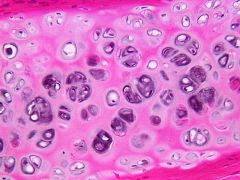
Hyaline cartilage
|
The most abundant and weakest cartilage. Provides smooth surfaces for movement at joints, as well as flexibility and support.
|
|
|
Hyaline cartilage (picture)
|
|
|
|
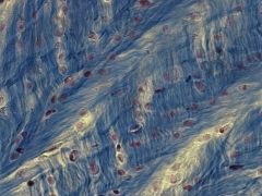
Fibrocartilage
|
The strongest type of cartilage (e.g. intervertebral discs). Supports and joins structures together.
|
|
|
Fibrocartilage (picture)
|
|
|
|
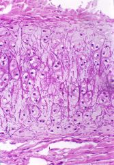
Elastic cartilage
|
Type of cartilage that provides strength and elasticity. Maintains the shape of certain structures.
|
|
|
Elastic cartilage (picture)
|
|
|
|
Chondrocytes
|
The cells of mature cartilage
|
|
|
Lacunae
|
Small, hollow spaces, such as that found in bones in which the osteocytes lie.
|
|
|
Perichondrium
|
A covering of dense irregular CT that surrounds the surface of most cartilage.
|
|
|
Bone tissue
|
Consists of osteocytes and a matrix containing mineral salts and collagen fibers. It is classified as either compact or spongy.
|
|
|
Functions of bone tissue
|
Stores calcium and phosphorus, houses red bone marrow and yellow bone marrow.
|
|
|
Red bone marrow
|
Produces blood cells
|
|
|
Yellow bone marrow
|
Stores triglycerides
|
|
|
Blood
|
Consists of cells, platelets, and plasma
|
|
|
Plasma
|
Liquid matrix in the blood
|
|
|
Lymph
|
The EC fluid that flows through the lymphatic vessels. Consists of several types of cells in a clear liquid matrix that is similar to blood plasma but with less protein.
|
|
|
Membranes
|
Flat sheets of pliable tissue that cover or line a part of the body.
|
|
|
Epithelial membrane
|
The combo of an epithelial layer and an underlying CT layer.
|
|
|
Mucous membranes
|
Line body cavities that open directly to the exterior. Secretes mucus which prevents the cavities from drying out.
|
|
|
Serous membranes
|
Line body cavities that do not open directly to the exterior. Also cover the viscera located within these cavities.
|
|
|
Mesothelium
|
The epithelial portion of serous membranes. It is the portion that secretes serous fluid.
|
|
|
Serous fluid
|
A watery lubricating fluid that reduces friction during movement.
|
|
|
Cutaneous membrane (skin)
|
Composed of the epidermis and dermis (dense irregular CT).
|
|
|
Synovial membranes
|
Non-epithelial membrane that lines the cavities of some joints. Secretes synovial fluid.
|
|
|
Muscle tissue
|
Consists of cells that are modified for contraction and thus provide motion, maintenance of posture, and heat production.
|
|
|
Muscle fibers
|
Cells in muscle tissue.
|
|
|
Skeletal muscle tissue
|
Attached to bones. Striated, multinucleated, and its movement is voluntary.
|
|
|
Cardiac muscle tissue
|
Forms most of the heart wall. Striated. Involuntary control. The individual muscle fibers are connected by intercalated discs. Centrally located nucleus.
|
|
|
Smooth muscle tissue
|
Found in the walls of hollow internal structures. Cells are non-striated and spindle-shaped and have a centrally located nucleus. Movement involuntary.
|
|
|
Nervous tissue
|
Composed of neurons and neuroglia.
|
|
|
Neurons
|
Consist of a cell body and dendrites and axons.
|
|
|
Functions of neurons
|
Detect stimuli
Convert that stimuli into nerve impulses Conduct nerve impulses to other neurons, muscle fiber, or glands |
|
|
Neuroglia
|
Include a broad spectrum of several types of cells which protect and support neurons.
|
|
|
Excitable cells
|
Carry electrical signals (actions potentials)
|
|
|
Neurotransmitters
|
Chemicals that cause excitable cells to generate electrical signals.
|
|
|
Tissue repair
|
The process that replaces worn out, damaged, or dead cells.
|
|
|
Renewal capacity of epithelial cells
|
Continuous renewal capacity.
|
|
|
Renewal capacity of CT
|
Some have a continuous renewal capacity, while others replenish cells less readily.
|
|
|
Renewal capacity of muscle fibers
|
Poor renewal capacity
|
|
|
Renewal capacity of nervous tissue
|
The poorest renewal capacity
|
|
|
Adhesions
|
Abnormal attachments between tissues. Don't always cause problems, but can decrease flexibility, cause intestinal obstructions, and make subsequent operations more difficult.
|
|
|
Conditions affecting tissue repair
|
Nutrition
Proper blood circulation |
|
|
Dermatology
|
The medical specialty that deals with diagnosing and treating skin disorders.
|
|
|
Integumentary system
|
Composed of skin, air, oil & sweat glands, nails, and sensory receptors.
|
|
|
Skin (cutaneous membrane)
|
The external covering of the body that consists of a superficial, thinner epidermis, and a deep, thicker dermis that is anchored to the SQ.
|
|
|
Epidermis
|
The superficial, thinner portion of skin, which is composed of epithelial cells, Contains keratinocytes, melanocytes, Langerhans cells, and Merkel cells.
|
|
|
Dermis
|
The deeper, thicker CT portion of the skin.
|
|
|
Subcutaneous layer (hypodermis)
|
Layer that consists of areolar and adipose tissue. Serves as a fat storage area, an area for blood vessel passage, and the location of some nerve endings. Not part of the skin.
|
|
|
Keratinocytes
|
The most abundant of the epidermal cells, produce keratin.
|
|
|
Keratin
|
A tough, fibrous protein that helps protect the skin and underlying tissues.
|
|
|
Lamellar granules
|
Produced by the keratinocytes, release a water-repellent sealant.
|
|
|
Keratinization
|
The process whereby the contents of keratinocytes are replaced with the protein keratin.
|
|
|
Stratum basale
|
The deepest layer of the epidermis, composed of a single row of keratinocytes.
|
|
|
Melanocytes
|
Epidermal cells that produce the pigment melanin.
|
|
|
Melanin
|
A yellow-red or brown-black pigment that contributes to skin color and absorbs damaging UV light.
|
|
|
Langerhans cells
|
Epidermal cells that participate in immune responses. Easily damaged by UV light.
|
|
|
Merkel cells
|
Epidermal cells that make contact with sensory neurons to detect touch sensations.
|
|
|
Epidermal ridges
|
Increase friction for better grasping ability and provide the basis for fingerprints.
|
|
|
Layers of the epidermis
|
Stratum basale
Stratum spinosum Stratum granulosum Stratum lucidum Stratum corneum |
|
|
Sebaceous glands (oil glands)
|
Exocrine glands in the dermis of skin, almost associated with a hair follicle. Secretes sebum.
|
|
|
Sudoriferous glands (sweat glands)
|
Their cells release sweat into hair follicles or onto the skin surface through pores.
|
|
|
Types of pigments
|
Melanin
Carotene Hemoglobin |
|
|
Hemoglobin
|
The oxygen-carrying pigment in red blood cells.
|
|
|
Carotene
|
A yellow-orange pigment that gives egg yolk and carrots their color.
|
|
|
Albinism
|
The inherited inability of an individual to produce melanin.
|
|
|
Vitiligo
|
The partial or complete loss of melanocytes from patches of skin.
|
|
|
Hairs (pili)
|
Present on most skin surfaces except the palms, palmar surfaces of the fingers, soles, and plantar surfaces of the toes.
|
|
|
Shaft
|
The superficial portion of hair that projects above the surface of the skin.
|
|
|
Root
|
Portion of the hair below the surface that penetrates into the dermis and sometimes into the SQ layer.
|
|
|
Hair follicle
|
Surrounding the root of the hair, composed of two layers of epidermal cells, surrounded by a CT sheath.
|
|
|
Bulb
|
Base of each hair follicle that is enlarged into an onion-shaped structure.
|
|
|
Arrector pili
|
Smooth muscles attached to hairs. Contraction pulls the hairs into a vertical position, resulting in "goose bumps."
|
|
|
Hair root plexuses
|
Nerve endings surrounding each hair follicle. Sensitive to touch.
|
|
|
Ceruminous glands
|
Present in the outer ear canal and secrete cerumen (ear wax).
|
|
|
Nails
|
Plates of tightly packed, hard, dead, keratinized cells of the epidermis.
|
|
|
Nail root
|
Portion of the nail that is not visible.
|
|
|
Lunula
|
The whitish semilunar area near the nail root.
|
|
|
Nail body
|
The portion of the nail that is visible.
|
|
|
Free edge (of the nail)
|
The part of the nail body that extends past the end of the finger or toe.
|
|
|
Nail matrix
|
The proximal portion of the epithelium deep to the nail root. The region in which the superficial cells divide to produce new nail cells.
|
|
|
Functions of skin
|
Thermoregulation
Protection Cutaneous sensations Excretion Synthesis of vitamin D |
|
|
Basal cell carcinomas
|
Account for the majority of skin cancers. The tumor arises form the cells in the stratum basale and rarely metastasize.
|
|
|
Squamous cells carcinomas
|
Account for 20% of skin cancer and arise from the stratum spinosum. Have a variable tendency to metastasize.
|
|
|
Malignant melanomas
|
Arise from melanocytes, accound for 2% of skin cancers. The most prevalent life threatening cancer in young women.
|
|
|
Warning signs of malignant melanoma (ABCDE)
|
A - asymmetry
B - border (irregular border) C - color (uneven coloration) D - diameter E - elvoving |
|
|
Risk factors of skin cancer
|
1. skin type
2. sun exposure 3. family history 4. age 5. immunological status |
|
|
Burn
|
A tissue damage caused by excessive heat, electricity, radioactivity, or corrosive chemicals that denature the proteins in the skin cells.
|
|
|
Systematic effects of a burn
|
Ex: dehydration and infection. A greater threat to life than the local effects of a burn.
|
|
|
First degree burn
|
Involves only the epidermis. Involves mild pain and redness, but no blisters.
|
|
|
Second degree burn
|
Destroys the epidermis and part of the dermis. Redness, blistering, edema, pain.
|
|
|
Third degree burn
|
Destroys the epidermis, dermis, and SQ. Most skin function is lost and region is numb.
|
|
|
Osteology
|
The study of bone structure and the treatment of bone disorders.
|
|
|
Functions of bone and the skeletal system
|
Support
Protection Assistance in movement Mineral homeostasis Blood cell production Triglyceride storage |
|
|
Hemopoiesis
|
Red bone marrow produces red blood cells, white blood cells, and platelets.
|
|
|
Yellow bone marrow
|
Consists of mainly adipose cells, which store triglycerides.
|
|
|
Types of bones
|
Long
Short Flat Sesamoid Irregular |
|
|
Long bones
|
Greater length than width
|
|
|
Short bones
|
Nearly equal length and width
|
|
|
Flat bones
|
Thin, parallel plates; provide extensive surfaces for muscles attachment
|
|
|
Sesamoid bones
|
Sesame seed shaped
|
|
|
Irregular bones
|
Complex, various shapes that don't fit into any other category of bone shapes.
|
|
|
Diaphysis
|
The shaft of the long bone.
|
|
|
Epiphyses
|
The ends of the bone. Articulate with adjacent bones.
|
|
|
Metaphyses
|
The areas between the epiphysis and diaphysis.
|
|
|
Epiphyseal plate
|
The site of bone elongation in growing bones.
|
|
|
Hyaline cartilage (articular cartilage)
|
At the ends of the bones. Reduces friction and absorbs shock at freely moveable joints. Located on articulating surfaces of bones.
|
|
|
Periosteum
|
A CT covering the surface of the bone which contains osteogenic cells.
|
|
|
Osteogenic cells
|
Promote bone growth in width, assist in fracture repair, and help nourish bone tissue.
|
|
|
Medullary cavity (canal)
|
The tunnel-like space within the diaphysis that contains yellow bone marrow, consisting of adipose CT.
|
|
|
Endosteum
|
The lining of the medullary cavity of the bone.
|
|
|
Types of bone cells
|
Osteogenic cells
Osteoblasts Osteocytes Osteoclasts |
|
|
Osteogenic cells
|
Undergo cell division and develop into osteoblasts.
|
|
|
Osteoblasts
|
Bone-building cells, promoting bone deposition.
|
|
|
Osteocytes
|
Mature bone cells that maintain bone tissue. They are derived from osteoblasts.
|
|
|
Osteoclasts
|
Derived from monocytes, and are therefore an entirely different class of bone cells. They serve to break down bone tissue, a process known as bone resorption.
|
|
|
Calcification
|
Deposition of mineral salts in a framework formed by collagen fibers in which the tissue hardens.
|
|
|
Compact bone tissue
|
Contains few spaces and is arranged in repeating structural units called osteons.
|
|
|
Central canal
|
A channel that contains blood vessels, nerves, and lymphatic vessels.
|
|
|
Concentric lamellae
|
Rings of hard, calcified EC matrix that resemble the growth rings of a tree.
|
|
|
Lacunae
|
Small spaces between the lamellae that contain osteocytes.
|
|
|
Canaliculi
|
Small channels radiating in all directions from the lacunae and are filled with EC fluid.
|
|
|
Volkmann canals
|
Allow a route for blood vessels, lymphatic vessels, and nerves of the medullary cavity, periosteum, and central canals to connect.
|
|
|
Spongy bone tissue
|
Bone tissue that contains units called trabeculae rather than osteons.
|
|
|
Trabeculae
|
Units of the spongy bone tissue. Irregular latticeworks of thin columns of bone.
|
|
|
Ossification
|
The process by which bone is formed.
|
|
|
Situations of bone formation
|
1. The initial formation of bones in an embryo and fetus.
2. The growth of bones from birth until the full adult size is reached. 3. Remodeling of bone. 4. Repair of fractures. |
|
|
Intramembranous ossification
|
The formation of bone directly from mesechyme arranged in sheet-like layers resembling membranes. Forms the flat bones of the skull and the mandible, and involves 4 steps.
|
|
|
Endochondral ossification
|
The formation of bone from hyaline cartilage models. This is how most of the bones are formed. Involves 6 steps.
|
|
|
Bone remodeling
|
The ongoing replacement of old bone tissue by new bone tissue.
|
|
|
Bone resorption
|
Old bone is destroyed by osteoclasts.
|
|
|
Bone deposition
|
New bone is constructed by osteoblasts.
|
|
|
Ingredients for healthy bone growth and remodeling
|
1. calcium, phosphorus, magnesium
2. vitamins A, C, D 3. hormones 4. weight-bearing exercises |
|
|
Human growth hormone
|
Hormone secreted from the anterior pituitary gland.
|
|
|
Insulin-like growth factors
|
Hormones secreted locally from bone, as well as systemically from the liver.
|
|
|
Thyroid hormones
|
Hormones secreted from the thyroid gland.
|
|
|
Insulin
|
Hormone secreted from the pancreas.
|
|
|
Estrogen
|
Hormone secreted from the ovaries.
|
|
|
Androgens
|
Hormones secreted from the testes, as well as from the adrenal cortex in both men and women.
|
|
|
Nutrient artery
|
Passes through the nutrient canal and sends branches into the central canals of the osteons to provide nutrients for osteocytes.
|
|
|
Fracture
|
Any break in a bone.
|
|
|
Simple fracture
|
A fracture in which the skin is intact.
|
|
|
Compound fracture
|
A bone fragment caused by a puncture through the skin.
|
|
|
Comminuted
|
A bone fracture in which there are several bone fragments.
|
|
|
Greenstick
|
A bone fracture in which a young bone bends and partially breaks.
|
|
|
Impacted
|
A bone fracture in which one bone fragment is firmly driven into another.
|
|
|
Stress fracture
|
A series of microscopic fissures in bone which forms without any evidence of injury to other tissues.
|
|
|
Steps of fracture repair
|
1. phagocytes come in and clean up any dead bone tissue.
2. chodroblasts form fibrocartilage at the fracture site that bridges the broken ends of the bone. 3. osteoblasts replace the fibrocartilage with spongy bone. 4. bone remodeling occurs via the work of osteoblasts and osteoclasts. |
|
|
Parathyroid hormone
|
An important hormone which increases blood calcium levels.
|
|
|
Calcitonin
|
A hormone that is secreted by the thyroid gland and decreases blood calcium levels.
|
|
|
Demineralization
|
Loss of calcium and other minerals from bone matrix, which may result in osteoporosis.
|
|
|
Osteoporosis
|
A decrease in the amount and strength of bone tissue. Bone resorption outpaces bone formation.
|
|
|
Rickets and osteomalacia
|
Disorders in which bones fail to calcify.
|
|
|
Joint
|
A point of contact between two or more bones, between cartilage and bones, or between teeth and bones.
|
|
|
Arthrology
|
The scientific study of joints.
|
|
|
Types of joint structures
|
Fibrous
Cartilaginous Synovial |
|
|
Synarthroses
|
Joint is immovable.
|
|
|
Amphiarthroses
|
Joint is partially moveable.
|
|
|
Diarthroses
|
Joint is freely moveable.
|
|
|
Suture
|
A fibrous joint that unites skull bones.
|
|
|
Interosseous membrane
|
A fibrous joint made of a broad sheet of ligament that allows some movement between adjacent bones.
|
|
|
Cartilaginous joint
|
A type of joint that lacks a synovial cavity, has the articulating bones connected by either fibrocartilage or hyaline cartilage, and allows little or no movement.
|
|
|
Fibrous joint
|
Type of joint that lacks a synovial cavity, has the articulating bones held together by fibrous CT, and permits little or no movement.
|
|
|
Synovial joint
|
Type of joint that has a synovial cavity between the articulating bones and are freely moveable.
|
|
|
Articulating cartilage
|
Covers the ends of bones at synovial joints. Reduces the friction between the bone ends and helps absorb shock.
|
|
|
Articular capsule
|
Surrounds a freely moveable joint, and encloses the synovial cavity and unites the articulating bones.
|
|
|
Articulating ligaments
|
Commonly associated with synovial joints. May be found inside or outside the joint capsule.
|
|
|
Articular discs (menisci)
|
Modify the shape of the joint surfaces of the articulating bones, help maintain the stability of the joint, and direct the flow of synovial fluid to areas of greatest friction.
|
|
|
Bursae
|
Synovial fluid filled sac-like structures that cushion the movement of one body part over another.
|
|
|
Tendon sheaths
|
Tube-like bursae that wrap around tendons where there is a considerable friction, such as the tendon of the biceps brachii at the shoulder joint.
|
|
|
Plantar joints
|
Permit mainly side-to-side and back-and-forth gliding movements. these joints are biaxial or tiaxial, and include the intercarpal, intertarsal, sternoclavicular, acromioclavicular, sterocostal, and vertevrocostal joints.
|