![]()
![]()
![]()
Use LEFT and RIGHT arrow keys to navigate between flashcards;
Use UP and DOWN arrow keys to flip the card;
H to show hint;
A reads text to speech;
34 Cards in this Set
- Front
- Back
- 3rd side (hint)
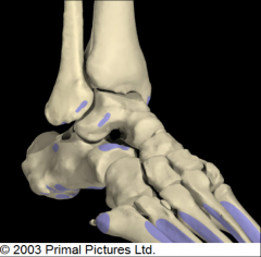
BONES OF THE ANKLE: ANTERIOR & LATERAL VIEW |
1. talus within mortise 2. talocural articulation 3. talus shape 4. subtalar joint |
|
|
|
1. TALUS WITHIN MORTISE |
- talus bone sits within mortise - mortise = space framed by distal aspect of tibia, medial malleolus, lateral, and malleolus |
|
|
|
2. TALOCURAL ARTICULATION |
- articulation b.t the talus & distal aspect of tibia DORSIFLEXION & PLANTARFLEXION occurs within the saggital plane about the frontal axis. INTERNAL & EXTERNAL ROTATION occurs within the transverse plane about the longitudinal axis. |
|
|
|
3. TALUS SHAPE |
- wedge shaped - anterior aspect = broad - posterior aspect = tapered - shape allows internal + external rotation while in plantarflexed position |
|
|
|
4. SUBTALAR JOINT |
- between calcaneous & talus INVERSION & EVERSION occurs within the frontal plane about the sagittal axis. |
|
|
|
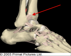
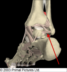
LATERAL LIGAMENTS OF ANKLE |
1. anterior tibio-fibular ligament 2. anterior talo-fibular ligament 3. calcaneal-fibular ligament 4. posterior talo-fibular ligament |
|
|
|
1. ANTERIOR TIBIO-FIBULAR LIGAMENT |
- prevents excessive spread within the frontal plane b.t tibia & fibula bones - walking: tibia/fibula spread a lil - if injured they shift too much = high ankle sprain |
|
|
|
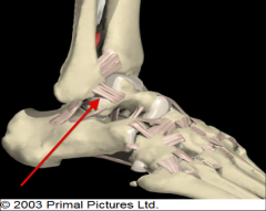
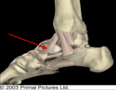
2. ANTERIOR TALO-FIBULAR LIGAMENT |
- prevents excessive anterior displacement of talus relative to fibula *AKA prevents foot from shifting too far forward on lower leg |
|
|
|
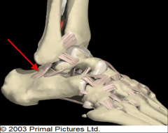
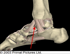
3. CALCANEAL-FIBULAR LIGAMENT |
- prevents foot from excessive inversion within frontal plane about sagittal axis - happens when you "roll" your ankle |
|
|
|
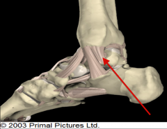
4. POSTERIOR TALO-FIBULAR LIGAMENT |
- prevents excessive posterior translation of talus relative to fibula *AKA prevents foot from shifting too far backwards on lower leg |
|
|
|
LATERAL & MEDIAL MALLEOLUS |
- lateral extends more distal than medial - so lateral is father down talus - so magnitude of inversion > eversion |
|
|
|
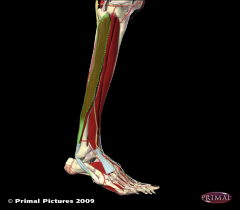
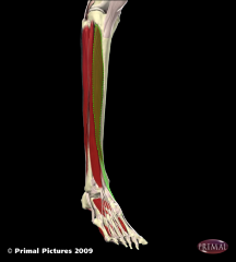
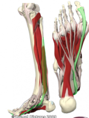
LATERAL LOWER LEG MUSCLES |
1. peroneous longus muscle 2. peroneous brevis muscle |
|
|
|
1. PERONEOUS LONGUS |
FUNCTION: - primarily plantarflexion of foot - eversion of foot WHERE: - lateral compartment of lower leg INNVERVATED BY: - superficial fibular nerve - L5, S1 |
|
|
|
2. PERONEOUS BREVIS |
FUNCTION: - plantarflexion of foot - eversion of foot WHERE: - lateral compartment of lower leg INNVERVATED BY: - superficial fibular nerve - L5, S1 |
|
|
|
MEDIAL LIGAMENTS OF ANKLE |
the deltoid ligament: 1. tibio-navicular 2. anterior tibio-talar 3. tibio-calcaneal 4. posterior tibio-talar |
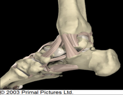
1. tib-navic 2. tib-calc 3. ant tibio-tal 4. post tibio-tal |
|
|
1. TIBIO-NAVICULAR LIGAMENT |
- prevents excessive plantarflexion w. external rotation |
"tibio near big toe" |
|
|
2. ANTERIOR TIBIO-TALAR LIGAMENT |
- prevents excessive anterior translation of talus on tibia *AKA keeps talus from moving up on leg - deep to tibio-navicular lig. - hard to see |
switch tal & tib |
|
|
3. TIBIO-CALCANEAL LIGAMENT |
- prevents excessive eversion * AKA keeps sole of foot from facing too far inwards - very strong |

"tibio-calcan-eversion" |
|
|
4. POSTERIOR TIBIO-TALAR LIGAMENT |
- prevents excessive posterior translation of talus on tibia - doesnt stop excess eversion cause that occurs at subtalar j |
switch tal & tib |
|
|
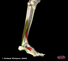
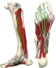
ANTERIOR LOWER LEG MUSCLES |
1. extensor hallicus longus 2. extensor digitorum longus 3. tibialis anterior |
|
|
|
1. EXTENSOR HALLICUS LONGUS |
FUNCTION: - extension of big toe - dorsiflexion of foot WHERE: - anterior compartment of lower leg INNERVATED BY: - deep fibular nerve - L5, S1 |
|
|
|
2. EXTENSOR DIGITORUM |
FUNCTION: - extension of digits 2-5 - dorsiflexion of foot WHERE: - anterior compartment of lower leg INNERVATED BY: - deep fibular nerve - L4, L5, S1 |
"extends digits" |
|
|
3. TIBIALIS ANTERIOR |
FUNCTION: - dorsiflexion of foot - supination of foot WHERE: - anterior compartment of lower leg INNERVATED BY: - deep fibular nerve - L4, L5 |
|
|
|
4 ARCHES IN FOOT & FUNCTION |
1. medial longitudinal arch 2. lateral longitudinal arch 3. transverse tarsal arch 4. transverse metatarsal arch - absorb/dissipate ground reaction force that's upwards through body while we stand/walk - if not it will proceed up leg... - shin-splints - inflammation |
|
|
|
1. MEDIAL LONGITUDINAL ARCH |
- most noticeable - runs length of foot - on medial aspect of foot |
"long = length" |
|
|
2. LATERAL LONGITUDINAL ARCH |
- also runs length of foot - on lateral aspect of foot |
"long = length" |
|
|
3. TRANSVERSE TARSAL ARCH |
- anterior to calcaneous - in tarsal bones of foot - cuneiforms, navic., cuboid - runs width of foot |
"trans - across tarsal bones" |
|
|
4. TRANSVERSE METATARSAL ARCH |
- in 5 metatarsal bones of foot - runs width of foot |
"meta - 5 bones before tarsals" |
|
|
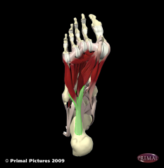
LIGAMENTS OF FOOT |
1. long plantar ligament |
|
|
|
1. LONG PLANTAR LIGAMENT |
FUNCTION: - stabilize medial longitudinal arch - supports lateral longitudinal arch WHERE: - intrinsic muscle! - originates & inserts on foot |
|
|
|
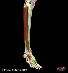
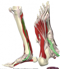
DEEP POSTERIOR MUSCLES OF LOWER LEG |
* run medial & under foot 1. flexor digitorum longus 2. flexor hallicus longus 3. tibialis posterior |
|
|
|
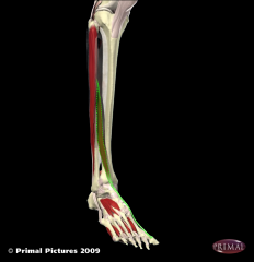
1. FLEXOR DIGITORUM LONGUS |
FUNCTION: - flex digits 2-4 - plantarflexion of foot WHERE: - deep posterior compartment of lower leg INNERVATION BY: |
|
|
|
2. FLEXOR HALLICUS LONGUS |
FUNCTION: - flexion of big toe - plantarflexion of foot WHERE: - deep posterior compartment of lower leg INNERVATION BY: - tibial nerve - L5, S1, S2 |
|
|
|
3. TIBIALIS POSTERIOR |
FUNCTION: - plantarflexion of foot - pronation of foot WHERE: - deep posterior compartment of lower leg INNERVATION BY: - tibial nerve - L4, L5 |
|