Reading...
![]()
Play button
![]()
Play button
![]()
Use LEFT and RIGHT arrow keys to navigate between flashcards;
Use UP and DOWN arrow keys to flip the card;
H to show hint;
A reads text to speech;
49 Cards in this Set
- Front
- Back
|
What are the 3 major nerves that supply the lower extremity?
|
femoral nerve, sciatic nerve, & obturator nerve
|
|
|
This nerve supplies the muscles of the anterior compartment of the thigh.
|
Femoral nerve
|
|
|
This nerve supplies the muscles of the medial (adductor) compartment of the thigh.
|
Obturator nerve
|
|
|
This is really 2 nerves that are bundled into 1 nerve in the thigh (tibial n. and common fibular n.).
|
Sciatic nerve
|
|
|
This nerve supplies the posterior compartment of the thigh, leg, & plantar surface of the foot.
|
Tibial nerve
|
|
|
This nerve supplies the anterior & lateral compartments of the leg and doral surface of the foot.
|

Common fibular nerve
|
|
|
The external iliac artery passes deep to the inguinal ligament and is then termed the _______________. This is the main artery to the lower extremity.
|
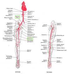
femoral artery
|
|
|
This artery supplies anterior of the thigh.
|

Femoral artery
|
|
|
This branch of the femoral artery supplies the posterior compartment of the thigh.
|
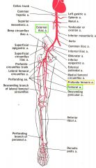
Profunda femoris artery
|
|
|
As the femoral artery passes 「adductor hiatus 」 behind the knee it is renamed___________ and supplies around the knee.
|
popliteal artery
|
|

As the popliteal artery enters the leg, it branches into the________ and________.
|

anterior tibial and posterior tibial arteries.
|
|
|
The anterior tibial artery ends as the ________ of the foot.
|
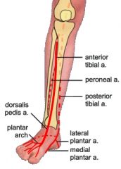
dorsalis pedis artery
|
|
|
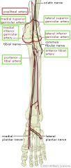
A branch of the posterior tibial artery that supplies the lateral compartmet of the lef.
|
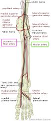
fibular artery
|
|
|
The posterior tibial artery ends as the _________.
|

medial and lateral plantar arteries of the foot.
|
|
|
Innervaterd by Anterior rami of lumbar nn This muscle acts conjointly in flexing thigh at hip joint & in stabilizing this joint.
|

Iliopsoas
psoas major m. psoas minor m. iliacus m. |
|
|
Innervated by Inferior gluteal n This muscle extends thigh & assists in its lateral rotation; steadies thigh & assists in rising from sitting position
|

Gluteus maximus
|
|
|
Innervated by Superior gluteal n Abducts & medially rotates thigh
|

Gluteal medius, Gluteal minimus m.
O: Iliac crest, between ant. & post. gluteal line I: Greater trochanter Sup. gluteal n. Extension / Med. rotation / Abduction |
|
Innvervated by Femoral nerve
This muscle flexes, abducts, & laterally rotates thigh at hip joint; flexes leg at knee joint |

Sartorius m.
|
|
These muscles are innervated by the femoral nerve. They extend leg at knee joint; rectus femoris also steadies hip joint & helps iliopsoas flex thigh
|

Quadriceps femoris
Rectus femoris m. , Vastus medialis m. Vastus intermedius m., Vastus lateralis m. |
|
|
These muscles are innervated by the sciatic nerve. They flex leg & rotate it laterally when knee is flexed; extends thigh
|
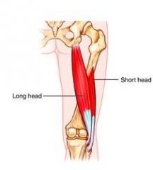
Biceps femoris
|
|
|
These nerves are innervated by the tibial division of the sciatic nerve. They extend thigh; flex leg & rotate it medially when knee is flexed
|

Semimembranous & Semitendinosus
|
|
|
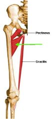
Innervated by obturator nerve.
Adducts thigh; flexes leg; helps rotate leg medially |

Gracilis
|
|

Innervated by Obturator neve. Adducts thigh.
|
Adductor longus m.
O: Inf. ramus of pubis, ramus of ischium I: Linea aspera Obturator n. (Ant.) Adduction (Thigh) Flexion (Thigh) Lat. rotation (Thigh) |
|
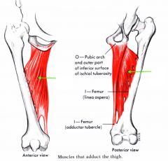
Innervated by Obturator nerve & tibial part of sciatic nerve. Adducts thigh; flexes thigh & extends thigh
|
Adductor magnus m.
O: Inf. ramus of pubis I: Linea aspera Obturator n. (Post.) Adduction (Thigh) Flexion (Thigh) Lat. rotation (Thigh) |
|
Innervated by obturator nerve.
Adducts thigh; to some extent flexes it |
Adductor brevis m.
O: Inf. ramus of pubis I: Inf. to lesser trochanter Obturator n. (Ant.) Adduction (Thigh) Flexion (Thigh) Lat. rotation (Thigh) |
|
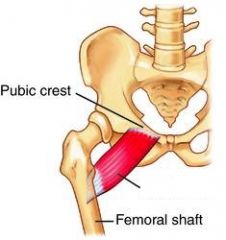
Adducts & flexes thigh; assist w. medial rotation of thigh
|
Pectineus m.
O: Sup. ramus of pubis (Pecteal line) I: Pecteal line of femur Femoral n. Adduction (Thigh), Flexion (Thigh), Lat. rotation (Thigh) |
|

Innervated by deep fibular nerve. Dorsiflexes ankle & inverts foot.
|
Tibialis anterior m.
O: Lat. condyle Lat. surface of tibia Interosseous memb. I: Med. cuneiform 1st metatarsal Deep peroneal n. Dorsiflexion (Foot), Inversion (Foot) |
|

Innervated by deep fibular nerve.
Extends lateral 4 digits & dorsiflexes ankle |
Extensor digitorum longus m.
O: Lat. condyle of tibia Ant. fibula Interosseous memb. I: Extensor expansion, Lat.4 digits Deep peroneal n. Dorsiflexion (Foot) Extension (4 toes) |
|

Innervated by deep fibular nerve.
Extends great toe & dorsiflexes ankle |
Extensor hallucis longus m.
O: Fibula, Interosseous memb. I: Distal phalanx of great toe Deep peroneal n. Dorsiflexion (Foot) Extension (Great toes) |
|

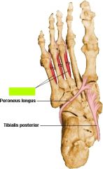
Innervated by Superficial fibular nerve. These muscles of the lateral leg everts foot & weakly plantarflexes ankle.
|
Fibularis longus & Fibularis brevis
|
|

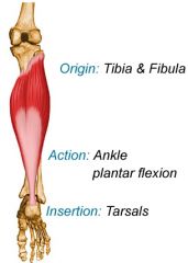
Innervated by Tibial nerve.
Plantarflexes ankle when knee is extended; raises heel during walking; flexes leg at knee joing |
Gastrocnemius m.
O: Lat. & med. condyle of femur I: Calcaneus Tibial n. Flexion (Leg) Plantar flexion (Foot) |
|
Innervated by Tibial nerve. Plantarflexes ankle independently of knee position; steadies leg on foot
|
Soleus m.
O: Soleal line (Tibia) Fibula I: Calcaneus Tibial n. Plantar flexion (Foot) |
|
Innervated by Tibial nerve. Weakly flexes knee & unlocks it; medially rotates ribia of unplanted limb
功能:屈膝關節、內旋小腿。 當膝關節在伸膝狀況卡住時,此肌肉有解圍的功能。 |
Popliteus m.
O: Lat. condyle of femur I: Post. aspect of upper tibia (Above soleal line) Tibial n. Lat. rotation (Thigh) Med. rotation (Leg) |
|

Innervated by Tibial nerve. Flexes great toe
|
Flexor hallucis longus
O: Post. surface of middle & distal end of fibula Around med. malleolus, Groove on caleaneus (Sustentaculum tali) I: Distal phalanx of great toe Tibial n. Flexion (Great toes) Plantar flexion (Foot) |
|

Innervated by Tibial nerve.
Flexes lateral four digits; plantarflexes ankle |
Flexor digitorum longus m.
O: Post. surface of middle end of tibia Around med. malleolus I: Base of distal phalanx of Lat.4 digits (Extensor expansion) Tibial n. Flexion (4 toes) Plantar flexion (Foot) |
|

Plantarflexes ankle; inverts foot
主要功能是踝關節蹠屈(plantar flexion),在走路與跑步的過程中扮演非常重要的角色 (關節可以進行背屈(dorsiflexion)、蹠屈(plantar flexion)、內翻(inversion)、外翻(eversion)) |
Tibialis posterior m
O: Post. surface of upper end of tibia and fibula Interosseous memb. Around med. malleolus I: Tuberosity of navicular Cuneiform (Med. + intermediate) Base of 2nd, 3rd, 4th metatarsal Tibial n. Inversion (Foot) Plantar flexion (Foot) |
|
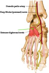
Innervated by deep fibular nerve.
Aides the extensor digitorum longus in extending the 4 medial toes |
Extensor hallucis brevis m.
O: Calcaneus & inf. extensor retinaculum I: Great toe (Prox. phalanx) Deep peroneal n. Extension (Great toe) |
|

Innervated by deep fibular nerve.
Aidses the extensor hallucis longus in extending the great toe |
Extensor digitorum brevis m.
O: Calcaneus & inf. extensor retinaculum I: 2nd, 3rd & 4th phalanges Deep peroneal n. Extension (2nd, 3rd & 4th Toes) |
|
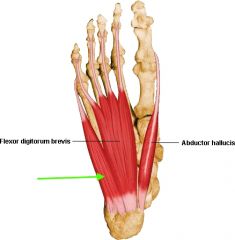
Innervated by medial plantar nerve.
Abducts & flexes 1st digit (great toe, hallux) |
Abductor hallucis m.
O: Calcaneus I: Great toe (Prox. phalanx) Med. plantar n. Abduction (Great toe) Flexion (Great toe) |
|

Innervated by medial plantar nerve.
Flexes lateral 4 digits. |
Flexor digitorum brevis m.
O: Calcaneus I: 2nd ~ 5th mid. phalanges Med. plantar n. Flexion (2nd ~ 5th toes) |
|
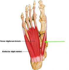
Innervated by Lateral plantar nerve.
Abducts & flexes little toe (5th digit) |
Abductor digiti minimi m.
O: Calcaneus I: Small toe (Prox. phalanx) Lat. plantar n. Abduction (Small toe) Flexion (Small toe) |
|

Innervated by Lateral plantar nerve.
Assists flexor digitorum longus in flexing lateral 4 digits |
Quadratus plantae m.
O: Calcaneus I: Tendon of flexor digitorum longus Lat. plantar n. Flexion (Toes) Flexor accessorius |
|

Innervated by medial plantar nerve. Flexes proximal phalanx of 1st digit.
|
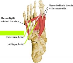
Flexor hallucis brevis m.
O: Cuboid Lat. cuneiform I: Great toe (Med. & lat. prox. phalanx) Med. plantar n. Flexion (Great toe) |
|
Innervated by lateral plantar nerve. Adducts 1st digit
|
Adductor hallucis m.
Transverse head: O: Metatarsophalangeal joint Oblique head: O: 3rd ~ 5th metatarsals I: Great toe (Lat. prox. phalanx) Lat. plantar n. Adduction (Great toe) Flexion (Great toe) |
|

Innervated by lateral plantar nerve. Flexes proximal phalanx of 5th digit, thereby assisting w. its flexion
|
Flexor digiti minimi brevis m.
O: 5th metatarsal I: Small toe (Lat. prox. phalanx) Lat. plantar n. Flexion (Small toe) |
|
Innervated by lateral plantar nerve. Adducts digits (3-5) & flex metatarsophalangeal joints
|

Plantar interossei m. (3)
O: 3rd ~ 5th metatarsals I: 3rd ~ 5th toes (Med. prox. phalanx) Lat. plantar n. Adduction (3rd ~ 5th toes) |
|
|
Abduct digits (2-4) & flex metatarsophalangeal joints
|
Dorsal interossei
|
|

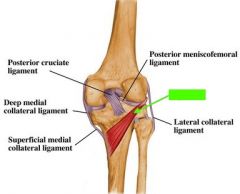
What is the function of the anterior cruciate ligament (ACL)?
|
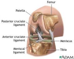
It prevents posterior displacement of the femur on the tibia & hyperextenstion of the knee joint.
前十字韌帶的功能,主要為限制脛骨向前位. 移,其次限制膝內翻及外翻彎曲,也同時限制. 脛骨內旋轉。 |
|

What is the function of the posterior cruciate ligament (PCL)?
|
It prevents the anteriior displacement of the tibia on the femur & helps prevent hyperflexion of the knee joint. It is the main stabilizing factor for the femur.
一是當作阻止脛骨向後移位的首要限制構造。 一是與前十字韌帶合作調節膝在伸直彎曲時轉回來機轉(knee Screw-home mechanism)。另外後十字韌帶也是維持對內外翻(varus, valgus), 及旋轉(rotatory) 穩定度的第二層保護網。 |