Reading...
![]()
Play button
![]()
Play button
![]()
Use LEFT and RIGHT arrow keys to navigate between flashcards;
Use UP and DOWN arrow keys to flip the card;
H to show hint;
A reads text to speech;
54 Cards in this Set
- Front
- Back
|
What type of joint is the sternoclavicular articulation?
|
a saddle type of synovial joint
|
|
|
What are the two articualtions fo the clavicle?
|
manubrium part of the sternum and acromion of the scapula
|
|

Identify numbers 1, 3, and 4.
|
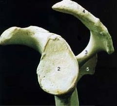
1. acromion
3. spine of the scapula 4. coracoid process |
|
|
What is the first bone in the body to ossify
|
clavicle
|
|
|
1) What does failure of the clavicle to fuse cause?
2) What is this confused with in injuries of the upper limb? |
1)separatation of the outer third and inner 2/3 of the clavicle
2)clavicle fracture |
|
|
The clavicle starts suuperiorly at the 2nd rib and goes to the ______rib.
|
7th rib
|
|
|
What is the consequence of the weak attachment of the clavicle to acromion?
|
site of common dislocations
|
|
|
T/F: the transfer of energy from the clavicle to the sternum increases the likely hood of fracture.
|
False
|
|
|
What vertebral level is the medial end of the scalular spine?
|
T3
|
|
|
what articulates in the glenoid fossa of the scapula?
|
the head of the humerus
|
|
|
What can happen with posterior dislocations of the clavicle?
|
it can can impinge on the trachea and cause shortness of breath
|
|
|
1. which pectoralis muscle serves as an accessory of respiration?
2. How is this done? |
1. pectoralis minor
2. if it's fixed by holding firmly to a table, it elevates the ribs |
|
|
what is the clinical relevance to lymphatics that lie deep to the pectoralis minor?
|
they cannot be palpated for cancer.
|
|
|
Give the rib number for the following:
1. True Ribs 2. False Ribs 3. Floating Ribs |
1. 1-7
2. 8-10 3. 11 & 12 |
|
|
How are false ribs connect to the sternum?
|
indirectly via the cartilage of the rib above it.
|
|
|
How do floating ribs attach to the sternum.
|
They don't attach at all
|
|
|
In terms of numbering, which vertebra is a rib associated (inferior or superior)?
|
superior
|
|
|
what does the tubercle of a rib articulate with?
|
the transverse process of its matched vertebra
|
|
|
1. What grooves are seen on the first ribs?
2. Do these grooves sit on the superior or inferior surface? |
1. Subclavian vein and artery.
2. superior |
|
|
Which rib is smooth, flat and has a tubercle for the serratus anterior muscle?
|
2nd rib
|
|
|
What is a rib incision?
|
it is when surgeons remove a piece of a rib for throacic access, where the periosteum is cut along the curve of the rib... the rib regenerate from the remaining periosteum
|
|
|
1. Which ribs are most commonly fracture?
2. Which is the weakest part of the rib? |
1. middle ribs
2. weakest part of the ribs is just anterior to the angle. |
|
|
What the name of the canal that the carries the intercostal VAD?
|
costal groove
|
|
|
what is a flail chest and what causes it?
|
1. when a sizeable part of the anterior or lateral throacic wall moves freely due to multiple rib fractures. (the section moves paradoxically-outward on inspiration and inward on experation.
It is very painful and impairs inspiration |
|
|
What is the role of the costal cartilages?
|
contribute to elasticity and prevents fracture of the sternum and the ribs... it calcifies in the elderly
|
|
|
1. At what vertebral levels does the manubrium rest?
|
1. T3-T4
|
|
|
1. At what vertebral level is the sternal angle (manubrialsternal joint)?
|
1. T-5
|
|
|
1. At what vertebral level is the xiphoisternal joint?
|
1. T-9
|
|
|
1. At what vertebral level does the body of the sternum rest?
|
T5-T9
|
|
|
What is the name of the articular surfaces on the sternum for the ribs?
|
costal notches
|
|
|
At what vertebral level is the xiphoid process
|
T-10
|
|
|
Are sternal fractures common?
|
no, but crush injuries can occur during traumatic compression of the throcic wall (stering wheel)
|
|
|
What type of fracture is seen in the sternum?
|
comminuted fracture (broken into bits)
|
|
|
What is a comminuted fracture
|
broken into several pieces
|
|
|
what is a median sternectomy?
|
sternum cut down th median plane for surgery, rejoined after surgery and held by wire sutures
|
|
|
What diagnostic test is the sternum useful for?
|
sternal biopsies and to obtain samples for bone marrow transplants
|
|
|
Which artery is compressed in thoracic outlet syndrome?
|
subclavian artery (between the clavicle and the first rib, which is worsened whent the angle between the neck and the shoulders is increased.)
|
|
|
At which vertebral levels are the following?
1. Superior thoracic aperture 2. Inferior throacic aperature |
1. Superior thoracic aperture: T1 (first rib and manubrium)
2. Inferior throacic aperature: T-12 |
|
|
In determining the names of the sternum, rib and cartilage joints, how are the following named?
1. Sternum 2. Ribs 3. Costal cartilatges 4. clavicle 5. give order for naming: 6 what is the name of the joint where the rib joins the vertebra |
1. Sternum: sterno
2. Ribs: chondral 3. Costal cartilatges: costo 4. clavicle: clavicular 5. Manubrio> sterno> costo > chondral > vertebral 6. costovertebral joint |
|
|
1. What is the site of rib dislocations?
2. What is a possible sequelae of rib dislocations? |
1. Sternocostal joint
2. damage to nerves, vessels and muscles |
|
|
1. where does does a dislocation occur in a "separation of the ribs?
|
1. at the costochondral joint
|
|
|
In passive expiration, what happens to the following?
1. intrathroacic pressure 2. intrathoracic volume 3. Elastic tissue of the lungs 4. intra-abdominal pressure 5. vertical diameter of diaphram 6. transverse diameter of the diaphram |
1. intrathroacic pressure: increases
2. intrathoracic volume: decrease 3. (once stretched) recoils 4. intra-abdominal pressure: decreases 5. vertical diameter of diaphram: decrease 6. transverse diameter of the diaphram: decrease |
|
|
Upon inspiration what happens to the middle parts of the ribs (which are most lateral)?
|
they rise
|
|
|
Because the ribs slop inferiorly off the vertebra, what effect does their elevation have on the manubrium/sternum?
|
inferior -->anterior movement
superior --> posterior movement |
|
|
What type of joint is the intervertebral joint?
|
symphysis (cartilaginous joint)
|
|
|
what type of joint is the costovertebral joint?
|
synovial plane joint
|
|
|
What type of joint is the sternoclavicular joint?
|
synovial saddle joint
|
|
|
what types of joints are the 2-7 sternocostal joints?
|
synovial plane joints
|
|
|
what type of joint is the first sternocostal joint?
|
cartilaginous joint
|
|
|
what type of joint is the costochondral joint?
|
cartilaginous joint
|
|
|
what type of joint are the interchondral joints
|
synovial plane joint
|
|
|
What type of joint is the sternomanubrial joint?
|
cartilaginous (symphysis)
|
|
|
what type of joint ist he xiphisternal joint?
|
cartilaginous (synchondrosis)
|
|
|
How does paralysis of the diaphram show up radiographically upon inspiriation?
|
paradoxically: thus if right side is paralyzed it wil move superiorly (being forced by the internal organs on that side, which were displaced by the organs on the opposite side that were displaced by the functioning side of the diaphram.)
|