![]()
![]()
![]()
Use LEFT and RIGHT arrow keys to navigate between flashcards;
Use UP and DOWN arrow keys to flip the card;
H to show hint;
A reads text to speech;
75 Cards in this Set
- Front
- Back
|
areolar tissue |
|
|
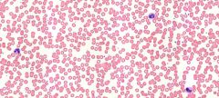
blood smear |
|
|
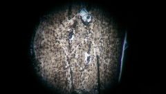
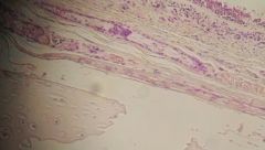
bone ground |
|
|
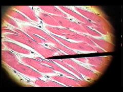
cardiac muscle |
|
|
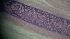
elastic ear cartilage |
|
|
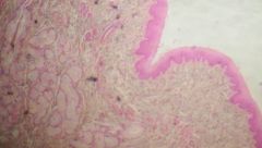
esophagus |
|
|
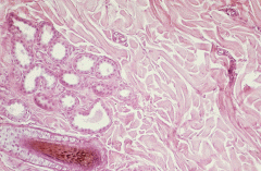
Human Scalp Follicle |
|
|
What are the four types of tissues in the body and describe their roles? |
1. Epithelial tissue covers exposed surfaces, lines internal pas- sageways and chambers,and forms glands. 2. Connective tissue fills internal spaces, provides structural support for other tissues, transports materials within the body, and stores energy. 3. Muscle tissue is specialized for contraction and includes the skeletal muscles of the body, the muscle of the heart, and the muscular walls of hollow organs. 4. Neural tissue carries information from one part of the body to another in the form of electrical impulses. |
|
|
What type of epithelial tissue are glands made from? |
They are usually made of simple cuboidal epithelium |
|
|
What are the types and functions of epithelial tissue? |
simple squamous epitheliumlocated in protected regions where absorption or diffusion takes place, or where a slick, slippery surface reduces friction The simple squa- mous epithelium that lines the body cavities enclosing the lungs, heart, and abdominal organs is called a mesothe- liumstratified squamous epitheliumgen- erally located where mechanical stresses are severesimple cuboidal epitheliumprovides limited protection and occurs where secretion or absorption takes place.Stratified cuboidal epitheliaThey are located along the ducts of sweat glands and in the larger ducts of the mammary glands.transitional epithelium tolerates repeated cycles of stretching and recoiling (returning to its previous shape) without damage. It is called transitional because the appearance of the epithelium changes as stretching occurssimple columnar epithelial typically found where absorption or secretion takes place, such as in the small in- testine stomach and large intestine, the secretions of simple columnar epithelia protect against chemi- cal stresses.Stratified columnar epithelia relatively rare, provi- ding protection along portions of the pharynx, epiglot- tis, anus, and urethra, as well as along a few large excretory ducts. The epithelium has either two layers or multiple layersciliated epithelium lines the respiratory tract moves mucus up from the lungs and toward the throat, as though on an escalator to prevent debris from entering |
|
|
Where are ciliated epithelium located? |
the ciliated epithelium that lines the respiratory tract moves mucus up from the lungs andtoward the throat, as though on an escalator.Where would you find a tight junction?tight Junctions form the closest contact between adjacent cells. Found in the apical region around the cell's circumference.Found in intestinal lining and digestive tract |
|
|
What are CAMs? |
Cell adhesion molecules- Large areas of opposing plasma membranes are interconnectedby transmembrane proteins |
|
|
What determines the classification of epithelium? |
Shape and layers
|
|
|
Provide an example of where you would locate Simple Squamous Epithelium and what are the functions of simple squamous epithelium? |
located in protected regions where absorption or diffusion takes place, or where a slick, slippery surface reduces friction, can be found in mesothelia lining pleural, pericardial, and peritoneal cavities; endothelia lining heart and blood |
|
|
Where are some locations where stratified squamous epithelium may be located and provide their function? |
FUNCTIONS: Provides physical protection against abrasion, pathogens, and chemical attackLOCATIONS: Surface of skin; lining of mouth, throat, esophagus, rectum, anus, and vagina |
|
|
In comparing the cuboidal epithelium which among them provides limited protection, where are they located? |
A simple cuboidal epithelium provides limited protection and occurs where secretion or absorption takes place. Such an epithelium lines portions of the kidney tubules |
|
|
Where are mesothelium located? |
lungs, heart, and abdominal organs |
|
|
What are the three body cavities and their pleura? |
(1) the pleura, which lines the pleural cavities and covers the lungs; (2) the pericardium, which lines the pericardial cavity and covers the heart; and (3) the peritoneum, which lines the peritoneal cavity and covers the surfaces of the enclosed organs |
|
|
Compare the diferrence between endocrine glands and exocrine glands? |
An endocrine gland produces endocrine (endo-, inside + krino, to separate) secretions, which are released directly into the sur- rounding interstitial fluid. These secretions, also called hor- mones, then enter the bloodstream for distribution throughout the body.Exocrine glands produce exocrine (exo-, outside) secretions, which are discharged onto an epithelial surface. Most exo- crine secretions reach the surface through tubular ducts, which empty onto the skin surface or onto an epithelium lining an internal passageway that communicates with the exterior. |
|
|
What are the three modes of secretion and provide an example? |
In merocrine secretion (MER-o. -krin; meros, part), the prod- uct is released from secretory vesicles by exocytosisExample is mucous secre- tions of the salivary glands coat food and reduce friction during swallowing.Apocrine secretion (AP-o. -krin; apo-, off) involves the loss of cytoplasm as well as the secretory productExample is milk production in the mammary glands involves a combination of merocrine and apocrine secretionsholocrine secretion, the entire cell becomes packed with secretory vesicles and then bursts, releasing the secretion, but killing the cell, example is sebaceous glands |
|
|
Which of the connective tissue is considered the to be the only cell that is always present in connective tissue proper? |
Fibroblasts (FI. -bro. -blasts) are the only cells that are always present in connective tissue proper |
|
|
Which cells are known as fat cells? |
Which cells are known as fat cells? |
|
|
What are mesenchymal cells? |
stem cells that are present in many connective tissues.Respond to infection by dividing to produce daughter cells that differentiate into fibroblasts, macrophages, or other connective tissue cells. |
|
|
What is the function of a macrophage? |
scavengers engulf damaged cells or pathogens that enter the tissue |
|
|
where would you find brown fat? |
Shoulders and on infants |
|
|
Give three examples of loose connective tissues. |
areolar tissue, adipose tissue, and reticular tissue |
|
|
Give examples of fluid connective tissues? |
Blood (plasma) and Lymph |
|
|
Provide three examples of dense connective tissue and their location and function? |
Elastic TissueLOCATIONS: Between vertebrae of the spinal column (ligamentum flavum and ligamentum nuchae); ligaments supporting penis; ligaments supporting transitional epithelia; in blood vessel wallsFUNCTIONS: Stabilizes positions of vertebrae and penis; cushions shocks; permits expansion and contraction of organsDense Irregular Connective TissueLOCATIONS: Capsules of visceral organs; periostea and perichondria; nerve and muscle sheaths; dermisFUNCTIONS: Provides strength to resist forces from many directions; helps prevent overexpansion |
|
|
Provide an example of the three type of cartilage and their locations and functions? |
Elastic TissueLOCATIONS: Between vertebrae of the spinal column (ligamentum flavum and ligamentum nuchae); ligaments supporting penis; ligaments supporting transitional epithelia; in blood vessel wallsFUNCTIONS: Stabilizes positions of vertebrae and penis; cushions shocks; permits expansion and contraction of organsDense Irregular Connective TissueLOCATIONS: Capsules of visceral organs; periostea and perichondria; nerve and muscle sheaths; dermisFUNCTIONS: Provides strength to resist forces from many directions; helps prevent overexpansionProvide an example of the three type of cartilage and their locations and functions?FibrocartilageLOCATIONS: Pads within knee joint; between pubic bones of pelvis; intervertebral discsFUNCTIONS: Resists compression; prevents bone-to-bone contact; limits movementElastic Cartilage LOCATIONS: Auricle of external ear; epiglottis; auditory canal; cuneiform cartilages of larynxFUNCTIONS: Provides support, but tolerates distortion without damage and returns to original shapeHyaline CartilageLOCATIONS: Between tips of ribs and bones of sternum; covering bone surfaces at synovial joints; supporting larynx (voice box), trachea, and bronchi; forming part of nasal septumFUNCTIONS: Provides stiff but somewhat flexible support; reduces frictionProvide the three types of muscle tissue, location and functions?Skeletal Muscle TissueLOCATIONS: Combined with connective tissues and neural tissue in skeletal musclesFUNCTIONS: Moves or stabilizes the position of the skeleton; guards entrances and exits to the digestive, respiratory, and urinary tracts; generates heat; protects internal organsCardiac Muscle TissueLOCATION: Heart FUNCTIONS: Circulates blood; maintains discs blood pressureSmooth Muscle TissueLOCATIONS: Found in the walls of blood vessels and in digestive, respiratory, urinary, and reproductive organsFUNCTIONS: Moves food, urine, and reproductive tract secretions; controls diameter of respiratory passageways; regulates diameter of blood vessels |
|
|
How does the integumentary system aid in the diagnoses of disorders in other systems? |
Changes in skin appearance are used to diagnose disorders in other systems , changes in excessive sweating/dryness could indicate issues with endocrine system such as hypothyroidism; changes in pigmentation results in cyanosis, a bluish coloration of the skin, do to a decline in oxygen in tissues, which can occur due to cardiovascular/asthma issues. Changes in pigmentation can also indicate jaundice, which is from the liver is unable to excrete bile, thus a yellowish pigment accumulates in cells; some tumors can affect the pituary gland that produces large amounts of melanocyte-stimulating hormone thus darkening the skin; occurs in Addison disease only with adrenocorticotropic hormone |
|
|
Name the two parts of the integument system |
The two parts of the integument system are the epidermis and the dermis. |
|
|
What are the accessory structures of the integument system? |
Hair and several other structures—hair follicles, sebaceous and sweat glands, and nails |
|
|
In skin repair what forms granulation tissue? |
The combination of blood clot, fibroblasts, and an extensive capillary network |
|
|
What is the purpose of a scab? |
To restore the integrity of the epidermis and prevent any microorganisms from entering the epidermis
|
|
|
A 5 year old child was hit in the head with a rock and the rock cut her going to a 90degree angle to her forehead what can be the result of this injury to her skin? |
A cut at right angles to a cleavage line of the forehead will be pulled open as severed elastic fibers recoil, resulting in greater scarring |
|
|
What are the two major connections that are important in the integumentary system? |
The integumentary system is made up of skin, glans, nails, and nerves. |
|
|
Which layer would you find loose connective tissue and the location of hypodermicinjections such as insulin? |
Hypodermis |
|
|
What are the functions of the integumentary system? |
Store lipids, produce keratin for protection and water repellant, productions of melanin toprotect against UV, maintain body temperature, synthesis of vitamin D3 to calcitriol, detect pressure, pain, temperature, protects underlying organs, minor excretion of salts water and wastes, coordination of immune system |
|
|
What is the protein that is produce that gives eye color? What does it mean if your eyes are light vs. dark in color? |
OCA2 provides instructions for making the protein called P protein which is located in melanocytes which are specialized cells that produce melanin. the pigmentation of the iris varies from light brown to black, depending on the concentration of melanin in the iris pigment epithelium (located on the back of the iris), the melanin content within the iris stroma (located at the front of the iris), and the cellular density of the stroma |
|
|
Name the phases in skin injury and repair? |
Inflammatory phase (bleeding occurs triggering mast cells), migratory phase (scab formation), proliferation phase (epidermal cells migrating over collagen fiber meshwork), Scarring phase (scab has been shed; fibroblasts contine to make scar tissue) |
|
|
Which cells are the one that will actually produce a scar? |
The bulk of the clot consists of an insoluble network of fibrin, a fibrous protein thatforms from blood proteins during the clotting response (trapped red blood cells) |
|
|
What makes us able to withstand long periods of being in water? |
The stratum corneum (top layer) which is water resistant prevents large amounts of water from leaking out; any damage to stratum corneum can cause insensible perspiration to skyrocket |
|
|
Which skin layer will you find the two components papillary and reticular layer |
the dermis |
|
|
Which fat soluble vitamin is synthesized in the skin using ultra violet light from the sun? |
Vitamin D3 |
|
|
Name the receptors found that are responsible for detecting fine touch, pressure, pain andtemperature? |
Sensory receptors including thermorecptos, merkel discs, —tactile (Meissner’s)corpuscles, located in dermal papillae—and receptors sensitive to deep pressure and vibration—lamellated (pacinian) corpus- cles, in the reticular layer. |
|
|
Which of the following two layers are considered to be made of stratified squamousepithelium? |
Epidermis |
|
|
What is the process known as that allows epithelium to receive nutrients and oxygen andrid of waste? |
Epidermal cells rely on the diffusion of nutrients and oxygen from capillaries within thedermis. |
|
|
Which layer of the epidermis is located above the basement membrane? |
Stratum Basale |
|
|
What is the epidermal ridge? |
The stratum basale forms epidermal ridges, which extend into the dermis and are adjacent to dermal projections called dermal papillae |
|
|
Which cells are found in the epidermis and what is their function? |
Basal cells (stem cells that divide to replace the more superficial keratinocytes that are shed at the epithe- lial surface) and Keratinocytes for protection and as water repellent (prevent water loss) |
|
|
How many layers of keratinocytes are found in the thick skin of the palms and soles ofthe hands and feet? |
5 layers |
|
|
Name each of the five layers of keratinocytes and their role in the tissue? For example |
1.Stratum – basale --- epidermal ridges – germinative layer of cells –stem cells Specialized cells merkel (touch) melanocytes (pigmentation)2. Stratum spinosum – promote immune cell response, increase thickness of epidermis, synthesize holecalciferol 3. Stratum granulosum – produce large amount of keratin, dehydrate and create tough interlocking layers (cells are killed)4. Stratum lucidum – flattened cells further, protection, prevent water loss5. Stratum corneum - prevent water loss, protection against microganisms |
|
|
What are the names of the two pigments that gives us our skin color? |
Carotene and melanin |
|
|
Which part of the cell will you find hemidesmosomes? |
Basal membrane |
|
|
How is cholecalciferol produced? |
When exposed to UV, the epidermis will synthesize cholecalciferol from acholesterol related steroid |
|
|
Which region of hair is attached to the integument? |
dermis |
|
|
Which cell layer does keratinization occur? |
Stratum corneum |
|
|
Give an example of a holocrine gland and where is it found and what is secreted? |
Give an example of a holocrine gland and where is it found and what is secreted? |
|
|
Which layer of the skin contains the cutaneous plexus? |
Hypodermis |
|
|
What is the cause of sagging and wrinkles? |
The dermis thins, and the elastic fiber network decreases in size. As a result, the integument becomes weaker and less resilient, and sagging and wrinkling occur. |
|
|
What are the two types of hair and compare/contrast them? |
Vellus; thin fine short hair usually found on body; Terminal long thick sometimes curlyusually found on head |
|
|
How does our capillaries contribute to our skin color? |
Hemoglobin attached to oxygen give off bright red color that give dermis a reddish tint; when dilated for example due to temperature more blood flows thus producing a strong red tint on skin |
|
|
How many days does it take for the skin’s life cycle to occur--- to move from the basale to the corneum layer? |
7-10 days |
|
|
Describe the main structural features of the epidermis, and explain the functionalsignificance of each. |
Sebaceous (se-BA-shus) glands, or oil glands, are holocrine glands that discharge an oily lipid secretion into hair folliclesSweat gland- temperature regulation, excretion, provide protectionHair follicles- protection from UV, cushion skull, sensory receptors Mammary and Ceruminous glands |
|
|
A person may become dehydrated if on a respiratory ventilator due to insensibleperspiration – define interstitial fluid loss? |
Fluid loss that’s not seen or felt usually occurs due to complication with stratum corneum; usually lost through evaporation |
|
|
Explain how dehydration can occur through osmosis? |
Diffusion from high concentration to lower concentration of water; especially when stratum corneum has been damages |
|
|
How does illness contribute to skin color? |
changes in pigmentation results in cyanosis, a bluish coloration of the skin, do to a decline in oxygen in tissues, which can occur due to cardiovascular/asthma issues. Changes in pigmentation can also indicate jaundice, which is from the liver is unable to excrete bile, thus a yellowish pigment accumulates in cells; some tumors can affect the pituary gland that produces lasrge amounts of melanocyte-stimulating hormone thus darkening the skin; occurs in Addison disease only with adrenocorticotropic hormone |
|
|
What type of glands are found especially on the palms and soles? |
Merocrine sweat gland |
|
|
Provide another name for the secretion of the ceruminous glands? |
Sweat glands combined with sebaceous glands |
|
|
What does an apocrine gland require to squeeze secretions onto the skin’s surface? |
myoepithelial cell |
|
|
Will you find the melanocyte above or below the basement membrane? |
above |
|
|
Please Goggle --ear grown on mouse in space – and give the piptide that is responsible and how it is produced? |
The Vacanti mouse was a laboratory mouse that had what looked like a human eargrown on its back. The "ear" was actually an ear-shaped cartilage structure grown by seeding cow cartilage cells into a biodegradable ear-shaped mold and then implanted under the skin of the mouse, then the cartilage naturally grew by itself |
|
|
How many types of sweat glands are found in the skin; give their names and what type ofsecretions are produced? |
Two, apocrine and merocrine; both produce sweat ; apocrine produces sticky cloudy and odorous sweat; merocrine produces watery sweat |
|
|
A burn unit patient will lose a lot of water and become dehydrated by sensibleperspiration which layer of the epidermis is damaged? |
Stratum corneum |
|
|
Which skin layer will you find the two components papillary and reticular layer? |
the dermis |