Reading...
![]()
Play button
![]()
Play button
![]()
Use LEFT and RIGHT arrow keys to navigate between flashcards;
Use UP and DOWN arrow keys to flip the card;
H to show hint;
A reads text to speech;
256 Cards in this Set
- Front
- Back
|
lateral epicondyle
|

Name part "a"
|
|
|
patellar surface
|

Name part "b"
|
|
|
medial epicondyle
|
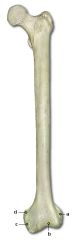
Name part "c"
|
|
|
adductor tubercle
|

Name part "d"
|
|
|
medial epicondyle
|

Name part "a"
|
|
|
coronoid fossa
|
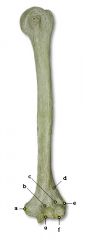
Name part "b"
|
|
|
Visceral Portion
|
The pertioneum covering the viscera
|
|
|
Parietal portion
|
The part of the peritoneum attched to the body wall.
|
|
|
Messenteries
|
Double layered folds which extend from the body wall to the viscera, holding these organs in place. Contain blood vessels and nerves that supply the viscera.
|
|
|
What is the most inferior boundary of the peritoneum?
|
Extend across the abdominal cavity at a level which is just superior to the plevic cavity. The top portion of the urinary bladder is covered with peritoneum.
|
|
|
What is the serous membrane of the abdominal cavity called?
|
Peritoneum
|
|
|
What membrane surrounds the heart?
|
Pericardium
|
|
|
Pleurisy
|
Inflammation of the membranes that line the chest cavity and surrounds the lungs.
|
|
|
What are the mebranes that line the pleural cavities called?
|
Parietal pleural
|
|
|
Body cavity membranes
|
Lines with serous membranes to provide a smooth surface for the enclosed internal organs. Moistened by a self-secreted fluid called serum.
|
|
|
What does the pelvic cavity contain?
|
Urinary bladder, sigmoid colon, rectum, uterus, and ovaries
|
|
|
What does the abdominal cavity contain?
|
Stomach, liver, gall bladder, pancreas, spleen, kidneys, and intestines
|
|
|
What are the portions of the abdominopelvic cavity?
|
Abdominal cavity
Pelvic cavity |
|
|
What portion of the ventral cavity is inferior to the diapragm?
|
Abdominopelvic Cavity
|
|
|
What do pleural cavities contain?
|
Right and left lungs and the pericardial caivity (in between) which contains the heart
|
|
|
What are the three portions of the thoracic cavity above the diaphragm?
|
A right pleural cavity, left pleural cavity and pericardial cavity
|
|
|
What seperates the upper and lower portions of the ventral cavity?
|
The diaphragm- a dome-shaped muscular structure
|
|
|
Viscera
|
term used to collectively refer to the organs of a cavity
|
|
|
What does the ventral cavity contain?
|
Thoracic and Abdominopelvic cavities
|
|
|
What does the Dorsal cavity include?
|
Cranial and Spinal cavities
|
|
|
What are the two major body cavities?
|
Dorsal cavity
Ventral cavity |
|
|
What percentage is calculated for a burn of the perineum?
|
1%
|
|
|
What percentage is calculated for a burn of the anterior and posterior surfaces of each lower limb?
|
9% anterior each limb
9% posterior each limb or 36% for both lower limbs |
|
|
What percentage is calculated for a burn of the anterior and posterior surfaces of the trunk?
|
4 times 9 or 36%
|
|
|
What percentage is calculated for a burn of the anterior and posterior surface of each upper limb?
|
9% anterior and 9% posterior
for each limb |
|
|
What percentage is calculated for a burn of the anterior and posterior surface of the neck and head?
|
9%
|
|
|
How is the seriousness of a burn determined?
|
By its depth, extent and area involved as well as the persons age and general health
+70% of body burned= more than 75% will die |
|
|
What are the characteristics of a 3rd degree burn?
|
Destruction of all skin layers
|
|
|
What are the characteristics of a 2nd degree burn?
|
Almost total destruction of epidermis with some extension into the dermis
|
|
|
What are the characteristics of a 1st degree burn?
|
Involves only th esurface epidermis
|
|
|
What are types of skin damage?
|
Infection, hormonal, insects, seborrhea (disorder of the sebaceous glands), Psoriasis (formation of silvery scales) and burns
|
|
|
How do melanocytes synthesize melanin?
|
From the amino acid tyrosine under the influence of the enzyme tyrosinase.
|
|
|
How is skin pigmentation determined?
|
Genetics through pigments. Melanin varies form yellow to black. Melanocytes release pigments, amount determining color.
|
|
|
What are the functions of skins?
|
Protection
Body Temperature Absortive and excretory organ Stimuli reception |
|
|
What are the appendages of the skin?
|
Hair, Nails, Glands
|
|
|
What are the layers of the skin?
|
Epidermis -outer epithelial layer
Dermis-contains blood vessels, nerves and lymphatics Subcutaneous-contains much adipose tissue |
|
|
What are the types of connective tissue?
|
Loose (areolar), Fibrous, Adipose, Cartilage, and Bone
|
|
|
Connective Tissue
|
Supports and binds together body tissues and provides protection and insulation for internal organs and compartmentalizes structures. Consists of cells, fibers, and ground substance. Ground substance and fibers make the matrix (outside cells). Rarely touch each other. Connective tissue (except cartilage) has a nerve and blood supply
|
|
|
What is the most abundant and widespread tissue of the body?
|
Connective Tissue
|
|
|
What areas of the body contain Pseudostratified columnar?
|
Portion of upper respiratory tract
|
|
|
What areas of the body contain transitional cells?
|
Urinary bladder, ureter
|
|
|
What areas of the body have stratified columnar cells?
|
Conjunctiva of the eye
|
|
|
What areas of the body have stratified cuboidal cells?
|
Sweat duct glands, male urethra
|
|
|
What areas of the body have stratified squamous cells?
|
Mouth, esophagus, tongue, superficial layer of the skin
|
|
|
What areas of the body have simple columnar cells?
|
GI tract, upper portion of respiratory tract (ciliated), uterus, Fallopian tube
|
|
|
What are some examples of simple cuboidal tissue?
|
Lines kidney tubules, ovarian surface
|
|
|
What are some examples of simple squamous tissue?
|
endothelium (heart, blood and lymph vessels)
mesothelium (serous membrane) |
|
|
What are the shapes of epithelial cells?
|
Squamous
Cuboidal Columnar Transitional |
|
|
Describe the squamous shape
|
cells are flat and attach to each other like floor tiles. They are very thin and thus substances move easily through them.
|
|
|
Describe the cuboidal shape
|
Cells are cube or hexagon shaped
|
|
|
Describe the Columnar shape
|
Cells are tall and cylindrical and may have cilia. They protect underlying tissue.
|
|
|
Describe the Tranistional shape
|
Cells may change from flat to columnar because of stretching, movement or expansion of various body areas.
|
|
|
What are the arrangements of Epithelial cells?
|
Simple- one cell layer
Stratified-several cell layers in thickness Psedostratified-one layer of mixture of cells |
|
|
Epithelial
|
Cover a surface or line a cavity and are classed according to shape. Cells generally are arranged in one or more layers depending on their function in a body area. Epitheleal tissue is avascular.
|
|
|
Name the 11 organ systems
|
Integumentary, Skeletal, Muscular, Nervous, Digestive, Circulatory, urinary, endocrine, Lymphatic, Reproductive, Respiratory
|
|
|
Median
|
The median place is the midline longitudinal plane dividing the head and torso into right and left halves. The median plane is the middle sagittal (mid-sagittal) plane.
|
|
|
Sagittal
|
The sagittal plane is the longtiudinal plane dividing the head and torso into left and right parts (not halves). It is parrallel to the median (not medial) plane.
|
|
|
Coronal or Frontal Plane
|
The coronal or frontal plane is a longitudinal plane dividing the body ( head, torso, limbs) or its parts into front and back halves or parts.
|
|
|
Transverse Plane or Cross Section
|
The transverse plane divided the body into upper and lower halves or parts (cross sections). It is perpendicular to the longitudinal planes. Transvers planes may be horizontal planes of the upright body.
|
|
|
Cranial, Superior, Rostral
|
These terms refer to a structure being closer to the head or higher than another structure of the body. These terms are not used with respect to the limbs.
|
|
|
Anterior, Ventral
|
These terms refer to a structure being more in front than another structure in the body. The term "anterior" is preferred.
|
|
|
Posterior, Dorsal
|
These terms refer to a structure being more in back than another structure in the body. The term "posterior" is preferred.
|
|
|
Medial
|
This term refers to a structure that is closer to the median plane than another structure in the body. "Medial" is not synonymous with median.
|
|
|
Lateral
|
This term refers to a structure that is further away from the median plane than another structure in the body
|
|
|
Proximal
|
Employed only with reference to the limbs, this term refers to a structure being closer to the median plane or root of the limb than another structure in the limb.
|
|
|
Distal
|
Employed only with reference to the limbs, this term refers to a structure being further away from the median plane or the root of the limb than another structure in the limb.
|
|
|
Caudal, Inferior
|
These terms refer to a structure being closer to the feet or the lower part of the body than another structure in the body. These terms are not used with respect to limbs.
|
|
|
Superficial
|
The term "superficial" is synonymous with external. Related to a reference point, a structure of the body is superficial.
|
|
|
Deep
|
The term "deep" is synonymous with internal. In relation to a refernce point, a structure further away from the surface is deep.
|
|
|
Ipsilateral
|
The term "ipsilateral" means "on the same side" (as the reference point).
|
|
|
Contralateral
|
"Contralateral" means "on the opposite side" (of the reference point).
|
|
|
Cranial Cavity
|
Occupied by the brain and its coverings, cranial nerves and blood vessels. The bony walls of the cranial cavity are lined by the dura mater.
|
|
|
Cranial Dura Mater
|
A tough, fibrous membrne that turns inward to form a meningeal layer that envelops the brain.
|
|
|
Vertebral Cavity
|
Houses the spinal cord, its coverings, related vessels, and nerve roots. Its dura mater is continuous with the cranial dura at the foramen mangus and it forms a sac whose bottom is at the level of the 2nd sacral vertebrate.
|
|
|
Define Tissue
|
A collection of specialized cells and cell products that performs a specific function.
|
|
|
What are four basic types of human tissue?
|
Epithelial, connective, muscle, and nervous
|
|
|
Define Epithelium
|
The covering of the internal and external organs of the body. Also the lining of vessels , body cavities, glands, and organs. It consists of cells bound together by connective material and varies in the number of layers and the kinds of cells.
|
|
|
What is a gland?
|
An organizes mass of cells that function as an organ to secrete or excrete substances (either exocrine or endocrine).
|
|
|
How are connective tissues classified?
|
On the basis of their physical properties; connective tissue proper, fluid connective tissues, and supporting connective tissues. The types are loose (areolar), fibrous, adipose, cartilage, and bone.
|
|
|
Define mucous membrane
|
A membrane lining all body passages that communicate with the air, such as the respiratory and alimentary glands that secrete mucus.
|
|
|
Define Serous Membrane
|
A thin membrane lining a closed body cavity and moistened with a serous fluid. Squamous epithelium and underlying loose connective tissue.
|
|
|
Define Cutaneous Membrane
|
(The Skin) One of the epithelieal membranes but unlike the others this membrane is exposed to air and is a dry membrane.
|
|
|
Define Synovial membrane
|
Contain no epithelial cells, only connective tissue. These membrabes line the fribrous capsules surrounding joints, where they provide a smooth surface and secrete a lubricating fluid. (ie the linging of the bursae in joints).
|
|
|
Define Athlete's Foot
|
An itchy, red, peeling condition of the skin between the toes, resulting from fungus infection,.
|
|
|
Define Contusion
|
An injury in which the skin is not broken, often characterized by ruptured blood vessels and discolorations; a bruise.
|
|
|
Define Cyst
|
An abnormal sac or enclosed cavity in the body that is filled with liquid or partially solid material.
|
|
|
Define Wart
|
A hard rough lump growing on the skin, caused by infection with certain viruses and occurring typically on the hands or feet.
|
|
|
Define Compound Fracture
|
A fracture in which broken bone fragments lacerate soft tissue and protrude through an open wound in the skin.
|
|
|
Define Stress Fracture
|
A fracture of bone caused by repeated application of a heavy load, such as the constant punding on a surface y runner, gymnasts, and dancers
|
|
|
Describe the structure of hair
|
Nonliving specialized epidermal derivatives. It consists of kerotonized cells, tightly cemented together, which arise from the matrix at the base of a follicle. A follicle is a tubular epidermal downgrowth that pentrates into the dermis and widens into a bulb (the hair root) at its deep end. The follicle, together with a lateral outgrowth called the sebaceous gland, fomr the pilosebaceous system. Rapid cell production in the matrix and differentiation in the regions immediately above, produces a hair shaft which protrudes from the follicle mouth at the skin surface.
|
|
|
What are the principle parts of a nail?
|
Nail body, Nail bed, Lateral Nail folds, Eponychium (or cuticle), Lunula, Lateral nail grooves, and free edge
|
|
|
What causes osteoporosis?
|
Bone loss can occur as one gets older. Changes in hormone levels can cause it. A deprivation of calcium, vitamin D and phosphorous can also cause it. There are also diseases that can cause bone loss.
|
|
|
Describe the role of bone in calcium homeostastis
|
Bone serves as an important storage point for calcium, as it contains 99% of the total body calcium. Calcium is released from bone by parthyroid hormone. Calcintonin stimulates incorporation of calcium in bone. Low calcium intake may be a risk factor in the development of osteoporosis because of the need for calcium balance.
|
|
|
What is osteomylitis?
|
A usually bacterial infection of bone and bone marrow in which the resuling inflammation can lead to a reduction of blood supply to the bone.
|
|
|
What is osteoarthritis?
|
A form of arthritis, occurring mainly in older persons, that is characterized by chronic degeneration of the cartilage of the joints.
|
|
|
What is rheumatoid arthritis?
|
Chronic, progressive autoimmune disease causing connective tissue inflammation, mostly in synovial joints. Membrane inflammation and thickeniong scars joint structures and destroys cartilage.
|
|
|
Define TMJ
(Temporomandibular Joint Syndrome) |
A disorder caused by faulty articulation of the temporomandibular joint and characterized by facial pain, headache, ringing ears, dizziness and stiffness of the neck.
|
|
|
What is rheumatism?
|
Any combination of muscle or joint pain, stiffness or discomfort arising from nonspecific disorders. It is generally used as a lay expression to indicate a chronic or recurrent condition affecting a certain area and precipitated by cold, dampness, or emotional stress.
|
|
|
Define Spina Bifida
|
A congenital defect in which the spinal column is imperfectly closed so that part of the meninges or spinal cord protrudes, often resulting in hydrocephalus and other neurological disorders.
|
|
|
Define Bursitis
|
Inflammation of a bursa (a Fluid-filled sac near or involving a joint or bony protrusion that help reduce friction between a tendon or bone or between bone and skin as we move), especially in the shoulder, elbow or knee joint.
|
|
|
Define Scoliosis
|
Abnormal lateral curvature of the spine
|
|
|
Define Myasthenia gravis
|
A disease characterized by progressive fatigue and generalized weakness of the skeletal muscles, especially those of the face, neck, arms, and legs, caused by impaired transmission of nerve impulses following an autoimmune attacke on acetycholine receptors.
|
|
|
Define Rigor Mortis
|
Rigidity of the body that occurs after death caused by chemical changes in the muscle tissue. The biochemical cause of rigor mortis is hydrolysis of ATP in the muscle tissue, the chemical energy source required for movement. Myosin molecules devoid of ATP become permanently adherent to actin filaments to form actomyosin complex, causing muscles to become rigid.
|
|
|
Describe the structure, function and regeneration of skeletal muscle.
|
Skeletal muscle is striated and can be made to contract or relax by voluntary control. Skeletal muscle fibers are incapable of dividing, but new muscle fibers are produced through the division of satelite cells, stem cells that persist in adult skeletal muscle tissue.
|
|
|
Describe the structure, function and regeneration of cardiac muscle
|
Cardiac muscle is a type of involuntary mononucleated striated muscle found exclusively within the heart. Its function is to "pump" bood through the circulatory system by contracting. Cardiac muscle is myogenic, meaning it stimulates its own contraction without a requisite electrical impulse coming from the CNS. Caridac muscle tissue has a very limited ability to repair itself. Some cardiac muscle cells do divide, repairs are incomplete and some heart function is usually lost.
|
|
|
Describe the structure, function, and regeneration of smooth muscle
|
Smooth muscle is locates in the walls of hollow internal structures such as blood vessles, the stomach, intestines, and urinary bladder. Smooth muscle fibers are usually involuntary and they are non-striated. Certain smooth muscle fibers retain their capacity for division and can grow by hyperplasia. Smooth muscle is myoganic. Smooth muscle cells can divide, hence smooth muscle tissue can regenerate after an injury.
|
|
|
What is plantat fasciitis?
|
A painful inflammatory condition caused by excessive wear to the plantar fascia of the foot.
|
|
|
What are shinsplints?
|
Any of a various painful conditions of the shins caused by inflammation of the surrounding muscles, frequently occurring among runners.
|
|
|
What are the "hamstring" Muscles?
|
Any of the tendons at the rear hollow of the human knee. Muscles involved would primarily be the semitendinous and bicep femoris
|
|
|
What muscles and/or tendons comprise the "rotator cuff"?
|
Supraspinatus muscle,
Infraspinatus muscle, Teres Minor Muscle, Subscapularis muscle |
|
|
What is meant when we hear that somone has pulled a "groin" muscle?
|
A common sports injury that is due to a strain of the muscles of the inner thigh (the adductor muscles).
|
|
|
What is carpal tunnel syndrome?
|
A condition characterized by pain and numbing or tingling sensations in the hand and caused by compression of a nerve in the carpal tunnel at the wrist.
|
|
|
What is a hernia?
|
The protrusion of an organ or other bodily structure through the wall that normally contains it; a rupture.
|
|
|
What is a muscle strain?
|
A pulled or torn muscle
|
|
|
What is a sprain?
|
An injury to ligaments when a joint is put through a greater range of motion than it can normally endure.
|
|
|
What is a muscle cramp?
|
A sudden, involuntary spasmodic muscular contraction causing severe pain.
|
|
|
How much inorganic materials does bone contain?
|
65% mainly calcium phosphate
|
|
|
How much organic materials does bone contain?
|
35% mainly cells and a fribrous protein known as collegen.
|
|
|
What does the skeleton provide a storehouse for?
|
Calcium and Phosphorous
|
|
|
What blood cells are produced in the bone marrow?
|
Erythrocytes (Red Blood Cells), certain leucocytes, and platelets
|
|
|
What function does bone marrow serve?
|
Serves as a reservoir of nutrients and acts as the area of production of several types of blood cells.
|
|
|
What highly mitotic tissue does bone contain?
|
Bone Marrow
|
|
|
What type of tissue is the skeleton composed of?
|
Osseous (bone tissue) and this tissue is made hard by the deposition of inorganic material via calcification
|
|
|
What explains the rigidity of bones?
|
The intercellular material (matrix) is calcified. Calcium salts impregnate the cement substance of the matrix. Embedded in the calcified matrix are collagenous fibrils. These serve to reinforce bone.
|
|
|
What is the arrangement of bones intercellular substance?
|
Concentric cylindrical layers of calcified matrix enclosed a central longitudinal canal that contains a blood vessel. Each layer of bone matrix is called a lamella, the central canal is an Haversian canal. Most haversain canals contain a large single capillary. Bone cells (osterocytes) occupy small spaces called lacunae between the lamella. Microscopic canals (canaliculi), radiate in all directions from the lacunae to connect them with the Haversian canals and provide routes for tissue fluid to reach bone cells.
|
|
|
Where in bone are blood vessels especially abundant?
|
In the red bone marrow
|
|
|
Blood vessels pass into bones from where?
|
The periosteum
|
|
|
What is the main artery of the diaphysys in long bones?
|
Nutrient artery
|
|
|
What structure does the nutrient artery pass through?
|
The Nutrient Foramen
|
|
|
How much nerve supply to bone compared to blood supply?
|
Minimal, but there are enough present to send information to higher centers in the brain when a bone is damaged (fracture or tumor).
|
|
|
What are the two main affects aging has on bone tissue?
|
Loss of calcium and minerals and a decline in the rate of protein synthesis which causes a decreased rate of productio of collegen, which is the most abundant protein in the mammalian body.
|
|
|
Where is collegen found?
(In fiber form) |
Connective tissue, bonem cartilage, tendons, and ligaments
|
|
|
What is the macroscopic anatomy of the long bones?
|
Long bones have a greater length than width, have a shaft (diaphyssis) composed of compact bone, and proximal and distal ends (epiphysis) composed of spongy bone. A central medullary cavity contains bone marrow and is lined with endosteum, a thin membrane.
|
|
|
What are examples of long bones?
|
Femur, humerus, ulna, radius, phalanges, tibia, and fibula
|
|
|
What are examples of short bones?
|
Carpals (wrist)
Tarsals (ankle) |
|
|
What is a description of short bones?
|
Length and width are not gratly different
|
|
|
What is a description of flat bones?
|
Consists of inner and outer layers of dense bone and a central layer of spongy bone
|
|
|
What are examples of flat bones?
|
Cranial bones and scapula
|
|
|
What is a description of Irregular Bones?
|
Complicated and varied shapes
|
|
|
What are some examples of Irregular bones?
|
Vertebrae, sphenoid, ethmoid, sacrum, coccyx, and mandible
|
|
|
How many bones compose the skeleton?
|
206 bones
|
|
|
What are the two portions of the skeleton?
|
Axial and Appendicular Portion
|
|
|
How many bones are the Axial skeleton?
|
80 bones
|
|
|
What does the axial skeleton consist of?
|
Skull, vertebrae and thorax (ribs and sternum).
|
|
|
How many bones make up the Appendicular portion of the skeleton?
|
126 Bones
|
|
|
What does the Appendicular portion of the skeleton include?
|
The upper and lower limb as well as the bones supporting the limbs
|
|
|
What two separate groups divide the skull bones?
|
Cranium (brain box) and Facial bones
|
|
|
What do the 8 cranial bones consist of?
|
1 frontal, 2 parietals,
1 occipital, 2 temporals, 1 sphenpoid, 1 ethmoid |
|
|
What do the 14 facial bones consist of?
|
2 nasals, 2 lacrimals,
2 zygomatics, 1 Vomer, 2 maxillae, 2 palatines, 1 mandible, 2 inferior conchae |
|
|
What do the vertebrae (vertebra column) consist of?
|
24 separate bones, known as the vertebrae, plus the sacrum and coccyx.
|
|
|
What is the Vertebral Division?
|
7 Cervical, 12 Thorasic,
5 Lumbarm Sacrum, and Coccyx (tailbone) |
|
|
What does the Thorax consist of?
|
It is a cone-shaped bony cage formed by the sternum, 12 pairs of ribs, and the costal cartilages
|
|
|
What does the sternum consist of?
|
Any upper manubrium, joined to a central body (gladiolus) and a lower xiphoid process.
|
|
|
What is significant about the manubrium?
|
The manubrium is subcutaneous on the chest and allows an easy site for the removal of bone marrow to aid in the diagnosis of some blood disorders.
|
|
|
What is a description of the ribs?
|
The 24 ribs (12 pairs) attach posteriorly to the thoracis vertebrae and curve forward and down. There are true ribs, false ribs, and floating ribs.
|
|
|
What are the true ribs?
|
The first 7 pair -true ribs are directly attached by their cartilage to the sternum.
|
|
|
What are the false ribs?
|
The five ribs that are not directly attached to the sternum.
|
|
|
What are the floating ribs?
|
The last 2 pair of ribs that have no anterior attachment.
|
|
|
What are the divisions of the Appendicular skeleton?
|
Upper Appendage,
Lower appendage, os coxae (innominate) bones of the pelvic girdle |
|
|
What do the Upper appendages consist of?
|
Each contains 32 bones and consists of the shoulder girdle (scapula and clavicle), humerus, radius, ulna, carpals, metacarpals, and phalanges.
|
|
|
What do the Lower appendages consist of?
|
Each lower appendage consists of 30 bones. The os coxae (hip bone) joins the sacrum at the sacroiliac joint and affords attachment for the lower limb. Each lower limb consists of a femur, patella, tibia, fibula, tarsals, metetarsals, and phalanges.
|
|
|
What does the os coxae consist of?
|
Thw os coxae is the bone of the pelvic girdle. The areas of the os coxae are the superior ilium, a posterior ischium and an anterior pubis. The suture lines meet in the acetabulum which articulates with the head of the femur. The sacrum plus the two os coxae comprise the pelvis.
|
|
|
What are the distinct differences in the male and female pelvis?
|
Female pelvis has an enlarges pelvic outlet and wider, more circular pelvic inlet. A relatively wider and lower pelvis with a pubic angle of 100 degrees. The male pubic angle is 90 degrees.
|
|
|
How is a joint or articulation formed?
|
A joint or articulation is formed where a bone joins another bone, or where a cartilage joins a bone.
|
|
|
What is the structure of a joint?
|
It depends mainly on the function it must serve. A union may be rigid or it may allow variable degrees of motion. Movement (if permitted) may occur in one, two or three planes of motion. Joints depend on closely fitting bony parts, ligaments or muscles. The closer the fit of the bones, the stronger the joint.
|
|
|
What are the Classifications of Joints?
|
Fibrous (immovable),
Cartilaginous (slightly movable), Synovial (freely movable) |
|
|
Describe a Fibrous Joint
|
have no joint cavity and the bones are held together by fibrous membranes.
|
|
|
What are some examples of Fibrous Joints?
|
The collagenous connective tissue holding the radius and ulna together and the sutures of the skull.
|
|
|
What are the types of skull sutures and what bones do they join together?
|
Coronal--parietal and frontal;
Lambdoidal--parietals and occipital; Squamous--parietal and temporal; Sagittal--divides skull into halves |
|
|
Describe a Cartilaginous Joint
|
Have no joint cavity and hyaline or fibrous cartilage joins the bones together.
|
|
|
What are some examples of a Cartilaginous Joint?
|
The upper 10 pairs of ribs,
symphysis pubis and the intervertebral discs between vertebral bodies. |
|
|
Describe Synovial Joints
|
have joint cavities, synovial fluid and support ing ligaments. The articulating surfaces are smooth. There are hinge, pivot, ovoid, and ball and socket joints.
|
|
|
Describe a Hinge Synovial Joint and some examples
|
back and forth motion
knee, elbow, ankle, and finger |
|
|
What is so complex about the knee joint?
|
It could actually be classed as two joints; the patello-femoralm jointm where the femur connects with the patella and the tibio-femoral joint where the femur hinges with the tibia. These bone are held in place by a system of passive restraints, the fibrous ligaments (hold the joint in place) and muscle tissue (dynamic restraint). The menisci are pads of cartilage to stablize the bones and provide shock absorbency.
|
|
|
What are the two categories injuries of the knee are classed?
|
Acute macro-traumatic and
micro-traumatic |
|
|
What are some examples of Acute macro-traumatic injuries of the knee?
|
Rupture of the ACL (anterior cruciate ligament), rupture of the LCL (lateral collateral ligaments, and torn lateral meniscus
|
|
|
What is an example of a micro-traumatic knee injury?
|
Overstress over an extended period of time.
|
|
|
Describe a Pivot joint and some examples
|
turning rotation such as the head and forearm
|
|
|
Describe a Ovoid Joint and an example
|
side to side and back and forth motion such as the wrist
|
|
|
Describe a Ball and socket joint and give some examples
|
side to side, back and forth as well as rotational movement such as the hip and shoulder.
|
|
|
What is a Crest?
|
narrow ridge of bone; usually prominent.
|
|
|
What is a Fissure?
|
Narrow slit-like opening
|
|
|
What is a Foramen?
|
round or oval opening through a bone
|
|
|
What is a Fossa?
|
shallow, basin-like depression in a bone
|
|
|
What is a groove?
|
Furrow
|
|
|
What is the head of a bone?
|
bony expansion caried on a narrow neck
|
|
|
What is a meatus?
|
canal-like passageway
|
|
|
What is a Ramus?
|
arm-like bar of bone
|
|
|
What is a Sinus?
|
air-filled cavity in certain cranial bones
|
|
|
What is a spine?
|
Sharp, slender, often pointed projection
|
|
|
What is a Trochanter?
|
A large, blunt, irregularly shaped process
|
|
|
What is a Tubercle?
|
A nodule or small rounded process
|
|
|
What is a Tuberosity?
|
A large rounded projection; may be rough.
|
|
|
What types of muscle are there?
|
Skeletal, Cardiac, and Smooth
|
|
|
How does skeletal muscle produce movement?
|
By exerting force in tendons, which then pull on bones or other structure (skin).
|
|
|
What is the orgin of the muscle?
|
The attachment of a tendon to the stationary bone is called the origin of the muscle
|
|
|
What is the belly of the muscle?
|
The area between the origin and insertion
|
|
|
What is the insertion of the muscle?
|
The attachment of the other tendon to the bone that moves
|
|
|
What is the function of a Flexor muscle?
|
decreas the angle of a joint (between two bones).
|
|
|
What is the function of a Extensor muscle?
|
return the part from flexion to normal anatomic positions. Increas the angle of a joint between two bones.
|
|
|
What is the function of a Abductor muscle?
|
moves the bone away from the midline (of body or part).
|
|
|
What is the function of a Adductor muscle?
|
moves the part toward the midline (of body or part)
|
|
|
What is the function of a Rotator muscle?
|
causes a part to pivot upon its axis
|
|
|
What is the function of a Levator muscle?
|
raise a part
|
|
|
What is the function of a Depressor muscle?
|
lower a part (downward or inferior movement)
|
|
|
What is the function of a Sphincter muscle?
|
reduces the size of a opening
|
|
|
What is the function of a Tensor muscle?
|
tense a part, make it more rigid
|
|
|
What is the function of a Supinator muscle?
|
Turn the hand palm upward or anterior
|
|
|
What is the function of a Pronator muscle?
|
Turn the hand palm downward or posterior.
|
|
|
What is the function of the Trapezius?
|
rasie or lower shoulder and shrug them.
|
|
|
What is the function of the Deltoid?
|
Adbucts upper arm
|
|
|
What is the function of the Triceps brachii?
|
extends lower arm
|
|
|
What is the function of the Biceps brachii?
|
flexes supinated forearm
|
|
|
What is the function of the Brachioradialis?
|
flexes forearm
|
|
|
What is the function of the Masseter?
|
elevates mandible
|
|
|
What is the function of the Latissiumus dorsi?
|
extends upper arm
|
|
|
What is the function of the External oblique?
|
compress abdomen, depress ribs
|
|
|
What is the function of the Gluteus maximus?
|
extends femur
|
|
|
What is the function of the Adductor magnus?
|
adducts femur, bringing it to midline
|
|
|
What is the function of the Rectus abdominis?
|
compress abdomen
|
|
|
What is the function of the Temporalis?
|
closes jaw
|
|
|
What is the function of the Adductor longus?
|
adducts femur
|
|
|
What is the function of the Gracilis?
|
Adducts thigh; flexes and adducts leg
|
|
|
What is the function of the Rectus femoris?
|
flexes thigh; extends lower leg
|
|
|
What is the function of the Biceps femoris?
|
extends thigh
|
|
|
What is the function of the Peroneus longus?
|
extend and everts foot
|
|
|
What is the function of the Gastrocnemius?
|
extends foot; flexes lower leg
|
|
|
What is the function of the Brachialis?
|
flexes pronated forearm
|
|
|
What is the function of the Sartorius?
|
flexes hip and knee
|
|
|
What is the function of the Tibialis anterior?
|
flexes and inverts foot
|
|
|
What is the function of the Pectoralis major?
|
flexes upper arm
|
|
|
What is the composition of muscle?
|
75% water
20% protein 5% Salts, ions, etc. |
|
|
Describe skeletal muscle
|
It is voluntary, can withstand external but not internal pressure, and is multinucleated and striated.
|
|
|
Describe Smooth muscle
|
Associated with the viscera, mainly those organ which are holow (GI tract, gall bladder, urinary bladder), Involuntary (slow acting), Nonstriated, spindle-shaped, dual source of innervation
|
|
|
Describe Cardiac Muscle
|
Involuntary, Striated, Multinucleated and spindle-shaped.
|
|
|
What cause skeletal muscle to be striated?
|
The fibers exhibit alternate light and dark stripes. As a tissue, skeletal muscle is composed of cigar-shaped cells about 25 mm long.
|
|
|
What is sarcoplasm?
|
The cytoplasm of a muscle cell
|
|
|
How many nuclei does a skeletal muscle cell have?
|
Many
|
|
|
What is a sacrolemma?
|
The cell membrane of a skeletal muscle cell.
|
|
|
What gives the muscle cell the ability to contract?
|
Its due to a complex organization of fibers within it. These myofibrils are longtiudinally oriented and so nearly fill the cell that other organelles are sandwiched into spaces between them.
|
|
|
What are I bands?
|
The light areas of myofibrils in the striated appearance of muscle.
|
|
|
What are A bands?
|
The dark areas of myofibrils in the striated appearance of muscle.
|
|
|
What are H bands?
|
Pale areas in the middle of the A band in a muscle cell
|
|
|
What is the Z line?
|
A thin dark line in the middle of the I band of a muscle cell
|
|
|
How is the sacromere defined?
|
The region from one Z line to the next defines a repeating unit called a sacromere
|
|
|
What are myofilaments?
|
Two kinds of parallel strands that compose the myofibrils within the muscle cell
|
|
|
What relatively thick filament of protein does the A band contain?
|
Myosin
|
|
|
What relatively thin filaments attached to the Z line at one and extends through the I band to partially overlap the myosin strand of the A band largely made up of?
|
The protein actin
|
|
|
In a cross section through a myofibril, how do the myofilaments appear?
|
Packed in a way that each myosin filament is surrounded by six filaments and each actin filament is surrounded by three myosin strands. Numerous cross-bridges extend from each myosin filament to the adjacent actins.
|
|
|
Who won the Nobel Prize for their work on the physiology of skeletal muscle?
|
Huxley and Hansen
|
|
|
Who carried out early research on muscle physiology and defected to the US during WWII?
|
Dr. Albert Szent-Gyorgi
|