![]()
![]()
![]()
Use LEFT and RIGHT arrow keys to navigate between flashcards;
Use UP and DOWN arrow keys to flip the card;
H to show hint;
A reads text to speech;
86 Cards in this Set
- Front
- Back
|
Which of the following statements are true? A. The trochlea articulates with the radius B. The capitulum articulates with the ulnar C. The condyle of the humerus consists of the trochlea and capitulum D. The condyle of the humerus is located at the superior end of the bone. |
C. The condyle of the humerus consists of the trochlea and capitulum |
|
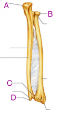
Which is the Olecranon? A. a B. b C. c D. d |

The Olecranon was A. a |
|

Which is the styloid process of the Ulnar? A. a B. b C. c D. d |

Styloid process is D. d |
|

Which is the Radial head? A. a B. b C. c D. d |

The Radial head is B. b |
|
|
What connects the margin of the ulnar shaft to radius? A. Ulnar head B. Radial notch C. Interosseous membrane D. Radius and phalanges |
C. Interosseous membrane |
|
|
The elbow joint is a: A. Hinge joint B. Hinge and pivot joint C. Ball and socket joint D. Saddle joint |
B. Hinge and pivot joint |
|
|
The muscles located on the anterior and medial surface of the arm are primarily responsible for: A. Extension B. Flexion C. Supination D. Pronation |
B. Flexion |
|
|
The four muscles responsible for flexion of the elbow are: |
1. Biceps Brachii 2. Brachioradialis 3. Brachialis (to a lesser extent, pronator teres) |
|
|
The muscles responsible for extension of the elbow are: |
Triceps brachii Anconeus |
|
|
The __________ inserts onto the radial tuberosity. A. Triceps Brachii B. Biceps Brachii C. Brachioradialis D. Anconeus |
B. Biceps Brachii |
|
|
What movements does the inferior radioulnar joint produce? |
Supernation Pronation |
|
|
The inferior radioulnar joint is: A. Uniaxial B. Biaxial C. Multiaxial D. None of the above |
A. Uniaxial |
|
|
The Pronator Teres is innervated by the: A. Median nerve B. Radial Nerve C. Ulnar Nerve D. Musculocutaneous nerve |
A. Median Nerve |
|
|
The Axial Skeleton consists of: A. Bones of the vertebral column, sternum, pelvis, skull and thorax B. Bones of the vertebral columns, patellar, sternum, skull and thorax C. Bones of the limbs and limb girdles D. Bone of the upper limb, lower lib and skull |
A. Bones of the vertebral column, sternum, pelvis, skull and thorax |
|

Which of the following is the Parietal bone? A. a B. b C. c D. d |
B. b is the Parietal bone |
|
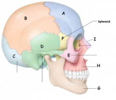
Which of the following is the Occipital bone? A. a B. b C. c D. d |
C. c is the Occipital bone |
|
|
Which of the following muscles are responsible for smiling? A. Zygomatic major B. Fronalis C. Orbicularis Occuli D. Mentalis |
A. Zygomatic major |
|
|
How many bones does the vertebral column have? A. 31 B. 35 C. 33 D. 32 |
C. 33 (7, 12, 5, 5, 4) |
|
|
Which of the following is incorrect. Primary Curvatures: A. Developed after birth B. Consists of the lumbar curvature and the Thoracic curvature C. Are convex posteriorly D. Consist of the Cervical curvature and the Lumbar curvature |
C. Are convex posteriorly |
|
|
Which of the following is correct. Lordosis is: A. Outward curving B. Inward curving C. Abnormal lateral curvatures D. None of the above |
B. Inward Curving A. Outward curving is Kyphosis C. Abnormal lateral curvatures is Scoliosis |
|
|
Which region lies in the area commonly known as the waist? A. Cervical B. Thoracic C. Lumbar D. Sacral |
C. Lumbar |
|
|
The __________ component of a typicall vertebra is associated with weight bearing. A. Pedicle B. Spinous process C. Lamina D. None of the above |
D. None of the above; the body would be the correct answer. |
|
|
__________ consists of a heart shaped body, steep inferiorly angled spinous process, small vertebrae foramen and facets on bodies and transverse processes. A. Sacral vertebrae B. Lumbar vertebrae C. Thoracic vertebrae D. Cervical vertebrae |
C. Thoracic vertebrae |
|
|
__________ consists of vertebrae fused into a single wedge shaped bone and pelvic sacral foramina. A. Sacral vertebrae B. Lumbar vertebrae C. Thoracic vertebrae D. Cervical vertebrae |
A. Sacral vertebrae |
|
|
Muscles that run along the side of the intervertebral joint causes A. Extension of the vertebral column B. Lateral flexion of the vertebral column C. Rotation of the vertebral column D. Flexion of the vertebral column |
B. Lateral flexion of the vertebral column |
|
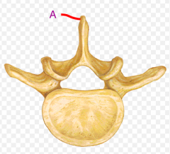
What is the name of the component labeled A: A. Body B. Transverse process C. Lamina D. Spinous process |
D. Spinous process |
|
|
What nerve innervates the Diaphragm? A. Intercostal nerve B. Phrenic nerve C. Facial nerve D. Regional spinal nerve |
B. Phrenic nerve |
|
|
The erector spine group is responsible for flexion of the vertebral column. A. True B. False |
B. False, the erector spine group are responsible for extension and lateral flexion not flexion of the vertebral column. |
|
|
The external intercostal muscles are responsible for inspiration/inhalation. A. True B. False |
A. True, the external intercostal muscles are responsible for inspiration/inhalation. |
|
|
The internal intercostal muscles are responsible for the elevation of the ribs. A. True B. False |
B. False, the internal intercostal muscles are responsible for forced expiration/exhalation and thus retract the ribs. |
|
|
The Thoracic Diaphragm is the primary muscles of inspiration/inhalation. A. True B. False |
A. True |
|
|
Contraction of the thoracic diaphragm causes the diaphragm to flatten/lower. A. True B. False |
A. True |
|
|
The diaphragm separates the thoracic and abdominal cavity. A. True B. False |
A. True |
|
|
The Pictorial girdle consists of the: A. Scapula and Humerus B. Radius and Ulnar C. Clavicle and Scapula D. Humerus and Radius |
C. Clavicle and Scapula |
|
|
The Rhomboids attach from the A. Thoracic vertebrae to the medial border of the Scapula B. Ribs to anterior Scapula surface C. Ribs to the posterior Scapular surface D. Thoracic vertebrae to lateral border of Scapula |
A. Thoracic vertebrae to the medial border of the scapula |
|
|
Which muscle can elevate, retract and perform upward/lateral rotation of the scapula? A. Serrates Anterior B. Rhomboids C. Teres Minor D. Trapezius |
D. Trapezius |
|
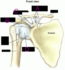
Which is the Coraco-humeral ligament? A. a B. b C. c D. d |
B. Is the Coraco-humeral ligament |
|
|
Which of the following is not a rotator cuff? A. Teres minor B. Subscapularis C. Teres Major D. Supraspinatus |
C. Teres Major |
|
|
Which of the following rotator cuff muscles are located posteriorly? A. Infraspinatus and Subscapularis B. Subscapularis and Supraspinatus C. Teres minor and Teres major D. Infraspinatus and Teres minor |
D. Infraspinatus and Teres minor |
|
|
Which of the following muscles can flex, adduct and medially rotate the arm? A. Deltoid B. Lattisimus dorsi C. Rhomboids D. Pectoralis Major |
A. Deltoid |
|
|
Which nerve innervates the Coracobracialis muscle A. Median nerve B. Brachial nerve C. Radial nerve D. Musculocutaneous nerve |
D. Musculocutaneous nerve |
|
|
Which muscles attaches from the scapular spine and clavicle and inserts into the deltoid tuberosity? A. Pectorialis major B. Teres major C. Deltoid D. Lattisimus dorsi |
C. Deltoid |
|
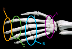
Name each of the following joints A. B. C. D. |
A. Carpometacarpal joint B. Metacarpophalangeal joint C. Proximal Interphalageal joint D. Distal Interphalageal joint |
|
|
The __________ are located within the carpal tunnel A. Flexor digitorum superficialis, flexor digitorum profundus, flexor pollicis longus, radial nerve B. Flexor digitorum superficialis, opponens pollicis, flexor pollicis longus and ulnar nerve C. Abductor digiti minimi, flexor digitorum profundus, flexor digitorum superficialis and ulnar nerve D. Flexor digitorum superficialis, flexor digitorum profundus, flexor pollicis longus and median nerve |
D. Flexor digitorum superficialis |
|
|
What feature of the hand helps increase friction to grasp objects firmly? A. Extensor retinaculum B. Flexor retinaculum C. Palmar aponeurosis D. Dorsal digital expansion |
C. Palmar aponeurosis |
|
|
Which functional region of the hand is located at the 5th digit muscles? A. Mid palm region B. Hypothenar region C. Thenar region D. None of the above |
D. Hypothenar region |
|
|
The palmar interossei performs __________ of the MCP joints A. Abduction B. Adduction C. Flexion D. Extension |
B. Adduction |
|
|
What are the three types of power grips? 1. 2. 3. |
1. Spherical grip 2. Cylindrical grip 3. Hook grip |
|
|
What are the four types of precision grips? 1. 2. 3. 4. |
1. Lateral pinch 2. Tip to tip/pincer 3. Pad to pad/pinch 4. Lumbrical grip |
|

What kind of grip is this? |
Lumbrical |
|
|
What movements does the hip joint produce? A. Flexion/extension, adduction and abduction B. Circumduction, flexion/extention C. Flexion/extension, adduction and abduction, circumduction D. Flexion/extention, adduction and abduction, medial/lateral rotation |
D. Flexion/extention, adduction and abduction, medial/lateral rotation. |
|
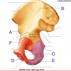
Which is the Ischial Tuberosity? A. a B. b C. c D. d E. e F. f |
D. d is the Ischial Tuberosity |
|
|
What muscles perform flexion at the knee joint? 1. 2. 3. 4. |
1. Biceps Femoris 2. Semitendinosus 3. Semimembranosus 4. Sartorius |
|
|
What are the extensions of the knee? 1. 2. 3. 4. |
1. Rectus Femoris 2. Vastus Lateralis 3. Vastus Medialis 4. Vastus Intermediairs |
|
|
What nerve innervates the Sartorius? A. Femoral nerve B. Obturator nerve C. Sciatic nerve D. Gluteal nerve |
A. Femoral nerve |
|
|
What are the three Tibiofibuar joints? 1. 2. 3. |
1. Proximal Tibiofibular joint 2. Intermedial Tibiofibular joint 3. Distal Tibiofibular joint |
|
|
What are the movement of the foot? A. Plantar flexion, dorsiflexion B. Eversion, Inversion C. Plantar flexion, eversion, inversion D. Plantar flexion, eversion, inversion, dorsiflexion |
D. Plantar flexion, eversion, inversion, dorsiflexion |
|
|
Name the bones of the foot C- T- N- C- C- |
Calcaneus Talus Navicular Cuboid Cuneiform - Medial - Intermediate - Lateral |
|
|
Which of the following is incorrect? A. The articular surfaces of the hip joint include the acetabulum and the femur B. The hip joint is a multi axial, ball and socket joint C. The iliofemoral ligament is located anterior and limits extension D. The ischiofemorial ligament is located anterior and limits extension |
D. The ischiofemorial ligament is located posterior not anterior |
|
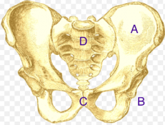
Name each area of the pelvis A. B. C. D. |
A. Ilium B. Ischium C. Pubis D. Sacrum |
|
|
The biceps femurs, semimembranosus and semitendinosus all arise from the ischial tuberosity A. True B. False |
A. True |
|
|
The Gluteus Minimus and Medius are innervated by the: A. Femoral nerve B. Sciatic nerve C. Gluteal nerve D. Obturator nerve |
C. Gluteal nerve |
|
|
The Tensor Fascia Late is innervated by the: A. Femoral nerve B. Sciatic nerve C. Gluteal nerve D. Obturator nerve |
C. Gluteal nerve |
|
|
Muscles that perform hip adduction include: A. Gracilis, Adductor magnus, adductor longus, adductor brevis B. Sartorius, Adductor magnus, Adductor longus, adductor brevis C. Sartorius, Gracillis, Adductor magnus, Adductor brevis D. Rectus femoris, Gracilis, Sartorius, Adductor Magnus |
A. Gracilis, Adductor magnus, Adductor longs and Adductor brevis |
|
|
Fill in the blanks with: Anterior, Posterior and Medial. The Femoral nerve is located __________. The Sciatic nerve is located __________. The Obturator nerve is located __________. The Patella is a __________ bone. |
The Femoral nerve is located Anterior. The Sciatic nerve is located Posterior. The Obturator nerve is located Medial. The Patella is a Anterior bone. |
|
|
Which of the following is incorrect? A. The knee joint is a hinge joint B. The knee joint is multiaxial, synovial joint C. The knee joint performs flexion and extension D. The patella articulates with the femur. |
B. The knee joint is uniaxial not multiaxial. |
|
|
The Fibular collateral ligament: A. Limits medial rotation B. Prevents abduction C. Prevents adduction D. None of the above |
C. Prevents adduction |
|
|
How many Carpal bones are there? A. 8 B. 7 C. 6 D. 9 |
A. 8 |
|
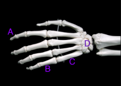
Which of the following is the metacarpal bone? A. a B. b C. c D. d |
C. c are the metacarpal bones |
|
|
The proximal row of Carpal bones consist of: A. Pisiform, Hamate, Triquetrum, Lunate B. Hamate, Capitate, Lunate, Scaphoid C. Trapezium, Trapezoid, Pisiform, Triquetrum D. Pisiform, Triquetrum, Lunate, Scaphoid |
D. Pisiform, Triquetrum, Lunate, Scaphoid |
|
|
The distal row of Carpal bones consist of: A. Hamate, Capitate, Trapezoid, Trapezium B. Hamate, Capitate, Lunate, Scaphoid C. Trapezium, Trapezoid, Pisiform, Triquetrum D. Pisiform, Triquestrum, Lunate, Capulate |
A. Hamate, Capitate, Trapezoid, Trapezium |
|
|
How many phalanges does the thumb have? A. 2 B. 4 C. 3 D. 1 |
A. 2 |
|
|
The metacarpal bones are numbered I-V from __________ to __________. A. Superior to inferior B. Lateral to medial C. Medial to lateral D. Proximal to distal |
B. Lateral to medial |
|
|
Which of the following is incorrect? A. The radiocarpal joint is a condyloid synovial joint B. The radiocarpal joint is between the proximal row of carpal bones (exl. pisiform) and the head of the radius and articular discs C. The radiocarpal joint consist of the proximal row of carpal bones and the inferior radioulnar joint D. None of the above |
C. The radiocarpal joint does not consist of the radioulnar joint. |
|
|
What movements can be performed at the wrist joint? A. Flexion/Extension B. Circumduction C. Flexion/Extension, Circumduction, Abduction/Adduction D. Flexion/Extension, Circumduction, Supination/Pronation |
C. Flexion/Extension, Circumduction, Abduction/Adduction |
|
|
The muscle responsible for flexion of the wrist consist of: A. Flexor Carpi Radialis, Flexor Carpi Ulnaris, Palmaris longus. B. Extensor Carpi Radialis, Extensor Carpi Ulnaris, Palmaris Longus C. Fleor Capri Radialis, Extensor Carpi Radialis longus and brevis D. Extensor Carpo Radialis, Extensor Carpi Ulnaris |
A. Flexor Carpo Radialis, Flexor Carpi Ulnaris, Palmaris longus |
|
|
What nerve innervates the Flexor Capri Radialis? A. Ulnar nerve B. Musculocutaneous nerve C. Median nerve D. Radial nerve |
C. Median nerve |
|
|
Which muscle attaches from the lateral epicondyle of the humerus to the carpals and 5th metacarpal? A. Flexor Carpi Ulnaris B. Extensor Carpi Ulnaris C. Extensor Carpi Radialis D. Palmaris Longus |
B. Extensor Capri Ulnaris |
|
|
The muscles responsible for adduction of the wrist consist of the: A. Flexor Carpi Radialis, Flexor Carpi Ulnaris B. Extensor Carpi Radialis, Extensor Carpi Ulnaris C. Flexor Carpi Radialis, Extensor Carpi Radialis D. Extensor Carpi Ulnaris, Flexor Carpi Ulnaris |
D. Extensor Carpi Ulnaris, Flexor Carpi Ulnaris |
|
|
What nerve innervate the flexor policies longus? A. Median nerve B. Radial nerve C. Ulnar nerve D. Musclulocutaneous nerve |
A. Median Nerve |
|
|
What nerve innervates the abductor policies longus? A. Median nerve B. Radial nerve C. Ulnar nerve D. Musclocutaneous nerve |
B. Radial Nerve
|
|
|
What muscle is responsible for abducting the thumb? A. Flexor Policis Longus B. Abductor Policis Longus C. Extensor Policis Longus D. Extensor Digitorum |
B. Abductor Policis Longus |
|
|
What is the extensor retinaculum? |
A ligament that holds the extensor tendons in place. |
|
|
What muscles crosses both the elbow and wrist joints? A. Brachioradialis B. Pronator Teres C. Extensor Polls Brevis D. Flexor Carpi Radialis |
D. Flexor Capri Radialis |
|
|
Which nerve innervates the extensor muscles of the forearm? A. Musculocutaneous B. Median C. Radial D. Ulnar |
C. Radial Nerve |
|
|
What set of intrinsic hand muscles abduct the digits in the metacarpalphalengeal joints? A. Flexor Digitorum Superficialis B. Lumbricals C. Palmar Interossei D. Dorsal Interossei |
D. Dorsal Interossei |