![]()
![]()
![]()
Use LEFT and RIGHT arrow keys to navigate between flashcards;
Use UP and DOWN arrow keys to flip the card;
H to show hint;
A reads text to speech;
37 Cards in this Set
- Front
- Back
|
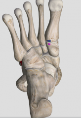
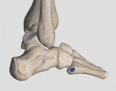
What are the dermatomes of the following; medial malleolus, lateral malleolus, inferior lateral foot, inferior medial foot. |
- medial: L4 - lateral: S1 - inferior lateral foot: S1 - inferior medial foot: L5 |
|
|
What are the contents of the anterior compartment of the leg? (6) |
- extensor/DF compartment - tibialis anterior muscle - extensor digitorum longus - extensor hallucis longus - anterior tibial artery and vein (2) - deep fibular nerve |
|
|
What are the contents from the posterior compartment of the leg? |
- neurovascular structures: posterior tibial artery and veins (2), posterior tibial nerve - superficial muscles: gastroc. plantaris, soleus - deep muscles: tibialis posteior, flexor hallucis longus, flexor digitorum longus |
|
|
The deep and superficial muscles of the posterior compartment of the leg are connected by ________. |
transverse intermuscular septum |
|
|
What are the contents of the lateral compartment of the leg? |
- neurovascular structures: superficial fibular nerve, fibular artery and veins (2) - muscles: fibularis longus and brevis |
|
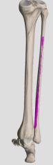
What originates here? |
- flexor hallucis longus - distal 2/3 of the fibula - interosseous membrane |
|
|
Where does the FHL insert? |
- distal phalanx of hallux |
|
|
What is the innervation and action of FHL? |
- inn. tibial nerve --> nerve to FHL (S2, S3) - action: plantar flexes foot, flexes hallux, supports medial longitudinal arch |
|
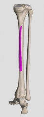
What originates here? |
- FDL - middle third of posterior surface of tibia, below soleal line |
|
|
What is the insertion of the FDL? |
plantar surface of the bases of distal phalanges |
|
|
What is the innervation and action of FDL? |
- tibial nerve --> nerve to FDL (S2, S3) - PF ankle joint - flexes interphalangeal joints of 4 lateral toes - supports longitudinal arches |
|
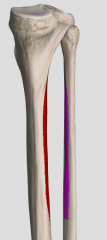
What muscle originates at these highlighted areas? |
- tibialis posterior - postero-lateral tibia (below soleal line) - postero-medial fibular shaft |
|
|
What are the insertions f the tibialis posterior? |
- NICLB - navicular - intermediate cuneiform - lateral cuneiform - bases of metatarsals 2, 3, 4 |
|
|
What is the innervation and action of TP? |
- tibial nerve --> nerve to tibialis posterior (L4, L5) - actions; PF, supinates foot, supports medial longitudinal arch |
|
|
The TP muscle is the deepest muscle of the posterior compartment of the leg. T/F |
True |
|
What inserts here? |
Fibularis longus - base of 1st metatarsal - medial cuneiform |
|
|
What is the origin of FL? |
- head and shaft of fibula |
|
|
What is the innervation of FL? |
superficial fibular nerve (L5, S1, S2 - same with fibularis brevis |
|
|
What arch does FL support? which does FB support? |
- FL supports transverse arch - FB supports lateral longitudinal arch |
|
What inserts here? |
Fibularis brevis - lateral side of the base of the 5th metatarsal |
|
|
The gastroc cannot produce plantar flexion when the knee is fully flexed. T/F |
True |
|
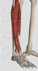
Label these muscles; what compartment(s) do they belong too? |
- fibularis longus - tibialis anterior - FB - EDL - EHL - they belong to the lateral and anterior compartment of the leg |
|
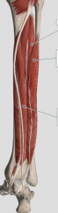
Label these muscles; what compartment(s) do they belong too? |
- belong to the deep posterior compartment of the leg - tibialis posterior - FHL - FDL |
|
|
Whats the main difference between the soleus and gastroc? |
- soleus is slow twitch muscle fibres, used for steadying leg on foot and slow walking - gastroc is fast twitch muscle fibres; provides force propulusion in walking, running, jumping |
|
|
Gastroc makes up the _____ part of the achilles tendon and the soleus makes up the _______. |
lateral; medial |
|
|
What is the largest branch of the popliteal artery? |
The posterior tibial artery; main supplier of the posterior leg and sole of foot |
|
|
When does the posterior tibial artery give off the fibular artery branch? |
at the aponeurotic arch of the soleus |
|
|
Starting from the popliteal artery (inferior of knee joint), what are the subsequent branches that come off of it?
|
- anterior tibial artery (after aponeurotic arch) - posterior tibial artery (after aponeurotic arch) - fibular artery - posterior medial malleoli arteries - posterior medial calcaneal arteries |
|
|
What branches come off the fibular artery? |
- posterior lateral malleoli arteries - posterior lateral calcaneal arteries |
|
|
What are the branches of the popliteal artery at the knee joint? |
- superior medial and lateral genicular arteries - middle genicular - inferior medial and lateral genicular - recurrent fibular |
|
|
Where does the sural nerve change its name? |
at the lateral malleolus, changes name to lateral dorsal cutaneous nerve |
|
|
What does the fibular artery supply? |
Muscles in the posterior and lateral compartment of the leg |
|
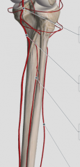
Label these arteries. |
- popliteal artery - posterior tibial artery - fibular artery - anterior tibial artery |
|
|
What part of the foot does the deep fibular nerve innervate? |
- Lateral big toe and medial second toe |
|
|
What are the contents of the posterior tarsal tunnel? |
- Tibialis posterior - FDL - tibial arteries and veins - tibial nerve - FHL |
|
|
What does the calcaneal tendon reflex test? |
- tests S1-S2 nerve roots |
|
|
What is the difference between proximal tarsal tunnel and distal tarsal tunnel syndrome? |
- proximal: entrapment of the tibial nerve or its calcaneal branches - distal: entrapment of the medial or lateral plantar nerve or its calcaneal branches h |