![]()
![]()
![]()
Use LEFT and RIGHT arrow keys to navigate between flashcards;
Use UP and DOWN arrow keys to flip the card;
H to show hint;
A reads text to speech;
93 Cards in this Set
- Front
- Back
|
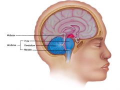
What are the FOUR major REGIONS of the brain? |
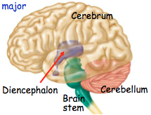
1) Brain Stem 2) Cerebellum 3) Cerebrum 4) Diencephalon |
|
|
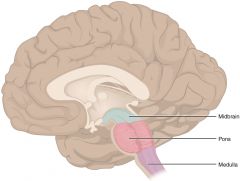
What 3 parts make up the Brain Stem? |

1) The Medulla
2) The PONS 4) The Midbrain |
|
|
What are the three parts that make up the Diencephalon? |
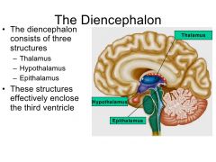
1) The Thalamus 2) The Hypothalamus 3) The Epithalamus |
|
|
What is the function of the medulla oblongata? Where is it located? |
The Medulla has 2 functions: control our heart rate through the cardiovascular center control our breathing, through the medullary rhythmicity area |
|
|
Where is Broca's Speech Area, and what is its main function? |
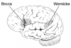
The Broca's Speech Area is located in the left frontal lobe of the brain, and it is in charge of directing the muscle movement involved in speech. |
|
|
Where is Wernicke's Area, and what is its main function? |

Wernicke's Area is located in the left temporal and parietal lobes, and is primarily involved in language comprehension/understanding. |
|
|
What is Aphasia? What causes it? |
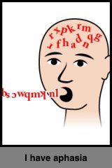
Aphasia is the impairment of language. It usually results from an injury to the Broca's Area and produces impaired speech |
|
|
What are the main functions of the PONS? Where is it located? |
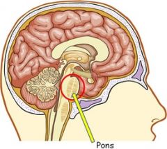
The PONS contains pontine nuclei which relay information from the motor areas of the cerebral cortex to the cerebellum. The PONS also aids in the control of breathing and also plays a key role in sleeping/dreaming. |
|
|
What are the main functions of the midbrain? Where is it located? |
The midbrain coordinates the movements of our head, eyes and trunk in response to visual/auditory stimuli. |
|
|
What is the main function of the cerebellum? Where is it located? |
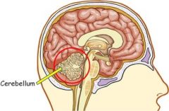
The main function of the cerebellum is the coordination of skilled movements and regulation of posture and balance. |
|
|
What is the main function of the thalamus? Where is it located? |
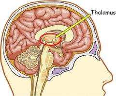
The thalamus is the relay station for ALL sensory info [except smell!] to the cerebral cortex. It does also contribute to motor functions through this transmission of info. Relays nerve impulses b/w areas of the cerebrum as well. Deals with memory, emotion, learning, movement (visual/auditory) and somatic sensations such as tickling/pain/body temp. |
|
|
What is the main function of the epithalamus? Where is it located? |

The epithalamus houses the Pineal gland (which controls melatonin secretion/sleep cycles) as well as Habenular nuclei which controls olfactory emotional memories/response. |
|
|
What is the main function of the Hypothalamus? Where is it located? |
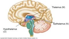
The Hypothalamus is the major regulator of homeostasis! That includes: - Control of ANS (HR, bladder, GI Tract) - Hormone Production - Emotion/Behavior Patterns - Regulation of eating/drinking - Thermoregulation - Circadian Rhythm and Consciousness |
|
|
What is the main function of the cerebrum? Where is it located? |
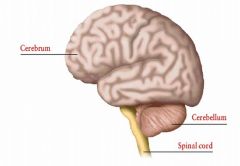
The main function of the cerebrum is to deal with higher brain functions, such as thought and action. It is also the largest part of brain. |
|
|
What is the cerebral cortex? What are its main functions and where is it located? |
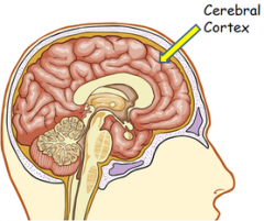
The cerebral cortex is the gray matter surrounding the cerebrum. It is around 2-4mm thick and contains billions of neurons. |
|
|
What are basal nuclei? What importance do they serve? Where are they located? What are some diseases associated with the injury of these nuclei? |
Basal Nuclei are paired masses of gray matter deep within each cerebral hemisphere. They influence the activity of the cerebral motor cortex, play a part in certain purposeful eye movements, and also play a role in the retrieval of stored cognitive, executive and emotional patterns from the cerebral cortex. Some of the diseases associated with Basal Nuclei are OCD, Parkinson's & Huntington's, and chronic anxiety. |
|
|
Where is the Primary Somatosensory Area located? |

The Primary Somatosensory Area is located in the postcentral gyrus (or, closer to the cerebellum) |
|
|
Where is the Primary Motor Area located? |
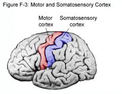
The Primary Motor Area is located in the precentral gyrus (or, in front of the somatosensory area). |
|
|
What kind of information does the Primary somatosensory area receive? |
The Primary somatosensory area receives information for: -touch, pain, pressure, temperature, tickle, proprioception (body position/movement). |
|
|
What kind of information does the Primary Visual Area receive? |
The Primary Visual Area receives visual information |
|
|
What results from an injury to the Broca's Area? |
Impaired speech results from an injury to the Broca's Area (but one is still able to understand words - just not able to speak what they want). |
|
|
What results from an injury to the Wernicke's Area? |
Impaired understanding of words results from an injury to Wernicke's Area. (one can still speak words and form sentences, they just do not make coherent sense). |
|
|
What are the primary functions of the Right Hemisphere? |
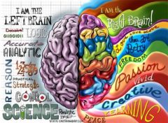
The Right Hemisphere is our more "musical" and "creative" side of the brain. It focuses on: Musical Awareness; space/pattern perception; insight/imagination/generating mental images of sight, sound, taste, and smell. |
|
|
What are the major functions of the Left Hemisphere? |

The Left Hemisphere is our more "logical" side of the brain. It deals with: Reasoning; spoken/written language; and numerical/scientific skills. |
|
|
What are the THREE layers of Cranial Meninges? |
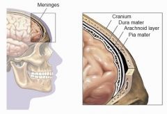
1) Dura Mater (MOST OUTER) 2) Arachnoid Mater (middle layer; spidery appearance) 3) Pia Mater (most inner layer; very thin/delicate) |
|
|
What is Pia Mater? |
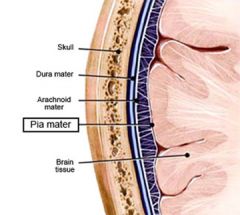
Pia Mater is the innermost layer of the cranial meninges. It is very thin and delicate, and because of those traits it provides the 'glossy appearance' of the brain. It is also highly vascular, and is the only layer to closely follows the brain's convolutions. |
|
|
What is Arachnoid Mater? |
Arachnoid Mater is the second layer of cranial meninges. It is a delicate, avascular layer and is very 'spidery' in its appearance. |
|
|
What are the TWO layers of the Dura Mater? |
1) External Periosteal Layer 2) Internal Meningeal Layer |
|
|
What is the External Periosteal Layer? |

The External Periosteal layer is the first layer of the Dura Mater and makes up the skull's inner periosteum (AKA endocranium). |
|
|
What is the Internal Meningeal Layer? |
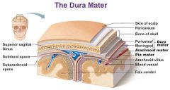
The Internal Meningeal Layer is the actual dura mater (the second layer of the dura mater) |
|
|
What do the extensions of the Dura Mater form? |
The extensions of the Dura Mater form hard, non-compliant membranes that divide the intracranial vault. These dural extensions then form 3 extensions: 1) Falx Cerebri 2) Falx Cerebelli 3) Tentorium Cerebelli |
|
|
What/Where is the Falx Cerebri? |

The Falx Cerebri is a sickle-shaped fold that divides the Left/Right hemispheres of the brain. It is located on the longitudinal cerebral fissure. |
|
|
What/Where is the Falx Cerebelli? |
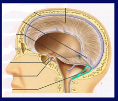
The Falx Cerebelli is a small, triangular shaped process that separates the cerebellar hemispheres. |
|
|
What/Where is the Tentorium Cerebelli? |
The Tentorium Cerebelli is a tent-like dural fold that separates the cerebrum from the cerebellum. |
|
|
Why is the Blood Brain Barrier significant? |
The selectivity of the tight junctions formed in the BBB restrict passage of harmful substances from the blood stream to the brain. |
|
|
What is potentially wrong with the tight junctions within our BBB? |
The tight junctions form such a selective barrier that at times, the active transport of glucose can be trying and there can be difficulty in treating infection within the brain because even medicine won't be allowed passage. |
|
|
What are the basic functions of our Cerebrospinal Fluid? |
Cerebrospinal Fluid offers help in - Cushioning/mechanical protection for the brain -Buoyancy and shock-absorbent -Homeostatic Controls (cerebral blood flow and breathing) -Transport system for hypothalamic processes -Circulation that exchanges nutrients |
|
|
What is hydrocephalus? |
Hydrocephalus is an accumulation of CSF due to an impaired CSF flow, absorption, and/or overproduction. It is characterized by increased intercranial pressure and thus could lead to brain damage/other complications/even death. |
|
|
What are the TWO types of hydrocephalus? |
-Internal Hydrocephalus deals with an obstruction inside the brain itself (i.e. a blockage in the ventricles) -External Hydrocephalus deals with an obstruction outside of the brain (i.e. within the subarachnoid). |
|
|
What is Bell's Palsy? |

Bell's Palsy is damage to the facial nerve either virally or bacterially. It results in facial muscle (and thus, expression) paralysis. Those suffering from this condition cannot even close their eyes, taste anything, or salivate. |
|
|
What does the Automatic Nervous System control? |
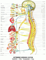
The ANS regulates the activity of smooth muscle, cardiac muscle, and certain glands that we don't normally think about. (Hence the name, autonomic). |
|
|
What is the main function of the ANS? |
The ANS works primarily to keep our homeostasis. |
|
|
Which body system releases ONLY Acetylcholine (ACh)? |
SOMATIC NERVOUS SYSTEM ONLY RELEASES ACh! |
|
|
Which body system can release both NE and ACh? |
AUTONOMIC NERVOUS SYSTEM CAN RELEASE BOTH NE AND ACh! |
|
|
What are the main differences between the SNS and the ANS? |
The SNS: - Sensory neurons input from receptors of special/somatic senses. - Sensations are consciously perceived - Somatic motor neurons innervate skeletal muscles to produce voluntary movement - the effect of a somatic motor neuron is always excitation MEANWHILE... - autonomic sensory neurons are associated with interoreceptors - sensory input is NOT consciously perceived - autonomic motor neurons regulate visceral activities thru both excitation & inhibition - All autonomic motor pathways consist of TWO parts (sympathetic/parasympathetic) |
|
|
What are the TWO parts of the ANS? |
1) Sympathetic Nervous System (fight/flight) 2) Parasympathetic Nervous System (rest/digest) |
|
|
What are the two neural pathways of the ANS? |
There are Postganglionic Fibers, and Preganglionic Fibers, both of which are used in the ANS system to transport/relay messages between the brain and the body part/organ. |
|
|
What can Postganglionic Fibers release? |
Postganglionic Fibers can release either ACh or NE |
|
|
What can Preganglionic Fibers release? |
Preganglionic Fibers can only release ACh |
|
|
What does the structure of the preganglionic/postganglionic fiber look like in the Sympathetic Nervous System? |
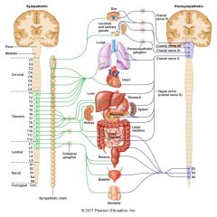
In the Sympathetic Nervous System, the preganglionic fibers are short, resulting in a more widespread response field. This is important because in our fight/flight mode our whole bodies are active and ready for action. |
|
|
What does the structure of the preganglionic/postganglionic fiber look like in the Parasympathetic Nervous System? |

In the Parasympathetic Nervous System, the structure of the postganglionic fibers are long, which results in a more localized response field. This is important because in our rest/digest mode our bodies are able to be more productive on more precise actions that have to do with being relaxed. |
|
|
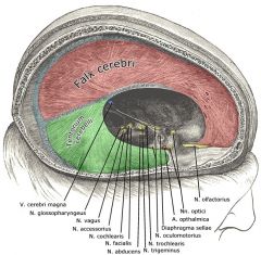
Which cranial nerve deals with smell? |
The Olfactory (I) nerve is a SENSORY nerve. It contains olfactory receptors. |
|
|
Which cranial nerve deals with vision? |
The Optic Nerve (II) is a SENSORY nerve. It contains visual signals initiated in the retina. |
|
|
Which cranial nerve(s) deals with eye movements? |
*Oculomotor Nerve (III) is a MOTOR nerve located in the midbrain & controls eye movement, pupil constriction, and focusing. **Trochlear Nerve (IV) is a motor nerve located in the midbrain and controls muscles that move the eyes extrinsically. ***Abducens Nerve (VI) is a motor nerve located in the PONS and is in charge on eye muscle ABDUCTION. ( **damage to this causes lazy eye). |
|
|
What is the Trigeminal Nerve? |
The Trigeminal Nerve (V) is a MIXED (both sensory and motor functions involved) nerve that originates in the PONS and has 3 branches dealing with the face: - Opthalamic Nerve (around the eyes) - Maxillary Nerve (around the nose/cheeks/upper jaw) - Mandibular Nerve (around the lower jaw/chin) |
|
|
What is the Facial Nerve? |
The Facial Nerve (VII) is a MIXED nerve that deals with functions related to the face: Sensory neurons relay info for taste buds, skin in our ear canal, and proprioceptors of the face & scalp. Motor neurons relay info for facial expression, salivation, middle ear/face/scalp/neck muscles. This part of the nerve is located in the PONS. |
|
|
What is the Vestibulochoclear Nerve? |
The Vestibulocochlear Nerve (VIII) is a SENSORY neuron that deals with hearing/balance. It has two branches: Vetisbular Branch that deals with balance (from inner ear to PONS and cerebellum) Cochlear Branch that deals with hearing from medulla to thalamus. |
|
|
What is the Vagus Nerve? |
The Vagus Nerve (X) is a MIXED nerve that deals with: Sensory neuron: skin of external ear; proprioception (neck/throat); baroreceptors (carotid sinus); chemoreceptors Visceral sensory neurons (hunger/fullness) These axons originate in the medulla and extend to the PONS |
|
|
What is the Glossopharyngeal Nerve? |
The Glossopharyngeal Nerve (IX) is a taste/swallowing MIXED nerve that deals with swallowing and saliva secretion. It originates in the medulla. |
|
|
What is the Accessory Nerve? |
The Accessory Nerve (XI) is a MOTOR nerve that supplies the somatic motor innervation to trapezius and sternocleidomastoid. It assists in head/neck movements. |
|
|
What is the Hypoglossal Nerve? |
The Hypoglossal Nerve (XII) is a MOTOR nerve that deals with tongue movement. It originates in the medulla and helps in speech & swallowing. |
|
|
What are the major responses to Parasympathetic Innervation? |
There is an acronym for the bodily functions that increase during Parasympathetic innervation: SLUDD. It stands for: Salivation Lacrimation (Tears) Urination Digestion Defecation There are also 3 bodily functions that decrease: Rate/force heart beat Airway size & rate of breathing Pupil Size |
|
|
Which body system can release both ACh or NE? |
Sympathetic! - however, NE can only be released @ the postganglionic fiber! |
|
|
Which body system only releases ACh? |
Parasympathetic! |
|
|
Which system stands for 'rest & digest'? |
The Parasympathetic Nervous System! Remember this because 'people sleep' (ParaSympathetic) |
|
|
Which system stands for 'fight or flight'? |
The Sympathetic Nervous System! Remember this because there's only one word: and then bam! you're ready for action! |
|
|
What are the TWO receptors that bind solely to ACh? |
Cholinergic Receptors Bind to ACh, and there are two of them: - Muscarinic (release slow metabolic response) - Nicotinic (release fast synaptic response) |
|
|
What receptors binds to NE? |
Adrenergic Receptors bind to NE, and there are two of them: - Alpha - Beta |
|
|
What are the two neurotransmitters found primarily in the brain? |
- Acetlycholine (ACh) - Norepinephrine (NE) |
|
|
What is the function of Acetlycholine? |
Acetlycholine is a neurotransmitter found in the PNS and the CNS and works as a neuromodulator. In the PNS, ACh activates muscles. |
|
|
What is the function of Norepinephrine? |
Norepinephrine is a neurotransmitter released from the sympathetic nervous system in response to stress. It is referred to as a stress hormone. |
|
|
Define Somatic |
Tactile, thermal, pain and proprioceptive sensations; i.e. anything dealing with the skin/outside of the body. |
|
|
Define Visceral |
Internal organ sensations/conditions |
|
|
Define Sensory Modality |
One sensory modality carried by a given neuron (touch/pain/vision) |
|
|
What are some special sensory modalities? |
- Smell - Taste - Vision - Hearing - Equilibrium |
|
|
What are the 4 Steps in the Process of Sensation? |
1.) Stimulation: appropriate stimulus in receptive field 2.) Transduction (Conversion): sensory receptor transduces energy in a stimulus into a graded potential 3.) Generation/Conduction: nerve impulses are propagated towards the CNS when threshold is reached 4.) Integration: CNS integrates the sensory input. Conscious sensations integrated into our cerebral cortex. |
|
|
Define Selectivity |
A given sensory receptor responds vigorously to a specific stimulus. Sensory receptors can transduce (convert) only 1 kind of stimulus |
|
|
What are the 3 Classifications of Sensory Receptors? |
1.) Microscopic Structure (contains 3 types of receptors) 2.) Receptor Location/Origin (contains 3 types of receptors) 3.) Type of Stimulus Detected (contains 6 types of receptors) |
|
|
What are the THREE types of receptors within the Microscopic Structure Classification of Sensory Receptors? |
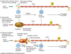
1.) Free Nerve Ending - have bare dendrites, lacking any structural specialization. These nerves deal with pain/temp/itch sensations 2.) Encapsulated Nerve Ending - dendrites are enclosed in connective tissue capsule. These nerves deal with pressure/vibration sensations. 3.) Separate Cells - synapse w/ sensory neurons. These nerves deal with hearing/taste/vision sensations. |
|
|
What are the THREE types of receptors within the Receptor Location/Origin Classification of Sensory Receptors? |
1.) Exteroreceptors - Found at/near external surfaces of the body. They respond to external stimuli 2.) Interoreceptors - Found in the viscera. These sensations are not consciously perceived. 3.) Proprioceptors - Found in muscles, tendons, joints, and the inner ear. They help to detect body position/movement of joints sensations (such as stretching, etc.) |
|
|
What are the SIX types of receptors within the Types of Stimulus Detected Classification of Sensory Receptors? |
1.) Mechanoreceptors - are sensitive to deformation/stretch/bending sensations 2.) Thermoreceptors - are sensitive to changes in temperature 3.) Nociceptors - respond to painful stimuli 4.) Photoreceptors - are activated by photons of light 5.) Chemoreceptors - detect chemicals in the mouth/nose and body fluid 6.) Osmoreceptors - detect the osmotic pressure of body fluids |
|
|
Explain Adaptation |
This is when the generator/receptor potential decreases in amplitude during a maintained, constant stimulus. For example: A woman puts on perfume in the morning. She goes downstairs, eats breakfast and cannot smell her perfume anymore. She puts more on. People next to her desk at work die from over-fumes of her sickeningly sweet and pungent scent. |
|
|
What is a rapid adaptor? |
A rapid adaptor involves pressure/touch/smell. These are the sensations easiest to overcome/adapt to. |
|
|
What is a slow adaptor? |
A slow adaptor involves pain/body position/chemical composition of blood. These are the sensations that are more challenging to adapt to/overcome. |
|
|
Explain what Nociceptors are. |
Nociceptors are pain-sensitive free nerve endings that are activated by tissue damage/intense thermal exposure/mechanical or chemical stimuli. They are found in EVERY TISSUE OF THE BODY EXCEPT FOR THE BRAIN. |
|
|
Define Referred Pain |
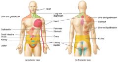
Referred Pain is the phenomenon when visceral pain is often felt in/just deep to the skin overlying the stimulated organ, or in a surface area distant from the simulated organ. For example, For people who have a heart attack, they first feel their pain in the left arm. |
|
|
What is a First Order Neuron? |
These are unipolar somatosensory neurons. It's cell body is located in the dorsal root of the ganglia while the other end terminates nearby in the gray horns of the cord. It takes the sensation straight to the CNS (no middle men!) |
|
|
What is a Second Order Neuron? |
These neurons conduct impulses from the brain stem/spinal cord to the thalamus. NOTE that these neurons ALWAYS DECUSSATE! |
|
|
What is a Third Order Neuron? |
These neurons conduct impulses from the thalamus to primary somatosensory area of the cortex (these neurons stay on the same side!) |
|
|
What are the Accessory Structures of the Eye? |
- Eyelids help keep the eye moist - Eyelashes help keep dirt out of the eyes - lacrimal apparatus is the area on charge of the production of tears |
|
|
What is the Fibrous Tunic |
The outermost layer of the eye. It consists of the sclera and cornea |
|
|
What is the Vascular Tunic? |
This is the middle layer of the wall of the eyeball. It is composed of the choroid, ciliary body and the iris |
|
|
What is the Nervous Tunic? |
This is the innermost layer of the wall of the eyeball. It houses the retina! |