![]()
![]()
![]()
Use LEFT and RIGHT arrow keys to navigate between flashcards;
Use UP and DOWN arrow keys to flip the card;
H to show hint;
A reads text to speech;
39 Cards in this Set
- Front
- Back
|
What makes up the quadratus muscles? |
- rectus femoris - vastus lateralis, medalis and intermedius |
|
|
What is the origin of vastus intermedius?
|
- anterior and lateral surface of the proximal 2/3 of the femoral shaft - proximal 2/3 of the lateral lip on the linea aspera |
|
|
What is the insertion of the vastus intermedius? |
- common tendon of the quadriceps femoris |
|
|
What innervates VI? |
nerve to VI of femoral L2,3,4 |
|
|
Origin of VM |
- inferior 1/3 of intertrochanteric line - medial lip of linea aspera - spiral line |
|
|
Insertion of VM |
- common tendon into tib tub - arches around medial border of patella at the medial patellar retinaculum |
|
|
What muscle is directly underneath the rectus femoris? |
vastus intermedius |
|
|
Origin of Rectus Femoris |
- AIIS (straight head) - superior part of acetabulum - fibrous capsule of hip joint |
|
|
Insertion of RF |
- common tendon via patellar ligament - tibial tuberosity - anterior proximal aspect of the tibial condyles |
|
|
Actions of RF |
- extends leg and flexes hip |
|
|
Origin of vastus lateralis? |
- greater trochanter - proximal 2/3 of intertrochanteric line - lateral lip of linea aspera |
|
|
Insertion of VL |
- common tendon into tib tub - IT band |
|
|
Origin of articularis genus |
anterior inferior part of femoral shaft |
|
|
Insertion of articularis genus |
synovial membrane of knee joint |
|
|
Action and innervation of articularis genus |
- retracts synovial suprapatellar flexion from entrapment between the articular surface during knee extension - inn. nerve to VI of femoral L2,3 |
|
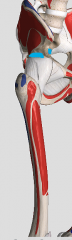
What muscle has its origin at the blue dots? |
Rectus femoris |
|
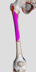
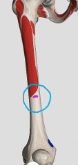
What muscles has its origin at the highlighted purple portion? |
vastus lateralis |
|
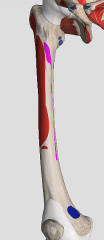
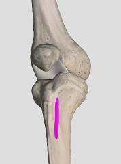
What muscle has its origin here? |
vastus intermedius |
|
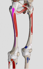
What muscle has its origin here? |
vastus medialis |
|
|
Origin of articularis genus |
|
|
What innervates the common tendon? |
femoral nerve and femoral artery and branches of the lateral and medial circumflex muscularis arteries |
|
|
What is the origin of the sartorius? |
- ASIS |
|
What inserts here? |
- sartorius - proximal medial surface of tibial shaft at pes anserine |
|
|
What are the actions of the sartorius? |
- flexes thigh - externally rotates the thigh and leg - flexes leg -abducts thigh + leg |
|
|
What are the lateral, superior and medial borders of the femoral triangle? |
- lateral: sartorius - superior: inguinal ligament - medial: adductor longus |
|
|
What makes up the floor of the femoral triangle? |
- iliopsoas m. - pectineus - adductor longus |
|
|
What are the contents of the femoral triangle? |
1. neuromuscular lacuna: iliopsoas muscle + femoral nerve 2. Vascular lacuna: femoral artery, femoral branch of GF nerve, femoral vein, femoral ring with deep inguinal LN |
|
|
What are the walls of the adductor canal? |
- lateral: vastus medialis - medial: adductor longus and magnus - anterior: vasto-adductorial membrane and sartorius muscle |
|
|
What are the contents of adductor canal from lateral to medial? |
- nerve to VM - saphenous nerve (L3/4) - femoral artery - femoral vein |
|
|
Where does the articular branch of the femoral nerve go to? |
Sartorius |
|
|
Femoral herniations are more common in females. T/F |
True; due to the wider pelvis in females |
|
|
Where does the intestine exit with a femoral herniation? |
the saphenous opening |
|
|
What is the iliopectineal arch? |
- aponeurotic extension of the inguinal ligament and iliopsoas fascia |
|
|
Medial to the iliopectineal arch, and deep to the inguinal ligament passes the ________ lacuna. |
vascular |
|
|
Lateral to the iliopectineal arch, and deep to the inguinal ligament passes the ________ lacuna. |
neuromuscular |
|
|
What is the inferior opening of the adductor canal? |
- the adductor hiatus - adductor magnus and hamstring part |
|
|
What is the anterior opening of the adductor canal? |
- opening in the vastoadductorial membrane - saphenous nerve and the descending genicular vessels leave |
|
|
How many perforating arteries arise from the deep artery of thigh? |
4 |
|
|
What does the acetabular artery from the medial circumflex artery supply? |
. |