Reading...
![]()
Play button
![]()
Play button
![]()
Use LEFT and RIGHT arrow keys to navigate between flashcards;
Use UP and DOWN arrow keys to flip the card;
H to show hint;
A reads text to speech;
17 Cards in this Set
- Front
- Back
- 3rd side (hint)
|
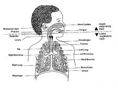
The upper respiratory tract or upper airway primarily refers to the parts of the respiratory system lying outside of the thorax or above the sternal angle.
|
Nose
nasal cavity paranasal sinuses Pharynx Nasopharynx Oropharynx Laryngopharynx |
|
|
|
Larynx (The larynx can be considered part of the upper respiratory tract,the lower respiratory tract, or both, depending on the source.)
|
.
|
|
|
|
The term lower respiratory tract refers to the portions of the
|
respiratory system from the trachea to the lungs.
|
|
|
|
lower respiratory tract refers to
|
the trachea (wind pipe)
the two bronchial tubes (one to each lung) the bronchioles, and the lungs. |
|
|
info
|
.
|
|
|
.
|
.
|
|
|
|
A Pancoast tumor, also called a
|
pulmonary sulcus tumor or superior sulcus tumor
|
|
|
|
Most Pancoast tumors are
|
non-small cell cancers.
|
|
|
|
A Pancoast tumor
It typically spreads to nearby tissues such as |
the ribs and vertebrae.
|
|
|
|
A Pancoast tumor
The growing tumor can cause compression of a |
brachiocephalic vein, subclavian artery, phrenic nerve, recurrent laryngeal nerve, vagus nerve, or, characteristically, compression of a sympathetic ganglion resulting in a range of symptoms known as Horner's syndrome.
|
|
|
|
Horner's syndrome.
|
compression of a sympathetic ganglion resulting in a range of symptoms
|
|
|
|
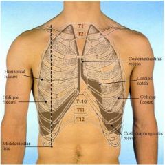
posterior border of the lung
|
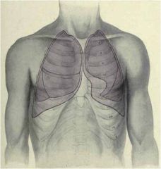
extends inferiorly from the level of the spinous process of CV. 7 to T.V.10 along the scapular line
|
|
|
|
oblique fissure
|
represented by a line that extends from the level of the spinous process of T.V.2 posteriorly to the 6th costal cartilage anteriorly
left lung: upper lobe is superior and anterior to this fissure; lower lobe is inferior and posterior to this fissure |
|
|
|
horizontal fissure
|
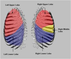
additional fissure in the right lung
represented by a line that extends anteriorly from the oblique fissure across the 4th costal cartilage right lung: upper lobe is superior to and middle lobe is inferior to this fissure; inferior lobe is posterior to the oblique fissure |
|
|
.
|
.
|
|
|
|
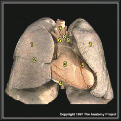
reflected as a cuff around the root of the lung and hangs down as a loose fold, called
|
pulmonary ligament,
what does it do? |
to allow for movement of the lung root during respiration
|
|
|
Lymphatic has 2 main cells =
|
Dendritic cells
Macrophages |
|