Reading...
![]()
Play button
![]()
Play button
![]()
Use LEFT and RIGHT arrow keys to navigate between flashcards;
Use UP and DOWN arrow keys to flip the card;
H to show hint;
A reads text to speech;
119 Cards in this Set
- Front
- Back
|
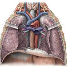
What is the thorax and what are its purposes?
|
Located between the root of the neck and the abdomen. It contains the heart, great vessels, lungs, and other important structures. It is a conduit for structures passing between the neck and abdomen and has an important role in breathing.
|
|
|
What are the three compartments of the thorax?
|
The mediastinum and 2 pulmonary cavities
|
|
|
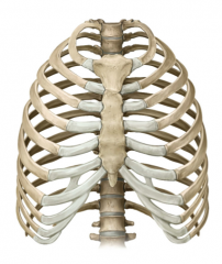
What are the boundaries of the thorax?
|
Anterior-Sternum and Costal Cartilages
Lateral-12 pairs of ribs Posterior-12 thoracic vertebrae |
|
|
What is the "Thoracic Inlet"?
|

The superior thoracic aperture bounded by the manubrium, first pair of ribs and the first thoracic vertebrae. It is closed by the suprapleural membrane of the lungs laterally. This is ironically where thoracic outlet syndrome occurs.
|
|
|
What is the "thoracic outlet"?
|

The inferior thoracic aperture is bounded by the xiphoid process, costal margin and ribs 11 and 12, and 12th thoracic vertebra. Closed by the respiratory diaphragm which ascends to the 5th intercostal space on the right and 6th on the left.
|
|
|
What is the respiratory diaphragm?
|
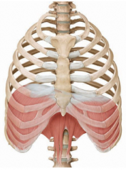
Primary respiratory muscle innervated by the phrenic nerve (C3,4,5 keeps the diaphragm alive) ascends to the fifth IC space on the right due to the liver and sixth on the left.
|
|
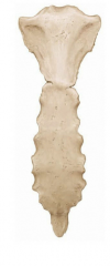
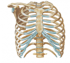
Name the three main components in the image
|

The Sternum
Manubrium "handle" (blue) Body (green) Xiphoid process (purple |
|
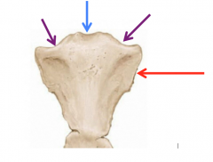
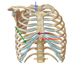
What are the landmarks highlighted in the image?
|

The Manubrium of the sternum
Jugular Notch (blue) Clavicular notch (purple) the attachment of the sternoclavicular joint Attachment for rib 1 (red) |
|
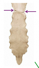
What is highlighted in the image?
|

The Sternal Angle (Angle of Louis) is the costal attachment for rib 2 and is level with the T4/5 vertebral level. The rest of the body contains attachments for ribs 2-7
|
|
|
What rib articulates with the Xiphoid Process?
|

7-attaches where the process meets the sternum
|
|
|
What is a median sternotomy?
|
When the sternum is divided during open heart procedures
|
|
|
Where is the sternum most commonly fractured?
|

At the manubriosternal joint-this is common in auto accidents.
|
|
|
What is the sternum biopsied?
|

Because it contains red bone marrow and therefore is a site for hematopoiesis.
|
|
|
Which ribs are true, false, and floating?
|
True: 1-7 (attaches to the sternum)
False 8-10 (attaches to the costal cartilages to the superior ribs) Floating: 11 & 12 (costal cartilage does not attach to anything) |
|

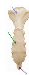
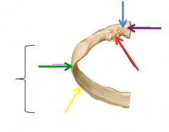
Name the regions highlighted in the image
|
Sternoclavicular joint (blue)
Costal Cartilage (green) Sternocostal Joint (purple) Xiphoid Process (red) |
|

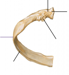
Name the regions highlighted in the area above
|
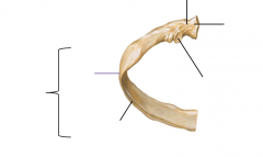
Neck (blue)
Head (purple) Tubercle (red) Angle (green) Costal Groove (yellow) Shaft (bracket) |
|
|
What does the costal groove contain?
|
This inferior groove contains a neurovascular bundle
|
|
|
What does the presence of a cervical rib cause?
|
"Throacic outlet syndrome" which can compress the subclavian artery and lower parts of the brachial plexus especially the C8 and T1 cord levels
|
|
|
Where do most rib fractures occur?
|
The weakest point just anterior to the angle; sharp edges can puncture lungs, liver, spleen (complication of CPR)
|
|
|
What are the common features of a thoracic vertebrae?
|
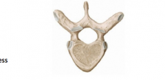
-Heart Shaped body
-Round vertebral foramen -Long, downward point spinous process |
|
|
How many facets do the thoracic vertebrae have that articulate with the ribs?
|
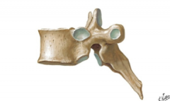
3
-Superior costal demifacet -Transverse costal facet -Inferior costal demifacet |
|
|
Rib 7 will articulate with which vertebrae?
|

The superior costal facet and transverse costal facet of T7 and the inferior costal facet of T6
|
|
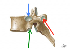
What are the regions highlighted in the image?
|

Superior costal facet (blue)
Inferior costal facet (green) Transverse costal facet (red) |
|
|
What part of the rib articulates with the transverse costal facet?
|

The tubercle
|
|
|
Name three ligaments that secure the rib posteriorly
|
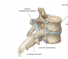
The superior costotransverse ligament
The lateral costotransverse ligament The costotransverse ligament |
|
|
What vertebral levels correspond to:
1) the superior manubrium 2) the sternal angle 3) the xiphoid process |
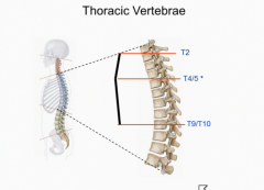
1) T2
2) T4/5 3) T9/10 |
|
|
The T4/5 level is also called what?
|

The transverse thoracic plane
|
|
|
The intercostal space between rib 4 and 5 is known as....
|

The 4th intercostal space-always named for the superior rib.
|
|
|
How many intercostal spaces exist?
|
|
|
|
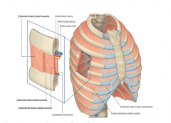
What three layers of muscle exist in an intercostal space?
|

External intercostal muscles
Internal intercostal muscles Innermost intercostal muscles |
|
|
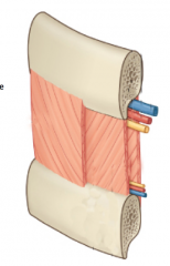
What direction do the fibers of the external intercostal muscles run?
|

They are oriented infer-medially like hands angled into pants pockets.
|
|
|
Which intercostal muscles are used during inspiration?
|

External Intercostal muscles
|
|
|
Which intercostal muscles have fibers oriented super-medially?
|

Internal and Innermost Intercostal muscles
|
|
|
Which intercostal muscles are used during expiration?
|

Internal Intercostal Muscles
|
|
|
Which intercostal muscles are incomplete?
|
The external intercostal muscles end anteriorly as a translucent membrane and the innermost intercostal muscles are incomplete anteriorly and posteriorly
|
|
|
Describe the intercostal neurovascular bundle
|

From superior to inferior they run vein, artery, nerve (VAN) in between the internal and innermost intercostal muscles. They are located at the inferior costal groove therefore when a chest tube is placed it is immediately superior to a rib to avoid nerve damage and excess bleeding.
|
|
|
If the intercostal neurovascular bundle runs inferiorly to a rib and superior to the intercostal muscles what supplies the inferior part of the intercostal muscles?
|

Clinically irrelevant collateral branches
|
|
|
What layers of facia are deep to the intercostal muscles?
|
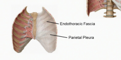
The endothoracic fascia which is loose CT and lines the entire inside of the thoracic cavity. Deep to that is the parietal pleura
|
|
|
What are the tranversus thoracis muscles?
|
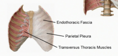
Not intercostal muscles because they attach the sternum to the ribs in a starburst shape. They anchor the internal thoracic artery (mammary artery) to the anterior chest wall.
|
|
|
Where do the intercostal arteries arise?
|
The posterior intercostal arteries arise from the thoracic aorta and the anterior intercostal arteries arise from the internal thoracic artery and the musculophrenic artery
|
|
|
What main artery supplies the internal thoracic artery?
|
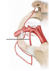
The subclavian artery
|
|
|
What are the two branches of the internal thoracic artery?
|
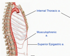
The musculophrenic and superior epigastric arteries
|
|
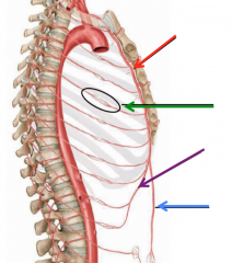
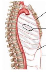
Name the regions highlighted in the image.
|

Internal thoracic artery (red)
Arterial Anastomosis (green) Musculophrenic artery (purple) Superior Epigastric artery (blue) |
|
|
What is an anastomosis?
|
Communication between two tubular structures-allows collateral circulation between the thoracic aorta and internal thoracic arteries
|
|
|
Where do the anterior and posterior intercostal veins drain?
|
The anterior intercostal veins drain into the internal thoracic and musculophrenic veins and the posterior intercostal veins drain into the azygous veins in the posterior mediastinum
|
|
|
What innervates the intercostal space?
|
Branches of VPR from levels T1-T11, The T12 branch is located below the 12th rib and therefore is known as the subcostal nerve
|
|
|
What kind of nerve supply do the intercostal nerves give?
|

Motor innervation (GSE) to the intercostal and transversus thoracic muscles and sensory innervation (GSA) to the anterior and lateral areas of skin.
|
|
|
What is a dermatome?
|
A strip of skin innervated by a single spinal nerve
|
|
|
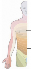
The nipple line exists in which dermatome
|

T4
|
|
|
The umbilicus line exists in which dermatome
|

T10
|
|
|
What is the midsternal line?
|

A line through the sternum in the midline
|
|
|
What is the midclavicular line?
|

A line through the midpoint of each clavicle
|
|
|
What is the midaxillary line?
|
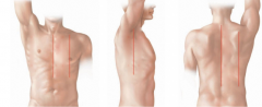
A line through the center of axilla
|
|
|
What is the midvertebral line?
|

A line through the spinous processes of the vertebral column
|
|
|
What is the scapular line?
|

A line through the inferior angle of the scapula
|
|
|
What are the two layers of pulmonary pleura?
|
Parietal (superficial) and visceral (deep). These layers are continuous with each other and form an airtight attachment with the hilum of the lung
|
|
|
What is the potential space between the visceral and parietal pulmonary pleura?
|
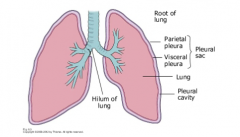
The pleural cavity-this contains a small amount of serous fluid to lubricate movements of lungs
|
|
|
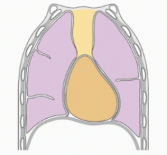
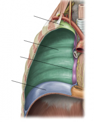
What are the four regions of the parietal pleura?
|

Cervical
Costal Mediastinal Diaphragmatic |
|
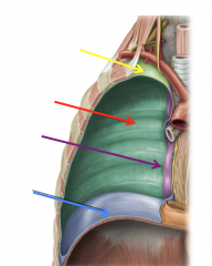
Name the regions highlighted in the image.
|

Cervical (yellow)
Costal (red) Mediastinal (purple) Diaphragmatic (blue) |
|
|
What provides the somatic innervation to the parietal pleura?
|
The phrenic and intercostal nerves-somatic innervation like touch is a conscious feeling. This is different from the visceral innervation of the gut.
|
|
|
What is pleuritis?
|
Inflammation of the pleura. Creates friction between parietal and visceral layers producing sharp, stabbing pain during respiration.
|
|
|
The pleura is composed of what type of cells?
|
Mesothelial cells. Mesothelioma is a malignant tumor of mesothelial cells associated almost exclusively with asbestos exposure.
|
|
|
What is the costodiaphragmatic recess?
|

Where the diaphragmatic pleura reflects from the perimeter of the diaphragm to meet the costal pleura.
|
|
|
What is the costomediastinal recess?
|

Where the mediastinal pleura reflects to meet the costal pleura. Larger on the left because of the heart.
|
|
|
Why is the costomediastinal recess larger on the left?
|
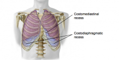
To accommodate room for the heart
|
|
|
At the midclavicular line what is the level of the lung and pleura?
|
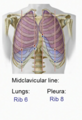
Lung: Rib 6
Pleura: Rib 8 |
|
|
At the Midaxillary Line what is the level of the lung and pleura?
|
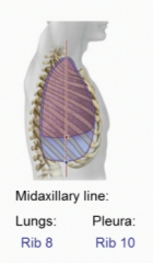
Lungs: Rib 8
Pleura: Rib 10 |
|
|
At the Scapular line what is the level of the lung and the pleura?
|
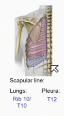
Lung: Rib 10/T10 Vertebra
Pleura: T12 |
|

A man suffers a gunshot wound to his right 4th intercostal space, there are no breath sounds on the right-diagnosis?
|

Pneumothorax-the lung has collapsed and fluid is filling the pleural cavity.
|
|
|
What is a pneumothorax?
|
Air in the pleural cavity-may result from injury to the chest wall or lung
|
|
|
What is a hemothorax?
|

Blood in the pleural cavity-in an upright patient, the blood accumulates in the costodiaphragmatic recess
|
|
|
What is a pleural effusion?
|

Fluid in the pleural cavity-may be caused by congestive heart failure, pneumonia, TB, or lung cancer
|
|
|
What is atelectasis?
|

Pulmonary collapse-air in the plural cavity reduces the negative pressure that normally keeps the lung inflated. The lung then collapses due to its inherent elasticity.
|
|
|
What is a tension pneumothorax?
|
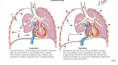
A life-theatening condition in which air accumulates and becomes trapped in the pleural cavity because the injured tissue acts as a one-way valve. Causes complete lung collapse and mediastinal shift toward opposite side. Compromises cardiac output, venous return, and compresses opposite lung
|
|
|
Why does a mediastinal shift cause distended neck veins?
|

Because it causes a chink in the SVC and IVC which compromises venous blood returning to the heart causing a back up to the jugular veins.
|
|
|
What is a thoracentesis?
|

Aka Thoracocentesis-procedure in which a needle is inserted into the pleural cavity to drain fluid, blood or pus. In an upright patient the fluid accumulates in the costodiaphragmatic recess.
|
|
|
What are the preferred sites of puncture for pneumothorax or a hemothroax?
|
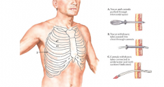
Pneumothorax: 2nd or 3rd intercostal space at the midclavicular line
Hemothorax: 4th to 6th intercostal space at the midaxillary line |
|
|
Why are needles inserted into the inferior part of the intercostal space?
|

To avoid the neurovascular bundle
|
|
|
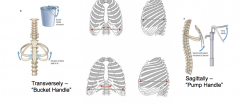
What are the dimensions of expansion in inspiration?
|

Vertically-contraction of the diaphragm
Transversely-contraction of EIM causes the bucket handle Sagitally-sternum moves like a pump handle and pulls the ribs up and out |
|
|
True or False: Inspiration and Expiration are both active movements
|
False-Inspiration is active causing different muscles to contract to expand the thoracic cavity whereas expiration is passive because air is forced out of the lungs by the elastic recoil of the alveoli.
|
|
|
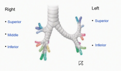
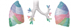
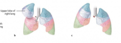
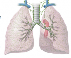
How many lobes and fissures exist in the right lung?
|

3 lobes (superior, middle, inferior)
2 fissures: Horizontal and oblique |
|
|
How many lobes and fissures exist in the left lung?
|

2 lobes: superior and inferior
1 fissure: Oblique |
|
|
What are the unique features of the left lung?
|
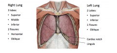
The cardiac notch that allows room for the heart and the lingula is a projection of the lung in front of the heart.
|
|
|
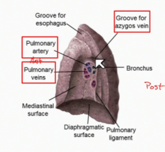
What is the difference between the hilum and the root of the lung?
|
The hilum is the space on the medial lung which allows for its attachments where the root is the vessels themselves including pulmonary arteries and veins, bronchi, bronchial vessels, etc
|
|
|
Which is superior in the root of the lung: pulmonary artery or vein?
|
The pulmonary artery
|
|
|
What is the pulmonary ligament?
|

An extension of the visceral pleura off the root of the lung.
|
|
|
Where is the cardiac impression?
|
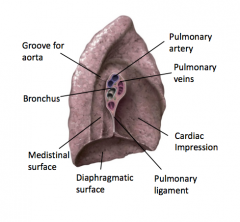
On the mediastinal surface of the left lung where there is an indentation to compensate for the size of the heart.
|
|
|
Where does the larynx become the Trachea?
|
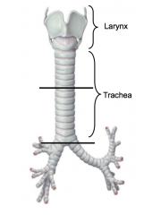
At the C6 vertebral level
|
|
|
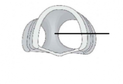
What composes the trachea?
|
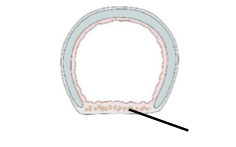
A C-shaped of hyaline cartilage and the trachealis muscle to allow expansion of the posterior esophagus
|
|
|
Where does the trachea bifurcate into left and right primary bronchi?
|
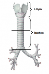
At the T4/5 Transverse Thoracic Plane (Sternal angle level)
|
|
|
What is the cartilage between the trachea bifurcation called?
|
Carina
|
|
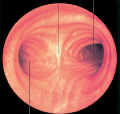
Where is the posterior and anterior in the image?
|

Posterior is down-the right main bronchus on the right because it is a more vertical path and the left is more closed off because it goes at a sharper angle to allow room for the heart.
|
|
|
Aspirated objects are more likely to enter which main bronchi?
|

The right primary bronchus because it has a steeper angle and is a wider space because it does not have to allow room for the heart like the left side.
|
|
|
Primary bronchi divide into...
|
Lobar bronchi-on the right superior, middle, and inferior and on the left superior and inferior to match the lobes of the lungs
|
|
|
Lobar bronchi divide into...
|

Segmental or tertiary bronchi that supply a segment of the lung. There are ten segments in each lung.
|
|
|
Why is it possible to remove one segment from the lung?
|
A segmentectomy (wedge resection) Because each segment has its own air and blood supply
|
|
|
What is the difference between a lobectomy and a pneumonectomy?
|
A lobectomy removes a lobe and a pneumonectomy removes an entire lung.
|
|
|
What are the conducting portions of the bronchial tree?
|
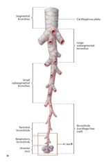
The tertiary and terminal bronchi which contain cartilage followed by the smooth muscle only of bronchioles and terminal bronchioles. These terminal bronchioles divide to start the respiratory portion of the bronchial tree starting with the respiratory bronchioles.
|
|
|
What are the respiratory portion components of the bronchial tree?
|
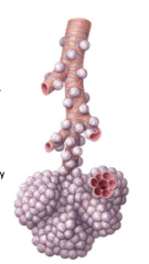
The respiratory bronchioles divide into alveoli. Gas exchange occurs mostly in alveoli. The smooth muscle of the respiratory bronchioles and the elastic fibers around the alveoli allow for that passive recoil that causes expiration.
|
|
|
What are the pulmonary vessels?
|
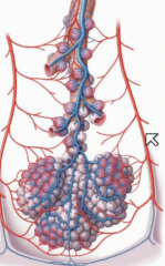
The pulmonary arteries carrying deoxygenated blood to the lungs distributed intrasegmentally and pulmonary veins carrying oxygenated blood toward the heart intersegmentally distributed among the BP segments.
|
|
|
What are the bronchial vessels?
|
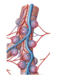
The vessels arising from the thoracic aorta or intercostal arteries supplying the lung tissue. These enter the hilum with the bronchi and supply oxygen and nutrients to the bronchial tree.
|
|
|
What is the order of lymph drainage from the lung?
|
The bronchopulmonary nodes to the tracheobronchial nodes to the paratrachial nodes to the bronchomediastinal trunks. On the right this trunk drains into the right lymphatic trunk and on the left it drains to the thoracic duct.
|
|
|
What is the pulmonary plexus?
|
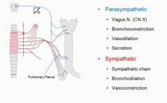
Parasympathetic and Sympathetic nerve supply to the lungs.
|
|
|
What is the parasympathetic innervation of the lungs?
|

The Vagus Nerve (CN X) which allows for Bronchoconstriction, Vasodilation, and Secretion of glands in the bronchial tree.
|
|
|
What is the sympathetic innervation of the lungs?
|
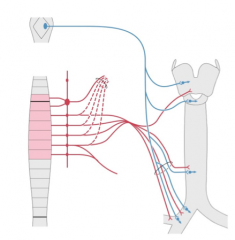
The sympathetic chain (lower cervical and upper thoracic levels) dilates the bronchioles (bronchodilation) vasoconstriction, and decreased mucous secretion.
|
|
|
Where do you listen to hear breath sounds from the right lung?
|
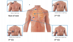
Apex-Root of the neck
Superior lobe- 2nd ICS Middle lobe-4th ICS Inferior lobe-6th ICS |
|
|
Where do you listen to hear breath sounds from the left lung?
|
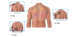
Apex-1st Rib
Superior Lobe-2nd ICS Inferior Lobe-7th ICS |
|
|
What is rhonchus?
|
A sound which indicates an obstruction in the central airways of the lung and can present as a rattling sound around the central airways.
|
|
|
What is Wheezing associated with?
|
A high-pitched sound due to obstruction of bronchioles; common in asthmatics
|
|
|
What is rales?
|
(Crackles) Crackling sounds in the lung caused by a "popping" open of alveoli and terminal bronchioles due to pneumonia, bronchitis, or pulmonary fibrosis.
|
|
|
Why do you listen to the left lung on the posterior side?
|

Because sounds are commonly obstructed by the sounds of the heart.
|
|
|
What is pneumonia?
|
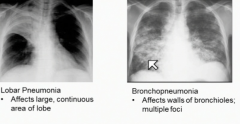
Inflammation of the lung tissue with pus formation (accumulation of white blood cells). There is lobar pneumonia which is a large continuous area whereas bronchopneumonia affects the walls of the bronchioles and has multiple foci.
|
|
|
Which pneumonia is bilateral?
|

Bronchopneumonia can be bilateral whereas lobar pneumonia is unilateral.
|
|
|
What is a pulmonary embolism?
|
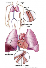
A blockage or blood clot of the pulmonary artery which causes difficulty breathing, tachycardia, chest pain, and cyanosis. This is commonly a complication of Deep Vein Thrombosis and is treated with anticoagulants or IVC filter.
|
|
|
What is COPD?
|

Chronic Obstructive Pulmonary Disease-Constriction or inflammation of the airways which is degenerative.
|
|
|
What are the three types of COPD?
|
Chronic Bronchitis, Emphysema (both caused by smoking), and Asthma.
|
|
|
What is chronic bronchitis?
|

Inflamed airways combined with excess mucous production. This is caused by smoking
|
|
|
What is emphysema?
|

A progressive diseased that destroys the lung tissue. Bronchioles collapse during exhalation trapping air in alveoli. Caused by smoking and a1-antitrypsin deficiency.
|
|
|
What is asthma?
|
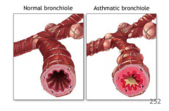
A chronic disease that results in narrowing of the airways due to inflammation or bronchoconstriction. This is a functional not structural problem therefore is reversible with sympathomimetics (mimics epinephrine).
|