Reading...
![]()
Play button
![]()
Play button
![]()
Use LEFT and RIGHT arrow keys to navigate between flashcards;
Use UP and DOWN arrow keys to flip the card;
H to show hint;
A reads text to speech;
75 Cards in this Set
- Front
- Back
|
Name four main facts of IV discs
|
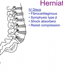
Fibrocartilaginous joints
Symphysis type joint Shock absorbers Resist compression |
|
|
What are some problems associated with IV discs?
|
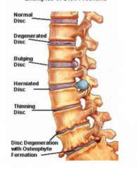
Degenerated disc
Bulging disc Herniated disc Thinning disc (loss of water, zygopophyseal joint damage) Disc degeneration with osteophyte formation |
|
|
Where do the majority of disc herniations occur?
|
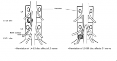
Between L4/L5 or L5/S1
|
|
|
What is the direction of most disc herniations?
|

Posteriolateral due to the PLL reinforcing the posterior side. This can crush a spinal nerve against a spondylophyte
|
|
|
A herniation between the L5 and S1 disc affects which nerve?
|

The first sacral nerve
|
|
|
Is a posteriolateral IV disc herniation bilateral?
|
No, it is unilateral due to the separation of each pair of spinal nerves
|
|
|
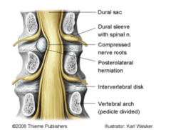
What is cauda equina syndrome?
|
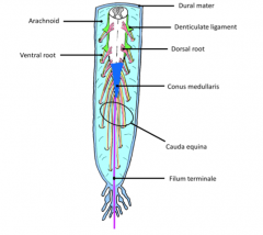
When the roots of the cauda equina are pinched by a lower lumbar disc herniation which causes saddle sensory loss and numbness or muscle weakness in the lower limb
|
|
|
What are the three muscles of the suboccipital triangle?
|
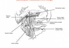
Rectus Capitis Posterior Major
Inferior Oblique Superior Oblique |
|
|
Which nerve does the suboccipital nerve stem from?
|

The Dorsal primary rami of C1-this innervates the suboccipital muscles
|
|
|
From where does the greater occipital nerve originate and which muscle does it wrap inferiorly?
|
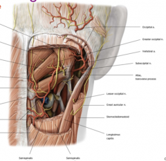
It originates from the DPR of C2 and wraps inferiorly around the inferior oblique. This innervates the posterior half of the skull
|
|
|
Where does the vertebral artery travel after exiting the transverse foramina of C6-C1?
|
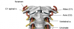
Along the posterior arch of Atlas and up through the foremen magnum.
|
|
|
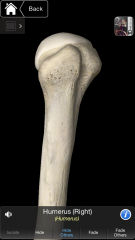
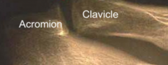
What are the bones of the shoulder girdle?
|
Clavicle
Superior border of Scapula Humerus |
|
|
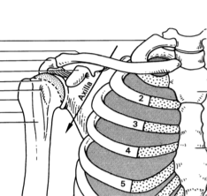
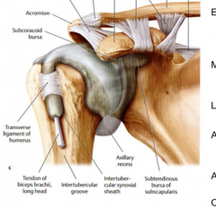
What is the axilla?
|
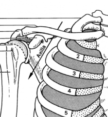
A passageway that allow nerves and vessels from the neck to travel to the upper limb. The apex is formed by the scapula, clavicle, and first rib
|
|
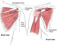
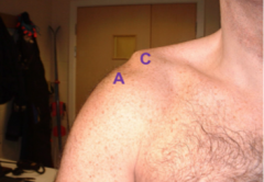
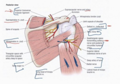
Is this a posterior or anterior image?
Is this right or left? |

Posterior
Right |
|
|
Where is the superior transverse scapular ligament?
|

It is a ligament in the superior notch of the scapula
|
|
|
Where can one find the suprascapular artery and nerve?
|
The artery travels above the superior transverse scapular ligament and the nerve travels below
ARMY OVER NAVY UNDER |
|
|
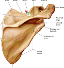
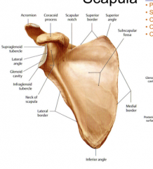
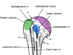
Name the three borders and angles of the scapula
|
Superior, Medial, Lateral Border
Superior, Inferior, Lateral Angle (glenoid cavity) |
|
|
What are the three fossae/depressions of the scapula?
|
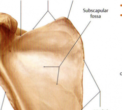
Supraspinous fossa
Infraspinous fossa Subscapular fossa |
|
|
Which muscle attaches to the subscapular fossa?
|
The subscapularis muscle
|
|
|
What is a bursa?
|
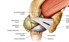
A synovial filled membranous sac "purse" that surrounds bony projections that facilitate the movement of tendons around the bony processes.
|
|
|
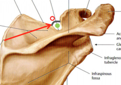
Describe the glenoid cavity?
|

The depression of the neck of the scapula that articulates with the humerus. The lip around the cavity is known as the labrum. It is a shallow cavity.
|
|
|
Where is the attachment of the tendon of the long head of biceps brachii?
|

The supraglenoid tubercle
|
|
|
The tendon of the long head of triceps attaches where?
|
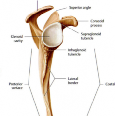
At the infraglenoid tubercle
|
|
|
What attaches to the anatomical neck of the humerus?
|
The distal end of the glenohumeral joint capsule
|
|

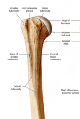
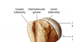
Name the structures highlighted in the image
|
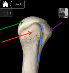
Greater Tubercle (red)
Intertubercular Groove/Sulcus (Bicipital Groove-tendon of the long head of bicep rests here) (blue) Lesser tubercle (purple) Head of humerus (green) |
|
|
What nerve and artery pass posterior along the surgical neck of the humerus?
|
Axillary Nerve
Posterior Circumflex Humeral Artery |
|
|
What is a "Foosh" fracture?
|

A fall on the outstretched hand can break the surgical neck of the humerus causing injury to the axillary nerve (innervates the deltoid and teres minor) and the posterior circumflex humeral artery
|
|
|
What is the deltoid tuberosity?
|
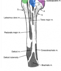
The distal attachment for the deltoid muscle on the lateral aspect of the humerus.
|
|
|
What is the lateral lip of the bicipital groove?
What is the medial lip of the bicipital groove? |

The Lateral lip is an extension of the greater tubercle and is where the pectoralis major attaches. The medial lip is a continuation of the lesser tubercle and is the attachment of the Teres Major
|
|
|
Where is the insertion of the Latissimus Dorsi?
|
The floor of the bicipital groove on the humerus
|
|
|
Where is the spiral/radial groove?
|

A groove on the anterior humerus that houses the radial nerve and the deep brachial artery
|
|
|
What does an injury to the radial nerve create?
|
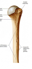
"wrist drop" The inability to raise the wrist and fingers. This occurs from a mid humeral break
|
|
|
What attaches to the superior, middle, and inferior facet of the greater tubercle of the humerus?
|
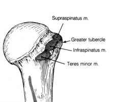
Respectively the supraspinatus, infraspinatus, and teres minor muscles.
|
|
|
Which muscle attaches to the lesser tubercle?
|
The subscapularis muscle
|
|
|
What are the rotator cuff muscles?
|
SITS
Supraspinatus, Infraspinatus, Teres Minor, Subscapularis |
|
|
What is the AC joint?
|

A synovial joint that connects the acromion process to the lateral clavicle-thsi is reinforced by the acromioclavicular ligament.
|
|
|
What is the coracoclavicular ligament?
|
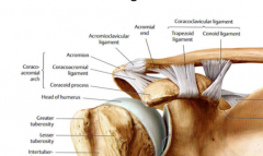
A strong ligament that suspends scapula and upper limb to the clavicle. It provides principle integrity and stability to AC joint and may be torn in severe shoulder separations.
|
|
|
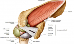
Which ligament bridges over the glenohumeral joint?
|

The coracoacromial ligament which resists upward displacement of the humerus from the glenoid cavity. Chronic, repetitive upward displacement of humerus may lead to impingement injury to rotator cuff and shoulder pain.
|
|
|
What are the joints of the upper limb?
|
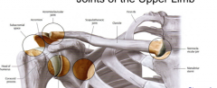
Sternoclavicular joint (clavicle-manubrium of sternum)-pivot type joint
Acromioclavicular joint (acromion process-clavicle) Glenohumeral joint (shoulder) Scapulothoracic joint "FAKE" the interface between the subscapularis muscle and the wall of the ribcage and the scapula |
|
|
What binds the clavicle to the first rib?
|
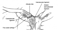
The costoclavicular ligament near the sternoclavicular joint. Damage to this ligament or joint could compromise the trachea if the clavicle is pushed posteriorly
|
|
|
What is shoulder separation?
|
Breakage of the acromioclavicular joint and a tear of the coracoclavicular ligament. This is different from shoulder dislocation. Also seen as a step off deformity
|
|
|
Why does the acromioclavicular joint not appear on an x-ray?
|
Because the fibrocartilage of the joint is non-mineralized and will not stop the x-ray
|
|
|
Where is the proximal attachment of the Glenohumeral joint capsule?
|
The neck of the scapula (proximal to the glenoid cavity)
|
|
|
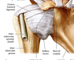
The long head of biceps tendon is surrounded by synovial membrane known as a synovial sheath-why?
|
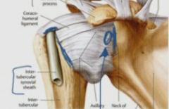
This allows enclosure of the glenohumeral joint capsule and smooth movement of the long head tendon within the bicipital groove.
|
|
|
What is the medial extension of the synovial membrane of the genohumeral joint cavity?
|

The bursa of the supscapularis muscle on the anterior aspect of the joint
|
|
|
TRUE or FALSE: The tendon of the long head of biceps is within the synovial cavity.
|
FALSE It is intracapsular but extrasynovial. Nothing lives within the synovial cavity except synovial fluid.
|
|
|
What muscle is responsible for initiating abduction of the upper limb?
|
Supraspinatus muscle (responsible for the first 15 degrees)
|
|
|
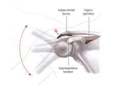
What is just inferior to the coracoacromial arch?
|

The subacromial bursa responsible for the smooth movement of the tendon of supraspinatus muscle. The coracoacromial ligament between the acromion and the coracoid processes creates the coracoacromial arch.
|
|
|
What muscles are responsible for the movement of the scapulothoracic joint?
|
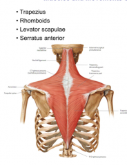
Trapezius, Rhomboids, Levator Scapulae, and Serratus Anterior
|
|
|
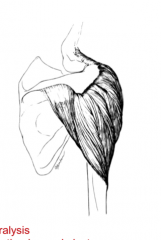
Which muscle attaches the scapula to the posterior thoracic wall?
|
The serratus anterior. This muscle is also very important in abduction of the shoulder.
|
|
|
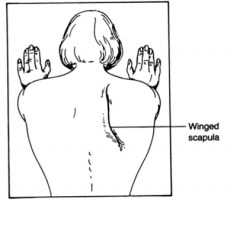
Which nerve innervates the serratus anterior muscle?
|

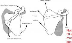
The long thoracic nerve which derives from C5, 6, and 7. When this superficial nerve is damaged paralyzing the serratus anterior the vertebral border of the scapula is pushed out (winged scapula) when someone performs a push motion.
|
|
|
Which muscles are responsible for the upward and downward rotation of the scapula?
|
Upward Rotation: Upper and lower fibers of Trapezius and Serratus anterior
Downward Rotation: Levator scapulae, Rhomboids, and gravity |
|
|
What must be done to test the movement of the GH joint?
|
The inferior angle of the scapula must be fixed in order to negate the movement at the scapulothoracic joint.
|
|
|
What muscles are responsible for flexion of the shoulder joint?
|
Pectoralis Major
Deltoid Biceps Brachii Coracobrachialis |
|
|
What muscles are responsible for the extension of the shoulder joint?
|
Latissimus Dorsi
Teres Major Deltoid Triceps Brachii Pectoralis major |
|
|
What muscles are responsible for the medial rotation of the shoulder joint?
|
Pectoralis major
latissimus dorsi Teres major Subscapularis |
|
|
What muscles are responsible for the lateral rotation of the shoulder joint?
|
Infraspinatus
Teres minor |
|
|
What muscles are responsible for the abduction of the shoulder joint?
|
Supraspinatus
Deltoid |
|
|
What muscles are responsible for the adduction of the shoulder joint?
|
Pectoralis Major
Latissimus Dorsi Teres Major |
|
|
What muscles are responsible for the circumduction of the shoulder joint?
|
Combination of muscles
|
|
|
What are the attachments of the Deltoid muscle?
|
The spine of the scapula, the acromion process, the lateral third of the clavicle, and the deltoid tuberosity
|
|
|
Why does the contraction of the deltoid muscle not pull the humerus directly up?
|

The rotator cuff muscles.
|
|
|
Why is the deltoid injury so devastating?
|

Most of GH function is lost because deltoid powers abduction, flexion, and extension.
|
|
|
What is the distal attachment of the posterior tricep muscle?
|
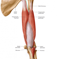
The olecranon (part of the elbow)
|
|
|
Which two muscles cross the GH (shoulder) and elbow joint?
|

The biceps and the triceps
|
|
|
Which structures descend from the axilla down through the spiral groove and between the lateral and long head of the triceps?
|

The radial nerve and the deep brachial artery
|
|
|
What are the movements of the latissimus dorsi?
|
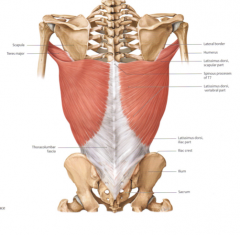
Extensor, medial rotation, and adduction of the arm. The crawl stroke is mediated by the lat dorsi
|
|
|
What two muscles are innervated by the suprascapular nerve?
|
The supraspinatus and the infraspinatus muscle
|
|
|
What muscles comprise the quadrangular space?
|

Lateral and Long head of Triceps
Teres Major Teres Minor |
|
|
What percentage of the Humeroscapular rhythm is due to the movement of the glenohumeral joint?
|
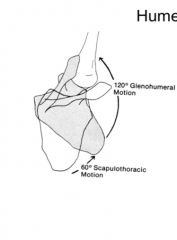
66.7%
|
|
|
What are the fused three layers surrounding the GH joint?
|
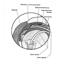
-Joint Capsule
-Tendon of the Supraspinatus -Subacromial Bursa |
|
|
What are the common sources of pain in the GH joint?
|
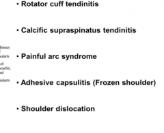
Rotator cuff tendinitis
Calcific Suprasinatus tendinitis Painful arc syndrome Adhesive capsulitis Shoulder dislocation |
|
|
What is painful arc syndrome?
|
The pain caused when the subacromal bursa or spraspinatus are inflamed and the movement from 60-180 degrees causes joint pain
|
|
|
Are most shoulder dislocations anterior or posterior?
|

Anterior
Imagine your arm behind the seat to your right and then a blow to the posterior proximal humerus. This is also called the subcoracoid dislocation. |
|
|
What nerve supplies the skin two inches below the acromion or the deltoid skin patch?
|
The axillary nerve
|