Reading...
![]()
Play button
![]()
Play button
![]()
Use LEFT and RIGHT arrow keys to navigate between flashcards;
Use UP and DOWN arrow keys to flip the card;
H to show hint;
A reads text to speech;
71 Cards in this Set
- Front
- Back
|
Who am I?
I contain the cell bodies of somatic motor/efferent neurons. |
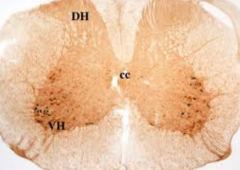
Ventral horns of the spinal cord
|
|
|
Who am I?
I am a potentially life-threatening condition whose symptoms include severe headache, neck stiffness, and intolerance to light |

Meningitis
|
|
|
Who am I?
I am the space from which a needle draws CSF during a lumbar puncture |
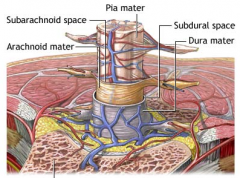
Subarachnoid space
|
|
|
Who am I?
I am a "bridge" of bone between the superior and inferior articular processes of lumbar vertebrae |
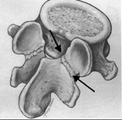
Pars interarticularis
|
|
|
Who am I?
I am the hydrated core of an IV disc. |
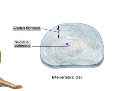
Nucleus Pulposus
|
|
|
Who am I?
I give rise to the future central nervous system |
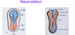
Neural Tube
|
|
|
Who am I?
I am an area/strip of skin supplied by a single spinal nerve |
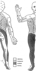
Dermatome
|
|
|
Who am I?
I give rise to specialized cells that form dermis, muscle, and skeletal tissue during embryonic development |
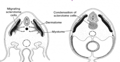
Somites
|
|
|
Who am I?
I contain cell bodies of sensory neurons associated with spinal nerves |
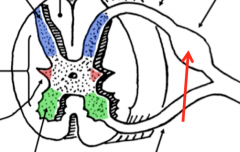
Dorsal Root Ganglion
|
|
|
Who am I?
I am a branch of a spinal nerve that supplies deep back muscles and a horizontal strip of skin on the back |
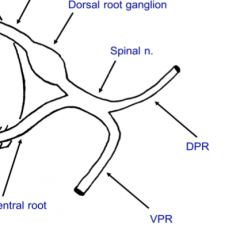
Dorsal Primary Ramus
|
|
|
Who am I?
I am the nerve that passes through the IV foramen between L3 and L4 vertebrae |
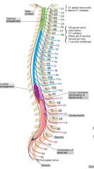
The L3 spinal nerve
|
|
|
Who am I?
I am the "body" of the atlas |
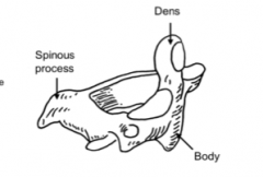
Dens/ Odontoid process
|
|
|
Who am I?
I am the nerve that supplies the rectus capitis posterior major muscle |
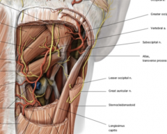
Suboccipital nerve
|
|
|
What are the common radiologic modalities?
|

-Conventional radiographs (x-rays)
-Computed tomography CT -Nuclear Medicine NM -Ultrasound US -Magnetic resonance imaging MRI |
|
|
Who won the first Nobel Prize?
|
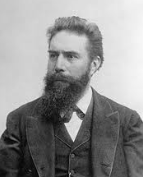
Wilheim Conrad Rontgen
|
|
|
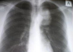
What is an X-ray?
|
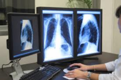
A shadow picture created with high energy photons. If photons hit the film they turn black but if bone attenuate or block the photons they are revealed on the film.
|
|
|
Can tissues be distinguished on x-ray film?
|

Yes the different densities of tissues such as bone, fat, and air are illustrated by differing opacities.
|
|
|
True or False: Conventional x-rays from both sides illustrate organs as the same size.
|
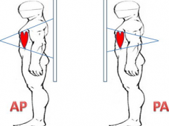
False-depending on what the x-ray beam hits first will increase the size of the shadow cast on the detector.
|
|
|
What is an orthogonal view?
|
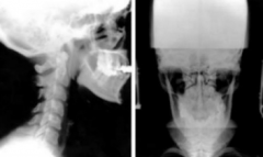
A film taken perpendicular to the first to give 3 dimensionality to organs or what is trying to be viewed
|
|
|
Who invented computer tomography?
|

Sir Godfrey Hounsfield developed the first commercial CT scanner and algebraic reconstruction technique to solve the problem of superimposition.
|
|
|
What are attenuation values?
|

The amount a tissue blocks high energy photons in computerized tomography
|
|
|
How many leaflets does the aortic valve have?
|
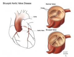
3-this is a typically tricuspid valve; however 0.5% of the population including Arnold Schwarzenegger have a bicuspid valve
|
|
|
What is radiopaque?
|
Impenetrable by X-rays. Certain dyes are radiopaque such as iodine that can highlight vessels in radiographs.
|
|
|
What are the basics of nuclear medicine?
|
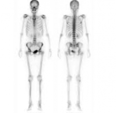
Something radioactive attached to a drug administered to a patient and tracked with gamma rays.
|
|
|
What is the major advantage of ultrasound technology?
|
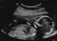
Sound waves which involves no ionizing radiation and can translate acoustic impedance into an image.
|
|
|
Who is Paul Lauterbur?
|

An American chemist whose work with the MRI made this technology possible
|
|
|
What are the milk lines?
|
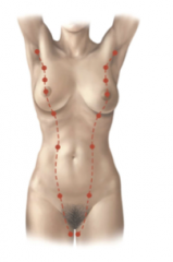
A ridge of ectoderm known as the mammary ridge that gives rise to breast in the pectoral region while the rest of the line dissipates. Accessory mammary glands and nipples can appear if there is not complete involution of the lines.
|
|
|
How do mammary glands develop?
|

In the sixth week of development the ectoderm builds up and invades the mesenchyme over the next week to form a primary bud. Over time these develop into multiple secondary buds which will be compound tubuloalveolar glands having several ducts
|
|
|
What are the ducts in mammary glands?
|
Lactiferous ducts
|
|
|
When do mammary glands begin to produce milk?
|
During the end of pregnancy when terminal ducts develop into functioning alveoli.
|
|
|
Do mammary glands exist in males?
|

Yes small lactiferous ducts which are nonactive. In males breasts remain rudimentary throughout life.
|
|
|
What ribs contain the breast?
|
Between the second and sixth rib, the nipple lies at the fourth intercostal space (nulliparous females) In multiparous females this can vary.
|
|
|
What is nulliparous?
|
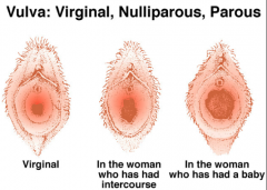
A female who has not had any children
|
|
|
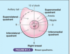
What is the axillary tail?
|

Breast tissue in the upper outer quadrant passing through facia and connects to the axilla
|
|
|
Which quadrant do most breast cancers occur?
|

The upper outer quadrant
|
|
|
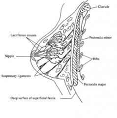
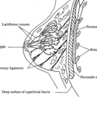
What are the components of the breast?
|
1. Nipple/areolar
2. Parenchymal/Glandular tissue (lactation apparatus) 3. Stromal Tissue (fat and loose connective tissue) |
|
|
What are the lactiferous sinuses?
|
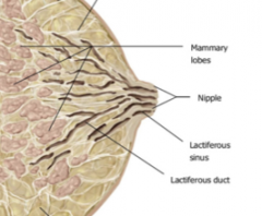
Reservoirs or ducts for milk superficial to the gland and just deep to the nipple
|
|
|
What are suspensory ligaments?
|
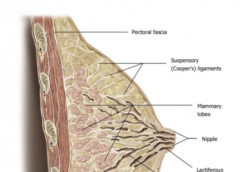
Bands of connective tissue that help support the meshwork and fat of the breast tissue.
|
|
|
What is the retromammary space?
|

A small line of connective tissue that separates the breast from the underlying pectoralis major
|
|
|
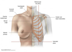
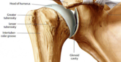
What are the greater and lesser tubercles of the humerus?
|
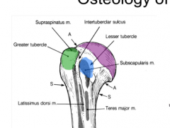
The bumps on the proximal end of the long bone surrounding the intertubercular groove or bicipital groove
|
|
|
What is the coracoid process?
|
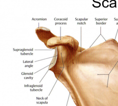
The attachment of the pectoralis minor located on the anterior side of the scapula
|
|
|
What are the two portions of the pectoralis major muscle?
|
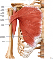
Clavicular head (attaches to the medial third of the clavicle)
Sternocostal head that attaches to the lateral border of the sternum and upper six ribs. |
|
|
Where does the tendon of the pectoralis muscle attach?
|
To the intertubercular groove of the humerus
|
|
|
What movements is the pectoralis major muscle responsible for?
|

Flexion, Adduction, and Medial Rotation
|
|
|
Which ribs does the pectoralis minor muscle attach to?
|
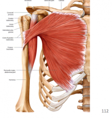
To the proximal parts of ribs 3-5 and distally to the coracoid process
|
|
|
What movements is the pectoralis minor responsible for?
|

Depression of the shoulder and an accessory muscle of respiration in asthmatics
|
|
|
What is the lateral pectoral nerve?
|
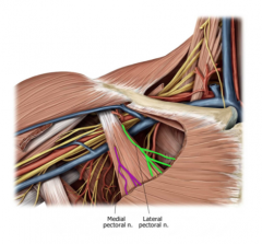
Fibers from spinal nerves C5, 6, and 7 which innervates the pectoralis major.
|
|
|
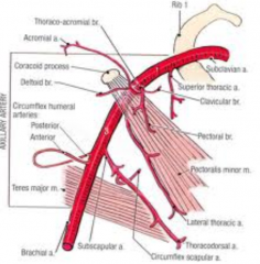
What blood vessel runs parallel to the lateral pectoral nerve?
|

The pectoral artery which is a branch of the thoracoacromial artery
|
|
|
What is the medial pectoral nerve
|

Contains fibers from C8 and T1 spinal nerves and is the nerve supply of the pectoralis minor and part of the pectoralis major
|
|
|
True or False: The lateral pectoral nerve is lateral to the medial pectoral nerve.
|
FALSE: These are named after their branches in the brachioplexus
|
|
|
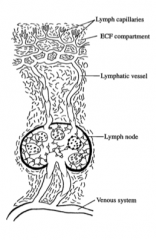
What is lymph edema?
|
Lymph or extracellular fluid that is lost by capillary beds and can be returned to the central circulatory system by the lymphatic system.
|
|
|
Where do lymph nodes return ECF to the circulatory system?
|
The venous system
|
|
|
What occurs when a lymph node is infected?
|

It is large soft, tender and painful
|
|
|
What is the case when a lymph node is large, hard/firm, and not tender i.e. painless?
|
Most likely malignant
|
|
|
What is the cisterna chyli?
|
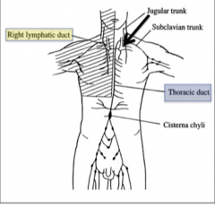
A lymph node in the proximal abdomen that drains into the thoracic duct (ascends along the bodies of the thoracic vertebrae
|
|
|
Where does the thoracic duct drain?
|
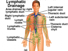
Between the left internal jugular and subclavian vein
|
|
|
What do the left and right bronchomediastinal trunk drain?
|
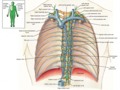
Visceral pleura from the lungs and lymph from the medial aspects of the breast.
|
|
|
Where do the right jugular trunk, subclavian trunk and bronchomediastinal trunk drain?
|
Into the right lymphatic duct which drains into the junction between the right internal jugular vein and the right subclavian vein.
|
|
|
What two veins combine to form the superior vena cava?
|
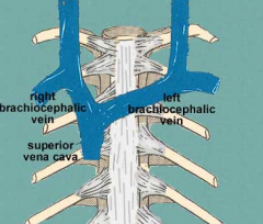
The union of the left and right brachiocephalic veins
|
|
|
What is the brachiocephalic vein?
|
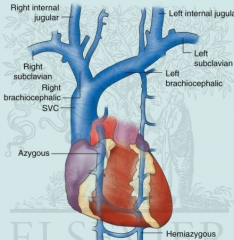
The union of the internal jugular and subclavian vein
|
|
|
What is the subclavian vein called once it passes the first rib?
|
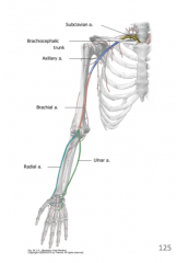
The axillary vein
|
|
|
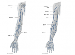
What two veins combine to form the axillary vein?
|

The basilic and the brachial vein
|
|
|
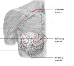
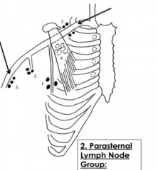
Which lymph nodes are associated with the internal thoracic/mammary veins?
|
The parasternal lymph nodes
|
|
|
What are the axillary lymph node groups?
|

1. pectoral/anterior nodes (75% lateral breast lymph)
2. subscapular/ posterior nodes 3. lateral/brachial nodes 4. central nodes (drains 1-3) 5. apical nodes (drains 4) |
|
|
What nerve passes through the central lymph nodes?
|
Intercostobrachial nerve which has roots from spinal nerve T2
|
|
|
What are the parasternal nodes?
|

Deep to the intercostal spaces on either side of the sternum receive the remaining 25% of breast lymph from the medial quadrants.
|
|
|
What is the most commonly diagnosed cancer in women?
|

Breast cancer
|
|
|
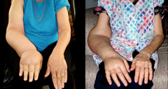
What are the common symptoms of breast cancer?
|
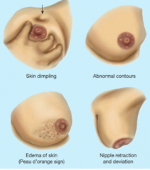
Painless lump or thickening, change in size, nipple discharge, dimpled skin, and peau d'orange (lymphatic blockage)
|
|
|
What are non-cancerous breast conditions?
|
Fibrocystic changes (increase in fibrous and glandular tissue), cysts, fibroadenomas (solid, fibrous, benign growths), infections (abscess), and trauma
|
|
|
What is mastitis?
|
Inflammation due to infections in the breast tissue
|
|
|
What are ductal carcinomas (DCIS)?
|
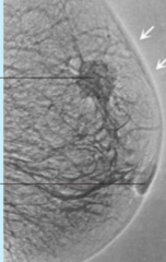
Malignant tumors that grow from the epithelial tissue in lactiferous ducts
|