Reading...
![]()
Play button
![]()
Play button
![]()
Use LEFT and RIGHT arrow keys to navigate between flashcards;
Use UP and DOWN arrow keys to flip the card;
H to show hint;
A reads text to speech;
83 Cards in this Set
- Front
- Back

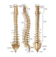
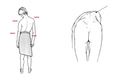
In what anatomical plane is the image in?
Name the primary and secondary curves |
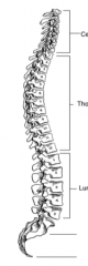
Sagittal plane
Primary: Thoracic and Sacral Secondary: Cervical and Lumbar |
|
|
True or False: The Vertebral Column is a semi-rigid structure
|
True
|
|

What anatomical plane is the image in?
|
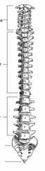
Coronal or Frontal plane
|
|
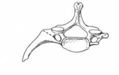
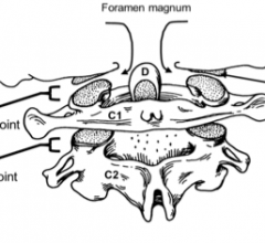
Name the bone in the image above
|

Atlas (C1)
|
|

Name the bone in the image above
|
Axis (C2)
|
|

Where is the first Intervertebral disc found?
Where is the last intervertebral disc found? |
Between C2 and C3 vertebrae
Between L5 and the Sacrum |
|
Name regions of the vertebral column (VC) from most mobile to least mobile.
|

Cervical (extension, flexion, and lateral bending)
Lumbar (extension, flexion, and lateral bending) Thoracic (rotation) -immobility due to rib attachment Sacrum |
|
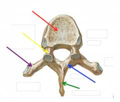
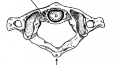
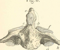
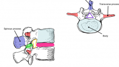
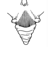
Name the regions of the bone highlighted by the arrows.
|

Thoracic Vertebra
Red: Arrow Yellow: Pedicle Purple: Transverse Process Green: Spinous Process Blue: Lamina |
|
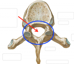
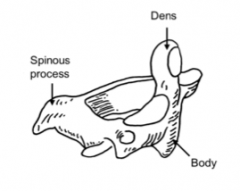
Name the region of the bone highlighted by the arrow.
What two structures compose the circled blue area, what is another name for this? |

The vertebral foramen
The pedicles and lamina create the vertebral arch |
|
|
What is the vertebral canal?
|
The space formed by the stacking of vertebral foramen that houses the spinal cord.
|
|
|
What are the components of the vertebral canal?
|
1) Spinal Cord
2) Meninges 3) Epidural/Extradural fat 4) Internal Vertebral Venous Plexus |
|
|
What are the articular processes?
|
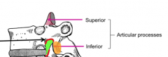
They are processes on the vertebrae coming off the pedicles that are covered in articular cartilage. There are two-superior and inferior-that form synovial joints known as articular facets that allow smooth movement of the vertebral column.
|
|
|
Describe the intervertebral foramen.
|
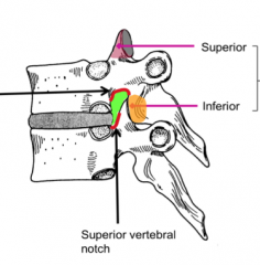
Each vertebra has an inferior and a superior notch so when stacked this space coming from the vertebral canal allows the exit of spinal nerves.
|
|
|
Name the primary curves of the vertebral column
|
Thoracic and Sacral-these are fully formed in newborns and persist in adults. Both curves open anteriorly
|
|
|
Name the secondary curves of the vertebral column.
|
The secondary curves form after birth and open posteriorly.
-Cervical (4 month) allows support of the head -Lumbar (12-18 months) allows for the distribution of weight when learning to sit up and stand. |
|
|
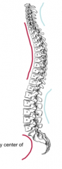
Where is the center of gravity in an adult human?
|
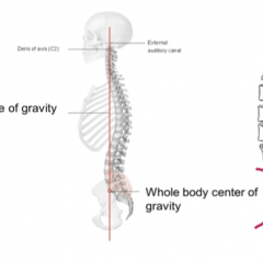
In front of the spinal column, which leads to the necessity of the lumbar and cervical posterior curves to compensate for weight bearing.
|
|
|
What is lordosis?
|
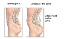
Lordosis is an exaggerated posterior curvature of the spine commonly found in the lumbar region. Common causes are pregnancy and obesity which overcome and can sprain the anti-gravity muscles of the lower back.
|
|
|
What is Kyphosis?
|
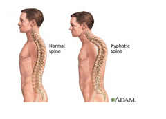
An exaggerated anterior curvature of the spine commonly found in the thoracic region. This hunchback is seen in elderly post-menopausal women due to a loss of estrogen bone mass decreases and compression fractures in the vertebrae leading to this curvature.
|
|
|
What is a lateral curvature of the spine?
|
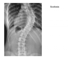
Scoliosis
Also known as "crooked back" this curvature with unknown source leads to uneven hip and shoulder levels. When performing Adam's test a common sight is a rib hump that projects on one side due to rotational movement of the scoliotic spine. |
|
|
What is a common complication of scoliosis?
|
Respiratory complications due to decreased thoracic capacity and therefore decreased lung capacity.
|
|
|
What is an arthrosis?
|
Another name for a joint where two skeletal elements unite. Joints are classified by the tissue in-between the joint.
|
|
|
What is the difference between diarthroses and synarthroses?
|
Diarthroses are synovial joints that allow free movement due to the synovial fluid between the skeletal elements. Synarthroses are non-synovial joints that are further classified as fibrous or cartilaginous joints.
|
|
|
What is the difference between fibrous and cartilaginous joints?
|
Fibrous joints contain fibrous connective tissue between skeletal elements that allow little to no movement such as the interosseous membranes of the forearm or the sutures of the skull.
Cartilaginous joints contain cartilage between the skeletal movement and commonly allow slight movement. |
|
|
Define the term syndesmoses.
|
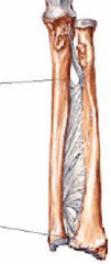
This type of fibrous joint allows slight movement between skeletal elements due to the interosseous ligament such as those in the forearms and legs.
|
|
|
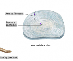
Describe the difference between synchondrosis and symphysis?
|
Synchrondosis is a cartilaginous joint that contains hyaline cartilage such as growth plates in the epiphysis of the long bones.
Symphysis is a cartilaginous joint that contains fibrocartilage such as the intervertebral discs and the pubic symphysis. |
|
|
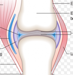
What are the principle features of synovial joints?
|
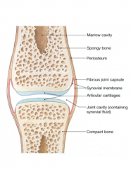
-Articular (hyaline) cartilage
-Fibrous joint capsule -Joint space with synovial fluid -Synovial membrane lining -Capsular ligaments -Sensory nerves and blood vessels to capsule and periosteum |
|
|
Why are blood vessels and nerves not present in hyaline cartilage?
|
This tissue is not innervated by nerves so that the movement of joints does not produce pain. Due to synovial fluid the joint capsule is supplied with nutrition and is able to distribute waste through diffusion.
|
|
|
What replaces hyaline cartilage of joints when it is worn down?
|
Fibrous tissue or exposed bone that can be very painful due to uneven surfaces and exposed innervated bone tissue.
|
|
|
What are the main factors of joint stability?
|
The shape of articular surfaces
Joint capsule and capsular ligaments Tone of muscles surrounding a joint |
|
|
What leads to the degeneration of articular cartilage?
|
Excessive use and loading or lack of motion which leads to a reduced diffusion of synovial fluid.
|
|
|
Describe osteoarthritis.
|
The most common form of arthritis is commonly known as wear and tear due to degenerative bone lost. Osteophytes form in response to loss of articular cartilage. This leads to joint pain and reduced mobility.
|
|
|
What is an osteophyte?
|
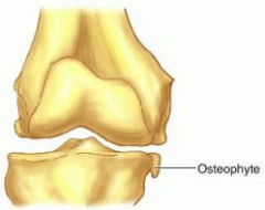
A reactive hypertrophic boney changes-a product of osteoarthritis
|
|
|
What is crepitus?
|
A grating noise associated with osteoarthritic joints
|
|
|
What is arthroscopy?
|
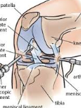
A surgical technique of visualizing interior of a synovial joint.
|
|
|
Name the types of intervertebral joints
|
The intervertebral disc is a symphysis joint of fibrocartilage.
The zygopophyseal joint (also known as facet joint) is formed from the articular processes and is synovial. |
|
|
What is a costovertebral joint?
|
Joints found in the thoracic region where the ribs connect with the vertebral column. These are synovial joints
|
|

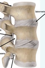
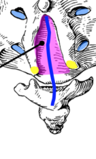
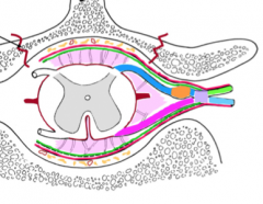
Name the ligaments of the vertebral column highlighted in the image above.
|
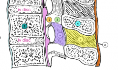
Anterior Longitudinal Ligament (ALL)-light blue
Posterior Longitudinal Ligament (PLL)-pink Ligamentum flavum-purple Interspinous ligament-yellow Supraspinous Ligament-Orange |
|
|
What is the purpose of the ALL?
|
This prevents hyperextension especially in the cervical region. Hyperextension of this region such as during a whiplash injury can cause a tear or a breakage of the bone of the body of a vertebra known as an Avulsion fracture. Anterior Longitudinal Ligament
|
|
|
Which ligament of the vertebral column is exaggerated to form the Ligamentum Nuchae?
|
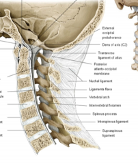
The supraspinous ligament
|
|
|
What does the ligamentum flavum connect?
|
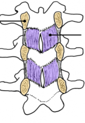
Proximal lamina of the vertebral column.
|
|
|
What is the vertebral venous plexus?
|

The venous system surrounding and within the vertebral column. These are freely anastomotic network.
|
|
|
Describe the veretebral venous plexus.
|
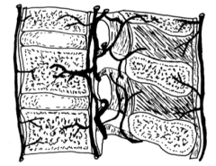
It extends from the base of the skull to the sacrum, drains blood from the vertebral column and spinal cord, lacks valves and therefore can have a bidirectional flow which can attribute to the metastatsis involved with cancer.
|
|
|
Describe the unique features of the cervical vertebrae (C3-C6)
|

The small bodies and large vertebral foramen are due to the decreased weight and large spinal cord. These also have the upturned lips of the uncinate processes that form uncovertebral joints (joints of Luschka) that are synovial lateral joints. The transverse processes have a posterior and anterior tubercle (rib homolog) around a transverse foramen that houses the vertebral artery.
|
|
|
Which vertebrae have transverse foramen?
|
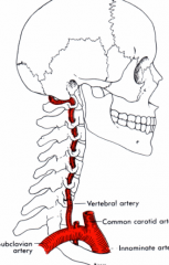
The cervical vertebrae do in order to house the vertebral artery.
|
|
|
Which vertebrae have bifid spinous processes?
|
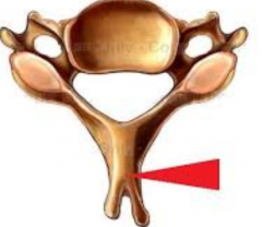
Cervical vertebrae
|
|
|
Where does the vertebral artery enter the vertebral column?
|
C6 through the Atlas where it continues through the foramen magnum to the brain.
|
|
|
What is unique about the C7 vertebrae?
|

It is the vertebra prominens because it has an especially long spinous process. It also has a cervical rib from the anterior tubercle in about 0.5-1% of the population.
|
|
|
What is thoracic outlet syndrome?
|
Nerves and blood vessels that cross superiorly to the top rib toward the upper limbs can become strained leading to numbness when a cervical rib is present.
|
|
|
What is atlas?
|
C1 vertebra that has a bump at the top of the posterior arch known as the posterior tubercle. Since this is essentially a ring of bone there is also an anterior arch which contains the dens of the axis.
|
|
|
What is the odontoid process?
|
The projection of the C2 (axis) vertebra (aka dens) that is anchored by the transverse ligament of the atlas.
|
|
|
What is the axis?
|
The second cervical vertebra (C2)
|
|
|
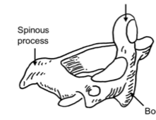
What bone does the articular processes of the atlas connect with to form the superior synovial joint of the vertebral column?
|
The occipital condyle
|
|
|
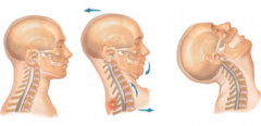
What is the name of the superior synovial joint of the vertebral column?
|
The atlanto-occipital joint. This is commonly used for the nodding or yes action of the head
|
|
|
What is the atlanto-axial joint?
|
The joint between the top two vertebrae that allows for rotation of the head in a no action.
|
|
|
True or False: The spinous process of the atlas is bifid
|
FALSE: The axis spinous processis bifid, the atlas has a posterior tubercle.
|
|
|
What is a potential complication of a dens fracture?
|
Due to the posterior location of the top of the spinal cord asphyxiation could follow from an inability to move the diaphragm. Even if this is prevented the patient will become a quadriplegic.
|
|
|
Describe a typical thoracic vertebrae.
|
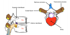
An increased body and a decreased vertebral foramen from the cervical vertebrae. There are posterior lateral transverse processes that have costal facets for a synovial joint with the ribs. All spinous processes point downward and the posterior lateral sides of the body have two demifacets to support the attachment of the rib at its head.
|
|
|
What determines the rib number?
|
The transverse process facet or the superior demifacet on the body of the thoracic vertebrae.
|
|
|
Describe a typical lumbar vertebra.
|
The body and vertebral foramen are larger with thin lateral transverse processes and broad flat spinous processes ideal for a lumbar puncture.
|
|
|
How many vertebrae are fused in the sacrum?
|
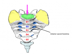
5
|
|
|
What are the wing-like extensions of the superior sacrum?
|
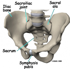
The ala which combine with the Ilium of the pelvic girdle to form the sacral-iliac joint.
|
|
|
Where is the apex of the sacrum?
|
S5
|
|
|
What is the significance of the S2?
|
The end of the dural sac of the spinal cord within the sacral canal.
|
|
|
What exits from the anterior sacral foramina?
|
Sacral Ventral Primary Rami
|
|
|
What is the coccyx?
|
The 3-5 fused vertebrae at the end of the vertebral column. This articulates with the sacrum.
|
|
|
What exits from the posterior sacral foramina?
|
Dorsal Primary Rami
|
|
|
What is the sacral hiatus?
|
The posterior inferior triangular gap headed by cornua "horns" providing an exit for the filum terminale externum also known as coccygeal ligament.
|
|
|
What is the coccygeal ligament composed of?
|
Pia mater
coccygeal ligament = filum terminale externum |
|
|
What are the main functional groups of the vertebral column?
|
The flexors and extensors
Flexors-sternocleidomastoid, anterior abdominal wall muscles Extensors-Deep back muscles "paraspinous muscles" specifically splenius, sub occipital, erector spine, and transversospinalis. |
|
|
Which nerve classification innervates the paraspinous muscles?
|
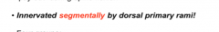
These deep back muscles are innervated segmentally by dorsal primary rami
|
|
|
Which muscle group lies deep to the erector spinae?
|
The transversospinalis group travel between the spinous and transverse processes of the vertebrae.
|
|
|
What are the splenius muscles?
|
The bandage muscles are part of the extensor muscles of the spine. The splenius attaches to the lower half of the ligmentum nuchae and the top six thoracic vertebrae. The capitus attaches to the base of the skull and the cervicis inserts into the transverse processes of C1-C4 just like the levator scapulae.
|
|
|
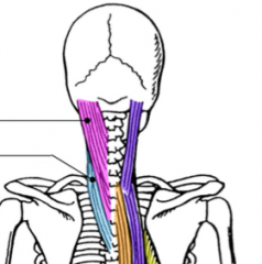
Name the muscles of the erector spinae group from lateral to medial?
|
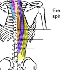
Iliocostalis, Longissimus, and Spinalis (I love Spaghetti)
|
|
|
Name the three spinal meninges from superficial to deep.
|
Dura mater, arachnoid mater, and pia mater.
|
|
|
What pressure exists between the arachnoid mater and the pia mater?
|

The cerebrospinal fluid of the subarachnoid space.
|
|
|
What lies superficial to the dura mater of the spinal cord?
|
The epidural fat and the internal vertebral venous plexus
|
|
|
What nerve comes off of the posterior side of the spinal cord?
|
The dorsal root which commonly has a collection of nerves known as the dorsal root ganglion (sensory)
|
|
|
What nerves come off the anterior side of the spinal cord?
|
The ventral root (motor)
|
|
|
What is the name for when the dorsal and ventral roots combine?
|
The spinal nerve which will then separate into two branches (rami) known as the dorsal and ventral primary rami.
|
|
|
What are the denticulate ligaments?
|
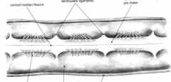
The lateral projections of pia mater that separate the ventral and dorsal roots
|
|
|
What is the conus medullaris?
|

The terminal end of the spinal cord which occurs in the L1/L2 level in adults and the L3 level in newborns and children
|
|
|
What is the cauda equina?
|
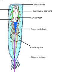
Descending nerve roots in the dural sac that form a horses tail like structure
|
|
|
What is the uncinate process?
|
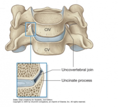
This is a curved lip of cervical vertebrae bodies that forms the uncovertebral joint with the superior vertebra.
|