![]()
![]()
![]()
Use LEFT and RIGHT arrow keys to navigate between flashcards;
Use UP and DOWN arrow keys to flip the card;
H to show hint;
A reads text to speech;
102 Cards in this Set
- Front
- Back
|
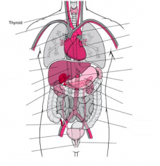
trachea superior vena cava lung liver inferior vena cava gall bladder kidney yreter appendix bladder urethra aorta heat esophagus diaphragm aorta spleen stomach pancreas small intestine large intestine rectum |
|
|
Where are the adrenal glands located? |
above/ on top of the kidneys |
|
|
What is the duodenum? |
the first part of the small intestine that is directly under/after the stomach |
|
|
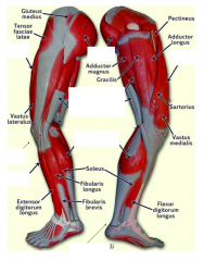
rectus femoris
patellar ligament tibialis anterior gluteus maximus hamstrings gastrocnemius achilles tendon rectus femoris |
|
|
What is the proper word for "abs"? |
rectus abdominis |
|
|
Where is the pectoralis major located? |
on the chest (in the breast area) |
|
|
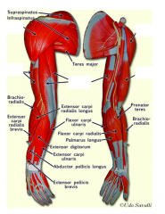
deltoid biceps brachii triceps brachii deltoid biceps brachii |
|
|
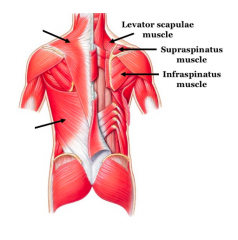
trapezius latissimus dorsi |
|
|
What is flexion? |
movement that decreases the angle between 2 articulating bones |
|
|
What is extension? |
movement that increases the angle between 2 articulating bones |
|
|
What is abduction? |
movement of a bone away from the midline |
|
|
What is adduction? |
movement of a bone towards the midline |
|
|
What 2 hormones does the pancreas secrete? |
insulin glycogen |
|
|
What is the function of the liver? |
to produce bile |
|
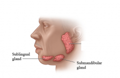
and function |
parotid gland secretes saliva |
|
|
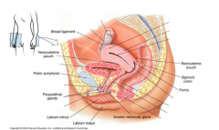
ovary fallopian tube urethra clitoris uterus cervix rectum (large intestine) vagina anus |
|
|
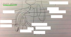
prostate penis urethra scrotum testes epididymus vas deferens seminal vesicles anus cowper's gland rectum (large intestine) urinary bladder |
|
|
What are the 4 classes of tissues? |
epithelial muscle connective cardiac |
|
|
What is an organ? |
groups of tissue that occur together and perform a specific function |
|
|
What is histology? |
the study of tissues |
|
|
What are the functions of epithelial tissue? |
protect, secrete, filter and absorb |
|
|
What are the locations of epithelial tissue? |
cover and line body cavities, the outer surface of the body (skin), the surface of internal organs, the lumina of ducts, vessels and tubes |
|
|
What are the characteristics of epithelial tissue? |
cells are closely joined together and have little intercellular matrix between them there is a free (apical) surface, and a basement membrane present in the lower surface |
|
|
What is a basement membrane? |
anchors to CT non-cellular, adhesive sheet consisting of glycoproteins and collagen fibres |
|
|
What is simple squamous epithelium? |
sigle layer of flattened celles resting on the basement membrane filtration and diffusion forms walls of alveoli and Bowman's capsules in the kidneys |
|
|
What is endothelium? |
simple squamous epithelium that liens this chambers of the heart and blood vessels |
|
|
What is mesothelium? |
simpel squamous epithelium that forms serous membranes that lien the ventral body cavities ad cover prams found in them |
|
|
What is simple cuboidal epithelium?
|
single layer of cube-shaped cells resting on a basement membrane nucleus is centrally located forms gland ducts and the walls of certain regions of kidney tubules |
|
|
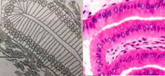
What is simple columnar epithelium? |
single layer of cells resting on a basement membrane nukes is located at the base of the cell mucus-secreating cells (goblet cells) are often present secretion and absorption lines stomach, small intestine and most of the large intestine |
|
|
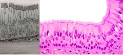
What is ciliated pseutostratified columnar epithelium? |
mixture of cells = appears stratified but is actually simple nuclei are located at varying distances from the basal surface liens nasal cavity, trachea and bronchi goblet cells secrete a sticky mucus that traps dust and other debris that might pass along airways |
|
|
What is stratified squamous epithelium? |
several layers of cells cells that are at the free edge are squamous-shaped and the ones at the basement membrane are either cuboidal or columnar found in areas that receive a lot of wear outer layers are shed off and replaced found in the lining of the mouth, esophagus, anus and vagina a special keratinized layer of dead surface cells is found in the epidermis of the skin |
|
|
What is connective tissue? |
abundant amounts of extracellular material (matrix) cells are spread out from eachother throughout the matrix 2 parts of the matrix = ground substance and fibres |
|
|
What is ground substance? |
fluid, gel-like, firm or rock-hard several types of protein fibres are embedded |
|
|
What is areolar CT? |
soft and pliable general packaging throughout the body fascia that is easily pulled apart furring blunt dissection contains different types of cells such as fibroblasts and macrophages 2 types of fibres = collagen and elastic |
|
|
What is a fibroblast? |
produce fibres in the matrix? |
|
|
What is a macrophage? |
phagocitizes bacteria |
|
|
What is adipose CT? |
commonly called fat a mature fat cell contains a large drop of fat and the cytoplasm is reduced to a thin layer surrounding the droplet long-term energy storage area found beneath the skin (insulation) act as a cushion and protects certain organs such as the kidneys and eyeballs |
|
|
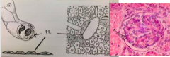
What is cartilage? |
consists of cell chondrocytes found in tiny cavities called lacunae and semisolid matrix, which is both strong and elastic found in articular surfaces of nones, in the walls fo the trachea, 10 ribs and sternum |
|
|
What is muscle tissue? |
responsible for movements materials thought the body with respect to one another, and for locomotion contractile = composed of cells called fibres that are elongated in the direction of the contraction |
|
|
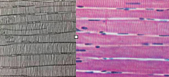
What is skeletal muscle? |
striated = have many nuclei per cell can be under voluntary control makes up the muscles of the body that are attached to the skeleton |
|
|
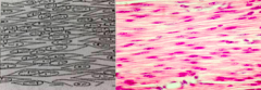
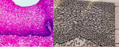
What is smooth muscle? |
long, spindle-shaped with a single centrally located nuclei no striations present found in the walls fo the gastrointestinal tract, arteries, veins, uterus and urinary bladder involuntary control |
|
|
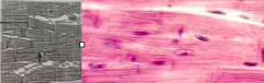
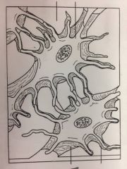
What is cardiac muscle? |
cells are striated cells are branched and interconnected by intercalated discs, which appear as dark bands scattered throughout the tissue forms much of the heart wall involuntary |
|
|
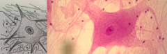
What is nervous tissue? |
within the nervous system contains 2 types of cells = nerve cells and glial cells give structural and functional support to neutrons found in the brain and spinal cord |
|
|
What are nerve cells? |
produce and transmit impulses neurons |
|
|
What are glial cells? |
smaller cells supporting cells called neuroglia |
|
|
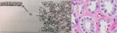
simple cuboidal epithelium |
|
|
cardiac muscle tissue: intercalated discs |
|
|
nervous tissue: glial cells cell body of a neuron |
|
|
smooth muscle tissue |
|
|
simple squamous epithelium |
|
|
simple columnar epithelium: cell connective tissue |
|
|
stratified squamous epithelium: nuclei basement membrane connective tissue |
|
|
ciliated pseudostratified columnar epithelium |
|
|
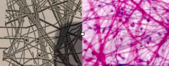
skeletal muscle tissue |
|
|
areolar muscle tissue: collagen fibres fibroblast nuclei elastic fibres |
|
|
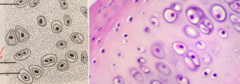
cartilage: chondrocytes matrix lacunae |
|
|
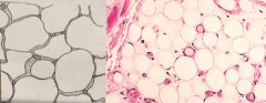
adipose tissue |
|
|
What is articulation? |
bones can only move at a joint any muscle must be attached to atlas 2 bones around the joint one bone is the movable bone relative to the other bone which is immovable |
|
|
What is the origin? |
the end of the muscle attached via a tendon to the least moveable bone
|
|
|
What is the insertion? |
the end of the muscle attached to the bone undergoing most movement |
|
|
How are joints classified? |
1. the presence or absence of a pace between articulating bones called the synovial point/cavity 2. the type of tissue tat develops between the articulating bones 3. the range of movement they permit |
|
|
What is a fibrous joint? |
relatively immoveable no joint cavity articulating bones are help together by fibrous CT |
|
|
What is a cartilaginous joint? |
slightly moveable
no joint cavity articulating bones are held together by cardiac tissue |
|
|
What are synovial joints? |
enclosed in a lubricated joint cavity
articulating bones are held together by ligaments and tendons connecting muscles to bones |
|
|
What is flexion? |
decreases the angle of a joint by bringing articulating bones closer together
|
|
|
What is extension? |
increases the angle of a joint by bringing articulating bones further away from one another (back to anatomical position) |
|
|
What is hyperextension? |
increases the angle of a joint beyond anatomical position
|
|
|
What is abduction? |
movement of a limb away from the midline axis
|
|
|
What is adduction? |
movement of a limb closer to the midline axis
|
|
|
What are the components of the axial skeleton? |
skull, sternum, rib cage, vertebrae |
|
|
What are the components of the appendicular skeleton? |
pelvic girdle, pictorial girdle and limbs (arms and legs)
|
|
|
What are the differences between a male and female pelvis? |
male = heart shaped, pubic arch < 90 degrees
female = oval shaped, pubic arch > 90 degrees |
|
|
What is diaphysis? |
smooth surface
composed of compact bone |
|
|
What is the periosteum? |
dense, fibrous CT covering the bone surface
fibres penetrate into the bone |
|
|
What is the nutrient formina? |
small opening into the bone that allow for passage of nutrient blood vessels into the bone for nourishment of living tissue
|
|
|
What are osteoblasts? |
secrete the bony matrix that increases the thickness of the long bone
|
|
|
What are osteoclasts? |
able to digest extracellular matrix of bone, and releasee calcium
this is called bone reabsorption |
|
|
What is epiphyses? |
knob-like ends of the long bone
composed of a thin layer of compact bone that encloses spongy bone |
|
|
What is articular cartilage? |
thin layer of hyaline cartilage
covers each epiphyseal surface |
|
|
What is ossification? |
process of bone formation
|
|
|
What is the epiphyseal plate? |
thin area of hyaline cartilage that provides longitudinal growth of the none during youth
after youth, epiphyseal lines are hardly visible |
|
|
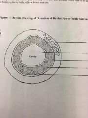
What is the medullary cavity? |
central cavity
a storage region for fat in the tissue known as yellow bone marrow (red in youth) |
|
|
What is hematopoiesis? |
process of blood formation
|
|
|
What is the edosteum? |
lines the diaphysis
|
|
|
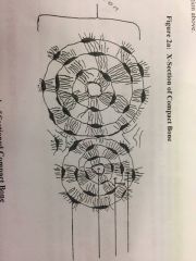
What are lamellae? |
concentric layers of hard, calcified, extracellular matrix
|
|
|
What are adipocytes? |
round, empty looking cells
|
|
|
What are the 3 types of joints? |
fibrous
synovial cartilaginous |
|
|
What are synovial joints? |
contain synovial fluid which is slippery
|
|
|
What is the synovial membrane? |
a thin, sleeve-like transparent membrane which links the heads of 2 bonestransports ions and molecules from the blood plasma into the cavity fo the omit to form synovial fluidsecretes hyaluronic acid to make it slippery
|
|
|
What is articular cartilage? |
covers the head son articulating bones
made of smooth, hyaline cartilage |
|
|
What is the articular capsule vs fibrous capsule ? |
sleeve of tissue
links 2 bone vs covers synovial membrane continuous with the periosteum of nones and joints |
|
|
What are ligaments? |
link bone to bone
|
|
|
What are tendons? |
link bone to muscle tissue
|
|
|
What are menisci? |
c-shpaed cartilaginous discs
stabilize --> act as a shock absorber medial vs lateral |
|
|
skeletal muscle
bone marrow endosteum --> single layer of cells compact bone periosteum |
|
|
canaliculi
lamellae central canal lacuna |
|
|
Volkmann canal
central canal |
|
|
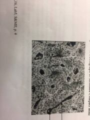
osteocyte
matrix canaliculi lacuna |
|
|
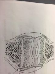
articular cartilage
joint cavity synovial membrane fibrous layer |
|
|
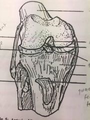
lateral collateral ligament
cartilage fibula femur posterior articulate ligament anterior cruciate ligament cartilage tibia medial collateral ligament quadriceps femoris tendon |
|
|
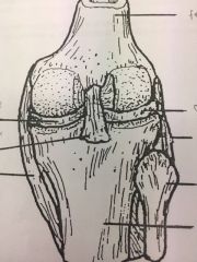
cartilage
posterior ligament medial collateral ligament femur cartilage lateral collateral ligament fibula tibia |
|
|
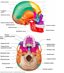
parental bone
temporal bone occipital bone zygomatic process frontal bone sphenoid bone ethmoid bone lacrimal bone nasal bone zygomatic maxilla mandible palatine zygomatic vomer |