![]()
![]()
![]()
Use LEFT and RIGHT arrow keys to navigate between flashcards;
Use UP and DOWN arrow keys to flip the card;
H to show hint;
A reads text to speech;
236 Cards in this Set
- Front
- Back
- 3rd side (hint)
|
Anatomical Terms & Homeostasis |
--- |
|
|
|
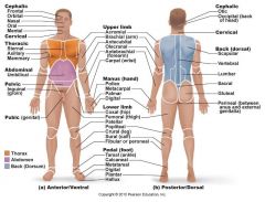
Surface Anatomy
|
Axial: Relating to head, neck and trunk, the axis of the body
Appendicular: Relating to limbs and their attachments to their attachments to the axis |
|
|
|
Anterior Body Landmarks |
Note the following regions
|
|
|
|
Abdominal |
Anterior body trunk region inferior to the ribs |
|
|
|
Acromial |
point of shoulder |
|
|
|
Antebrachial |
forearm |
|
|
|
Antecubital |
anterior surface of the elbow |
|
|
|
Axillary |
armpit |
|
|
|
Brachial |
Arm |
|
|
|
Buccal |
Cheek |
|
|
|
Carpal |
wrist |
|
|
|
Cephalic |
head |
|
|
|
Cervical |
neck region |
|
|
|
Coxal |
Hip |
|
|
|
Crural |
Leg |
|
|
|
Digital |
Fingers or toes |
|
|
|
Femoral |
Thigh |
|
|
|
Fibular (peroneal) |
Side of the leg |
|
|
|
Frontal |
Forehead |
|
|
|
Hallux |
Great toe |
|
|
|
Inguinal |
groin area |
|
|
|
Mammary |
breast region |
|
|
|
Mangus |
hand |
|
|
|
Mental |
chin |
|
|
|
Nasal |
nose |
|
|
|
Oral |
mouth |
|
|
|
Orbital |
Bony eye socket (orbit) |
|
|
|
Palmar |
palm of the hand |
|
|
|
Patellar |
anterior knee (kneecap) region |
|
|
|
Pedal |
foot |
|
|
|
Pelvic |
pelvis region |
|
|
|
Pollex |
thumb |
|
|
|
Pubic |
genital region |
|
|
|
Sternal |
region of the breastbone |
|
|
|
Tarsal |
ankle |
|
|
|
Thoracic |
chest |
|
|
|
Umbilical |
Navel |
|
|
|
Posterior Body Landmarks |
-- |
|
|
|
Acromial |
point of the shoulder |
|
|
|
Brachial |
arm |
|
|
|
Calcaneal |
heel of the foot |
|
|
|
Cephalic |
head |
|
|
|
Dorsum |
back |
|
|
|
Femoral |
thigh |
|
|
|
Gluteal |
buttocks or rump |
|
|
|
Lumbar |
Area of the back between the ribs and hips; loin |
|
|
|
Manus |
hand |
|
|
|
Occipital |
posterior aspect of the head or the base of the skull |
|
|
|
Olecranal |
posterior aspect of the elbow |
|
|
|
Otic |
ear |
|
|
|
Pedal |
foot |
|
|
|
Perineal |
region between the anus and external genitalia |
|
|
|
Plantar |
sole of the foot |
|
|
|
Popiteal |
back of the knee |
|
|
|
Sacral |
region between the hips (pverlying the sacrum) |
|
|
|
Scapular |
scapula or shoulder blade area |
|
|
|
Sural |
calf or posterior surface of the leg |
|
|
|
Vertebral |
area of the spinal column |
|
|
|
Surface Anatomy Diagram |
|
|
|
|
Body Orientation and Direction |
Study the terms below for visual aid |
|
|
|
Superior/Inferior (above/below) |
-Superior: structures always appear above other structures
-Inferior: structures always below other structures |
|
|
|
Anterior/Posterior (front/back) |
Anterior: structures more forward
Posterior: structures toward the backside of the back |
|
|
|
Medial/Lateral (toward the midline/away from the midline or median plane) |
Medial: The sternum (breastbone) is medial to the ribs
Lateral: The ear is lateral to the nose |
|
|
|
Cephalad (cranial) / Caudal (toward the head/ toward the tail) |
In humans, they are used interchangeably with superior and inferior |
|
|
|
Dorsal/Ventral (backside/belly side) |
In humans, the terms ventral and dorsal are used interchangeably with the terms anterior and posterior |
|
|
|
Proximal/ Distal (nearer the trunk or attached end/ farther from the trunk or point of attachment) |
These terms are used to primarily locate various regions of the body of limbs. |
|
|
|
Superficial (external) / deep (internal) (toward or at the body surface/ away from the body surface) |
These terms locate body organs according to their relative closeness to the body surface |
|
|
|
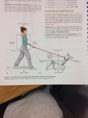
Diagram of Body Orientation and Direction
|
|
|
|
|
Body Plane and Sections
|
-section: cut
-plane: imaginary surface or line |
|
|
|
Planes |
Three Planes |
|
|
|
Sagittal Plane |
runs longitudinally and divides the body into right and left parts -Median(midsagittal plane): if it divides the body into equal parts or right down the midline of the body |
|
|
|
Frontal Plane (Coronal Plane) |
longitudinal plane that divides the body(or organ) into anterior and posterior parts |
|
|
|
Transverse Plane (Cross Sections) |
runs horizontally dividing the body into superior and inferior parts
|
|
|
|
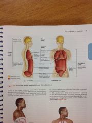
Dorsal Body Cavity |
-Cranial 1.brain -Vertebral 1.spinal cavity |
|
|
|
Ventral Body Cavity |
-Thoracic: 1. heart (pericardium) 2. Lungs (pleural) -Abdominopelvic: 1. Abdominal 2. Pelvic |
|
|
|
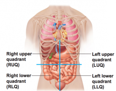
Abdominopelvic Quadrants |
1. Quadrant: divides the abdominal surface and the adbominopelvic cavity into four approximately equal regions -Right Upper -Right Lower -Left Upper -Left Lower |
|
|
|
Abdominopelvic Quadrant Diagram |
|
|
|
|
Abdominopelvic Regions |
1. Region: divides the abdominal surface and abdominopelvic cavity into nine separate regions by four planes -Hypochondriac Regions: Flanking the epigastric region laterally and overlying the lower ribs (RH: iver, Gallbladder; LH: diaphragm, Spleen) - Epigastric: immediately superior to the umbilical region; overlies most of the stomach (stomach) -Lumbar Regions: Between the ribs and the flaring portions of the hip bones; lateral to the umbilical region (RL: ascending colon of large intestine; LL: descending colon of large intestine) -Iliac Regions: lateral to the hyogastric region and overlying the superior parts of the hip bones (RI: cecum, appendix; LI: Initial part of the sigmoid colon) -Hypogastric Region: immediately inferior to the umbilical region; encompasses the pubic area
|
|
|
|
Abdominopelvic Regions Diagram |
|
|
|
|
Diffusion and Osmosis |
---- |
|
|
|
Selective (Differential) Permeability |
plasma membrane is selective about what passes through it |
|
|
|
Passive Processes |
concentration or pressure differences drive the movement -kinetic energy: is the driving force for diffusion. |
|
|
|
Diffusion |
Diffusion: movement of molecules from a region of their higher concentration to a region of a lower concentration -Concentration Gradient: difference in concentration -Simple diffusion: unassisted diffusion of solutes (dissolved substances) through a selectively permeable membrane -Facilitated Diffusion: passive transport process. |
|
|
|
Osmosis |
flow of water across a selectively permeable membrane -water moves down its concentration gradient |
|
|
|
Solutions |
1. Isotonic: cells retain their normal size and shape 2. Hypertonic: cells lose water by osmosis and shrink 3. Hypotonic: cells take on water by osmosis until they become bloated and burst (lyses) |
|
|
|
Filtration |
Filtration: passive process in which water and solute are forced through a membrane by hydrostatic (fluid) pressure -not selective -depends on pressure gradient and on the size of membrane pores |
|
|
|
Active Processes |
Active Processes: whenever a cell uses the bond energy of ATP to move substances across its boundries -Two types: 1. Active Transport 2. Vesicular Transport |
|
|
|
Active Transport |
Active Transport: requires carrier proteins that combine specifically with the transported substance -may be primary -driven directly by hydrolysis of ATP |
|
|
|
Vesicular Transport |
Vesicular Transport: fluids containing large particles and macromolecules are transported across cellular membranes inside membranous sacs (vesicles) 1. Endocytosis: vesicular transport moves substances into the cell 2. Exocytosis: vesicular transport moves substances out of the cell
|
|
|
|
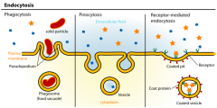
Three types of Endocytosis |
1. Phagocytosis (cell eating): the cells engulfs some relatively large or solid material such as a clump of bacteria, cell debris or inanimate particles -not routinely done 2. Pinocytosis/ Fluid-phase endocytosis (cell drinking): the cell "gulps" a drop of extracellular fluid containing dissolved molecules -non specific -routine activity of cells -receptor-mediated endocytosis: main mechanism for specific endocytosis 3. Exocytosis: vesicular transport process that ejects substances from the cell into the extracellular fluid -Secretory vesicle: protein-coated vesicle |
|
|
|
Diagram of Endocytosis |
|
|
|
|
The Microscope |
what did Biologist gain from the microscope being invented? -valuable tool to observe and study structures like cells that are too small to be seen by the unaided eye |
|
|
|
Care and Structure of the Compound Microscope |
Compound Microscope: precision instrument and should always be handled with care |
|
|
|
Rules for Microscope |
1. Transport 2. Cleaning 3. Use 4. Storage
|
|
|
|
Transport |
Transport: -hold it in an upright position with one hand on its arm and the other supporting its base -avoid swinging the instrument during its transport and jarring the instrument when setting it down |
|
|
|
Cleaning |
-use only special grit-free lens paper to clean the lenses -use circular motion to wipe the lenses -clean all lenses before and after use |
|
|
|
Use |
-always begin the focusing process with the lowest-power objective lens in position, changing to the higher-power lenses as necessary -use the coarse adjustment knob only with the lowest-power lens -Always use a cover slip with wet mount preparations |
|
|
|
Storage |
-before putting the microscope in the storage cabinet, remove the slide from the stage, rotate the lowest-power objective lens into position, wrap the cord neatly around the base, and replace the dust cover or return the microscope to the appropriate storage area -Never remove any parts from the microscope; inform you instructor of any mechanical problems that arise. |
|
|
|
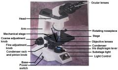
Identifying the Parts of a Microscope |
--- |
|
|
|
Base |
-supports the microscope |
|
|
|
Substage light or mirror |
Located on the base -light controls are located on the microscope base |
|
|
|
Stage |
the platform the slide rests on while being viewed -Mechanical Stage (spring clips): hold the slide in position for viewing |
|
|
|
Condenser |
small substage lens that concentrates the light on the specimen -Rack and Pinion knob: raises and lowers the condenser to vary light delivery |
|
|
|
Iris Diaphragm Lever |
arm attached to the base of the condenser that regulates the amount of light passing through the condenser |
|
|
|
Course Adjustment Knob |
Used to focus on the specimen |
|
|
|
Fine Adjustment Knob |
used for precise focusing once course focusing has been completed |
|
|
|
Head (Body Tube) |
Supports the objective lens system, which is mounted on a movable nosepiece, and the ocular lens or lenses |
|
|
|
Arm |
vertical portion of the microscope connecting the base and head |
|
|
|
Ocular (eyepiece) |
Depending on the microscope, there are one or two lenses at the superior end of the head or body tube. -Observations are made through the ocular(s) -An ocular lens has a magnification of 10X
|
|
|
|
Nosepeice |
rotating mechanism at the base of the head
|
|
|
|
Objective Lenses |
adjusting lens system that permits the use of a scanning lens (low-power lens), a high-power lens (oil immersion lens). -different magnifying and resolving powers |
|
|
|
Magnification and Resolution |
Magnification is achieve through the interplay of two lenses 1. Ocular Lens: the real image is magnified by the ocular lens to produce the virtual image seen by your eye 2. Objective Lens: objective lens magnifies the specimen to produce a real image that is projected to the ocular
Resolution (resolving power): the ability to discriminate two close objects as separate, is not. -Resolving Power: determined by the amount and physical properties of the visible light that enters the microscope |
|
|
|
Total Magnification |
any specimen being viewed is equal to the power of the ocular lens multiplied by the power of the objective lens used |
|
|
|
Diagram of Compound Microscope and its Parts |
|
|
|
|
Viewing Objects through the Microscope |
picture |
|
|
|
Field |
the area you see through the microscope |
|
|
|
Working Distance |
length of the bottom of the objective lens from the specime |
|
|
|
Parfocal |
the slide should be in focus (or nearly so) at the higher magnification once you have properly focused |
|
|
|
Classification of Tissues |
-- |
|
|
|
Tissues |
Tissues: Groups of cells that are similar in structure and function 1. Epithelial 2. Connective 3. Nervous 4. Muscle |
|
|
|
Organ |
Organ: the tissues organize to perform a specific body functions (heart, kidneys, lungs)
|
|
|
|
Histology |
Study of tissues |
|
|
|
Epithelial Tissue (Epithelium) |
sheet of cells that covers a body surface or lines a body cavity 1. Covering and lining epithelium 2. glandular epithelium |
|
|
|
Functions |
1. Protection 2. Absorption 3. Filtration 4. Excretion 5. Secretion 6. Sensory Reception |
|
|
|
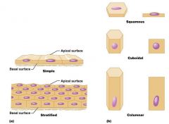
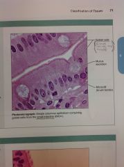
The covering and lining Epithelia are classified according to what two criteria: |
1. Arrangement or relative number of layers -Simple Epithelial (single) -Stratified Epithelial (multiple) 2. Cell Shape -Squamous (flat) -Cuboidal (cube -Columnar -Transitional (able to change shape and stratification) |
|
|
|
Simple Epithelial |
-consisting of one layer of cells attached to the basement membrane |
|
|
|
Stratified Epithelial |
consisting of two or more layers of cells |
|
|
|
Squamous |
Scalelike |
|
|
|
Cuboidal |
cubelike |
|
|
|
Columnar |
column-shaped |
|
|
|
Diagram of Classification of Epithelia |
|
|
|
|
Two Categorized Types of Epithelia |
1. Pseudostratified Epithelium (single player but looks like multiple) 2. Transitional Epithelium (able to change shape and stratification) |
|
|
|
Pseudostratified Epithelium |
simple columnar epithelium (one layer cells) -Cells vary in height and nuclei lie in different layers above the basement membrane, it gives the false appearance of being stratified |
|
|
|
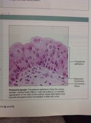
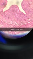
Transitional Epithelium |
particular stratified squamous epithelium formed of rounded (plump) -cells with the ability to slide over one another to allow the organ to be stretched -Found only in urinary bladder |
|
|
|
Two types of Epithelia Glands |
1. Endocrine Gland 2. Exocrine Gland |
|
|
|
Endocrine Gland |
lose their surface connection (duct) as they develop= ductless glands -secrete hormones into the extracellular fluid -the hormones then enter the blood or lymphatic vessels that weave through the glands |
|
|
|
Exocrine Glands |
retain their ducts and their secretion empty through these ducts either to the body surface or into body cavities -Sweat, oil glands, liver and pancreas |
|
|
|
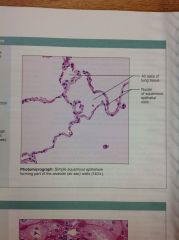
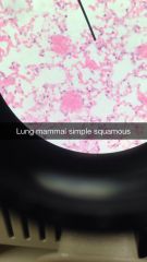
Simple Squamous Epithelium
|
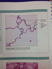
Lining of capillaries, lung aveoli, bowmans capsule, mesothelium
|
|
|
|
Simple Cuboidal Epithelium
|
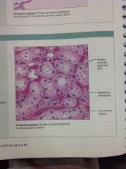
Kidney tubules, salivary, pancreatic duct
|
|
|
|
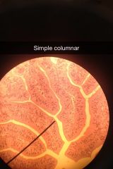
Simple Columnar Epithelium
|
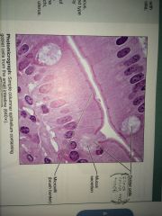
Lining of GI tract
|
|
|
|
Pseudostratified Columnar Epithelium
|
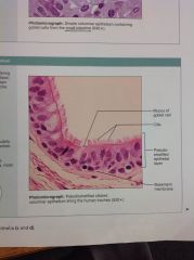
Associated with respiratory tract
|
|
|
|
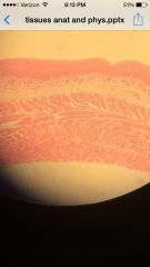
Stratified Squamous Epithelium
|
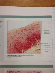
Skin epidermis
|
|
|
|
Transitional Epithelium
|
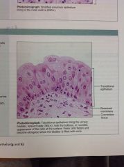
-Function: stretches readily and permits distension of urinary organ by contained urine -Location: Line the ureters, urinary bladder and part of the urethra |
|
|
|
Areolar Connective Tissue Diagram
|
|
|
|
|
Connective Tissue |
Perform a variety of functions, but they primarily: Protect, Support and Bind together other tissues of the body -found in all parts of the body as discrete structures or as part of various body organs -most abundant and widely distributed of the tissue type
|
|
|
|
Loose connective |
cells are fibroblasts (expect for fat- adipocytes) |
|
|
|
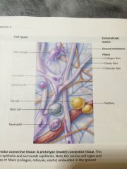
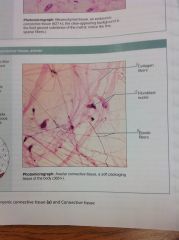
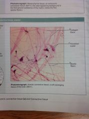
Areolar Connective Tissue |
found when tissue must be held in place but needs much space for fluid and vessels - most abundant type in body -present nearly everywhere -collagen dominates but has elastin fibers as well |
|
|
|
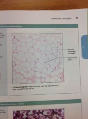
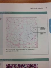
Adipose (fat) Tissue |
storage and metabolism fat |
|
|
|
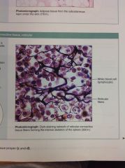
Reticular |
forms a supporting network -collagen, elastic and glycoproteins are present -found in stroma of organs and bone marrow |
|
|
|
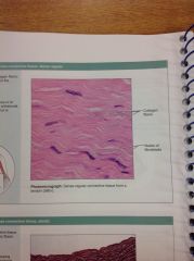
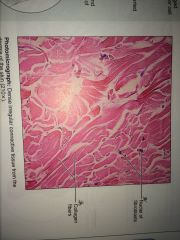
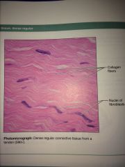
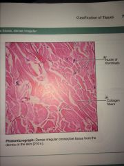
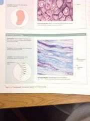
Dense Tissue |
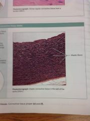
found when great strength and rigidity needed -collagen is densely packed in either a regular(parallel) or irregular arrangments -found in tendons, ligaments, organ capsules, fascia and sclera of eye *Elastic CT a type of dense regular connective tissue is found when great flexibility is needed, elastin fibers dominate -found in lung arteries, dermis of skin, lungs and bladder |
|
|
|
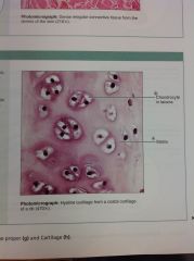
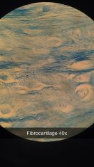
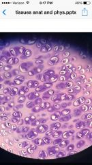
Cartilage |
nonvascular supporting tissue -cells are called chondrocytes sitting in a lacuna -semi-fluid extracellular matrix -3 types: 1. Fibrocartilage: collagen (rigid, strong); found in pubic symphysis, intervertebral discs 2. Elastic Cartilage: elastin (flexible); found in epiglottis, pinna 3. Hyaline Cartilage: more ground substance than fibers (mostly water), most abundant cartilage of the body; found in nasal septum, intercostal cartilages, larynx, trachea and articular surface |
|
|
|
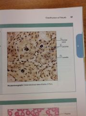
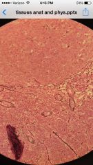
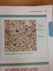
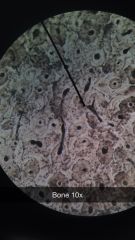
Bone |
vascular supporting tissue -cells called osteocytes -provides support to body -stores minerals and the marrow produces blood cells -*Haversian canal system: cells set in concentric circles surrounding their blood supply. |
|
|
|
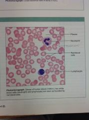
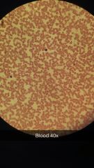
Blood |
supports metabolically -no fibers present -high water content 1. Red Blood Cells: cells without nuclei, biconcave discs 2. White Blood Cells: large defensive cells 3. Platelets (Thrombocytes): cell fragments function in repair and clotting
|
|
|
|
Connective Tissue Proper |
large amount of matrix: Connective Tissue Proper: includes areolar, adipose, reticular and dense (fibrous) connective tissue, cartilage, bone and blood. -all of these derive from an embryonic tissue called mesenchyme
|
|
|
|
Connective Tissue Proper: loose connective tissue, areolar
|
|
|
|
|
Connective Tissue Proper: loose connective tissue, adipose
|
|
|
|
|
Connective Tissue Proper: loose connective tissue, reticular
|
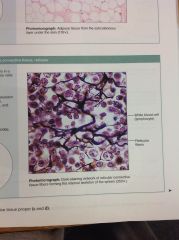
Stroma of organs and bone marrow
|
|
|
|
Connective Tissue Proper: dense connective tissue, dense regular
|
|
|
|
|
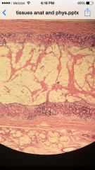
Connective Tissue Proper: dense connective tissue, dense irregular
|
|
|
|
|
Dense irregular
|
|
|
|
|
Cartilage: Hyaline
|
|
|
|
|
Bones (osseous tissue)
|
|
|
|
|
Blood
|
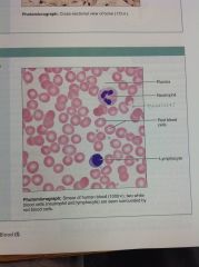
Blood
-Function: Transport of respiratory gases, nutrients, wastes and |
|
|
|
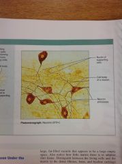
Nervous Tissue
|

|
|
|
|
Two Major Cell Populations that make up Nervous Tissue |
1. Neuroglia: special supporting cells that protect, support and insulate the more delicate neurons 2. Neurons: highly specialized to receive stimuli (excitability) and to generate electrical signals that may be sent to all parts of the body (conductivity) |
|
|
|
Muscle Tissue |
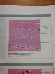
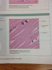
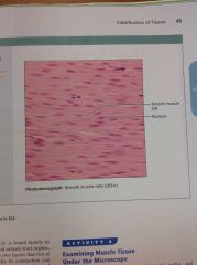
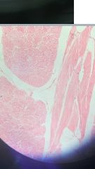
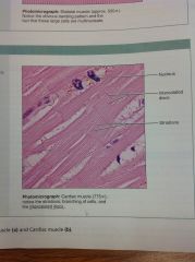
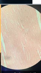
highly specialized to contract and produces most types of body movement -Three Basic Types: 1. Skeletal: the "meat" or flesh, of the body, is attached to the skeleton -under voluntary control and its contraction moves limbs and other external body parts 2. Cardiac: found only in the heart -intercalated discs: uninucleate cells that interdigitate (fit together) at junctions -involuntary control 3. Smooth (visceral muscle): found mainly in the walls of hallow organs -Two layers that run at right angles to each other |
|
|
|
Skeletal Muscle
|
|
|
|
|
Cardiac Muscle
|
|
|
|
|
Smooth Muscle
|
|
|
|
|
The Integumentary System |
Skin (Integument) insulates and cushions the underlying body tissues and protects the entire body from abrasion, exposure to harmful chemicals, temperature extremes and bacterial invasion -The hardened uppermost layer of the skin prevents water loss from the body surface
|
|
|
|
Basic Structure of the Skin |
Two distinct regions: 1. Epidermis: composed of epithelium 2. Dermis: underling connective tissue -Hypodermis (superficial fascia): immediately deep to the dermis. It is not considered to be part of the skin. Primarily of adipose tissue |
|
|
|
Epidermis |
Structurally: avascular epidermis is keratinized stratified squamous epithelium consisting of four distant cell types and four to five distinct layers |
|
|
|
Cells of the Epidermis |
1. Keratinocytes: the most abundant epidermal cells -main function is to produce keratin fibrils -Keratin: fibrous protein that gives the epidermis its durability and protective capabilities. -Tightly connected to each other by desosomes 2. Melanocytes: spidery black cells that produce brown-to-black pigment called melanin -the skin tans because melanin production increases when the skin is exposed to sunlight -Melanin provides a protective pigment umbrella over the nuclei of the cells in the deeper epidermal layers -a concentration of melanin in one spot is called a freckle 3. Dendritic cells (Langerhans cells): cells play a role in immunity 4. Tactile (Merkel) cells: Occasional spiky hemispheres that, in combination with sensory nerve endings, form sensitive touch receptors called tactile (merkel) discs located at the epidermal-dermal junction |
|
|
|
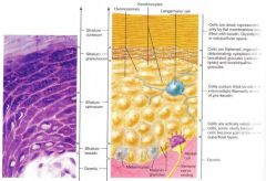
Layers of the Epidermis |
Epidermis consists of four layers in thin skin, which covers most of the body -Thick skin, found on the palms of the hands and soles of the feet, contains an additional layer, the stratum lucidum -From deep to superficial: 1.Stratum Basale 2. Stratum spinosum 3. Stratum granulosum 4. Stratum lucidum 5. Stratum corneum |
|
|
|
Stratum Basale (basal layer) |
single row of cells immediately adjacent to the dermis -cell are constantly undergoing mitotic cell division to produce millions of new cells daily |
|
|
|
Stratum Spinosum (Spiny Layer) |
Stratum consisting of several cell layers immediately superficial to the basal layer. -cells contain thick web-like bundles of intermediate filament made of pre-keratin protein -appear spiky because as the skin tissue is prepared for histological exam, they shrink but their desmosomes hold tight. -Cells divide pretty rapidly in this layer, but less so than in stratum basale -only one that receives adequate nourishment via diffusion of nutrients from dermis |
|
|
|
Stratum Granulosum (granular layer) |
thin layer named for the abundant granules its cells contain -Two types: 1. Lamellar granules: contain waterproofing glycolipid that is secreted into the exracellular space 2. Keratohyaline granules: combine with intermediate filaments in the more superficial layers to form keratin fibers |
|
|
|
Stratum Lucidum(clear layer) |
very thin translucent band of flattened dead keratinocyted with indistinct boundaries. -Not present in regions of thin skin |
|
|
|
Stratum Conreum (horny layer) |
outermost epidermal layer consists of some 20-30 cell layers and accounts for some bulk for the epidermal thickness -they are dead and flattened scalelike remnants are fully keratinized -constantly rubbed off and being replaces |
|
|
|
Dermis |
dense irregular connective tissue making up the dermis consist of two regions: 1. Papillary Area 2. Reticular Area |
|
|
|
Papillary Layer |
the more superficial dermal region composed of areolar connective tissue -very uneven and has fingerlike projections from its superior surface, dermal papillae, which attach it to the epidermis above (produce fingerprints, pain and touch receptors)
|
|
|
|
Reticular Layer |
the deepest skin layer -composed of dense irregular connective tissue and contains many arteries and veins, sweat and sebaceous glands, and pressure receptors
|
|
|
|
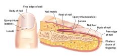
Nail Diagram |
|
|
|
|
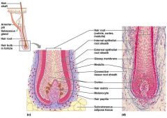
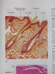
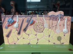
Hairs and Associated Structures
|
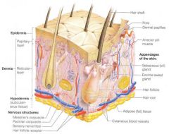
-Hair: structure consisting of medulla, a central region surrounded first by the cortex and then by a protective cuticle:
1. Root: portion of the hair enclosed within the follicle 2. Shaft: portion projecting from the scalp surface 3. Hair bulb: collection of well-nourished germinal epithelial cells 4. Follicle: structure formed from both epidermal and dermal cells. -enclosed by a thickened basement membrane, the glossy membrane and a peripheral connective tissue sheath 5. Arrector pili muscle: small bands of smooth muscle cells connect each hair follicle to the papillary layer of the dermis *goose bumps* |
|
|
|
Structure of hair and hair follicle diagram
|
|
|
|
|
Cutaneous Glands
|
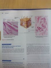
Two categories:
1. Sebaceous (Oil) Glands 2. Sweat (Sudoriferous) Glands |
|
|
|
Sebaceous Glands |
found nearly all over the skin, except for the palms of the hands and the soles of the feet -outlets for the glands of epithelial openings called pores. -Sweat glands are categorized by the composition of their secretions 1. Exocrine (merocrine sweat) glands: produce clear perspiration consisting primarily of water, salts and urea. 2. Apocrine glands: found predominantly in the axillary and genital areas, they secrete the basic components of eccrine sweat plus proteins and fat rich substances
|
|
|
|
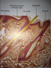
Photomicrographs of skin
|
|
|
|
|
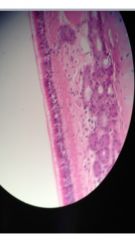
The main structural features in epidermis of thin skin
|
|
|
|
|
Homeostasis |
-- |
|
|
|
Differentially permeable |
not all substances penetrate the plasma membrane the plasma equally |
|
|
|
Solutions |
all of the liquids found in organisms |
|
|
|
Solute |
the particles found in organism -salt, proteins |
|
|
|
Thermal Kinetic Energy |
the energy associated with a constant random motion of cells |
|
|
|
Kinetic |
the energy seen in moving bodies |
|
|
|
Potential |
energy that is stores or inactive |
|
|
|
Brownian Movement |
movement of invisible particles |
|
|
|
Concentration Gradient |
unequal distribution of molecules |
|
|
|
Diffusion |
the movement of molecules down their concentration gradient |
|
|
|
Passive Diffusion |
movement is caused by the passive diffusion to occur |
|
|
|
Osmosis |
diffusion of water through a differentially permeable membrane from a region in which it is highly concentrated to a region in which its concentration is lower |
|
|
|
Electrolytes |
substances that break up into separate ions when dissolved in water |
|
|
|
Net movement |
which way the concentration is moving |
|
|
|
Active Transport |
solute molecules may move against a concentration gradient |
|
|
|
Parfocal |
when the object is in focus under low power it is also in focus under medium and high power |
|
|
|
Field of View |
angle of visible field |
|
|
|
Depth of Field |
amount of distance between the nearest and farthest objects that appear in acceptably sharp focus |
|
|
|
Focal Plane |
represents the area in a camera where light is focused. |
|
|
|
Slides! |
-- |
|
|
Simple Squamous
|
|
|
|
Simple Columnar
|
|
|
|
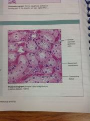
Simple Cubodial
|
|
|
|
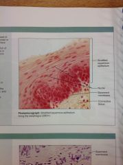
Stratified Squamous
|
|
|
|
Transitional Epithelium
|
|
|
|
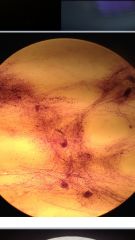
Connective Tissue Proper; Loose Connective: Areolar
|
|
|
|
Connective Tissue proper; loose connective, adipose
|
|
|
|
Connective Tissue proper; loose connective, reticular
|
|
|
|
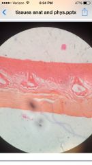
Dense Connective: Regular
|
|
|
|
Dense Connective: Irregular
|
|
|
|
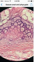
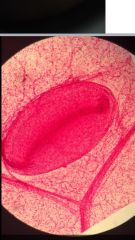
Cartilage: Fibrocartilage
|
|
|
|
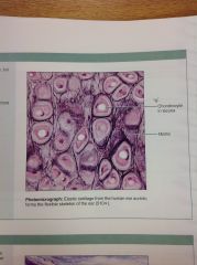
Cartilage: Elastic 40X
|
|
|
|
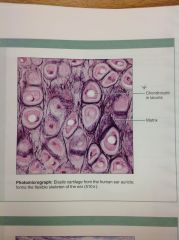
Cartilage: Elastic 10X
|
|
|
|
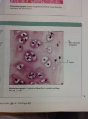
Cartilage; hyaline
|
|
|
|
Bone
|
|
|
|
Blood
|
|
|
|
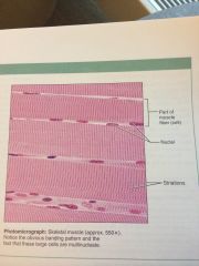
Skeletal Muscle 40X
|
|
|
|
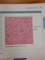
Smooth Muscle 40X
|
|
|
|
Cardiac Muscle
|
|
|
|
Nervous Tissue
|
|
|
|
|
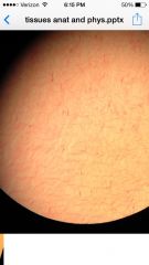
Skin (unpigmented + pigmented)
|
|
|
|
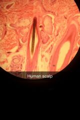
Skin (Human Scalp)
|
|
|
|
|
Pacinian Corpsucle
|
|
|
|
|
Hair Follicle Model |
|
|
|
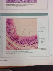
Pseudostratified columnar epithelium
|
|
|
|
|
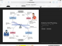
Homeostasis
|
ability of an organism to maintain constancy in the internal environmet
|
|
|
|
Homeostasis
|
|
|