![]()
![]()
![]()
Use LEFT and RIGHT arrow keys to navigate between flashcards;
Use UP and DOWN arrow keys to flip the card;
H to show hint;
A reads text to speech;
63 Cards in this Set
- Front
- Back
|
Parotid GLand |
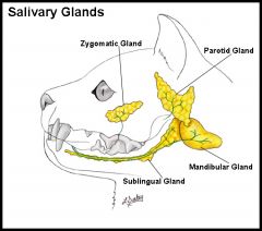
large salivary gland having a lobulated texture. |
|
|
Parotid Duct |

flat duct which runs from the front of the parotid gland across the masseter muscle and penetrates the upper lip to open into the oral cavity. |
|
|
Submaxillary |
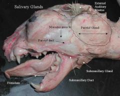
second largest salivary gland. |
|
|
Sublingual Gland |
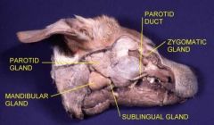
oblong salivary gland which abuts against the front of the submaxillary and its duct. |
|
|
Lymph Nodes |
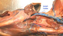
portions of the circulatory system |
|
|
Lips |
opening into oral cavity |
|
|
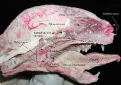
Hard Palate |
forms roof of oral cavity, ridges are known as palatine rugae |
|
|
Soft Palate |
fleshy extension from the hard palate. The uvula is missing in the cat |
|
|
Vestibule |
portion of the oral cavity between the teeth and cheeks. The pockets where people put dip. |
|
|
Tongue |
muscular structure in the floor of the oral cavity and partially the throat area. |
|
|
Papillae |
the spikey things on the tongue. more pronounced on the cat than on the human. |
|
|
Frenulum |
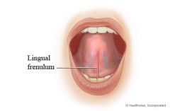
|
|
|
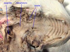
oropharynx |
ventral to the soft palate |
|
|
palatine tonsils |
a pair of specially named lymph nodes |
|
|
nasopharynx |

lies dorsal to the soft palate |
|
|
auditory tubes |

slitlike openings |
|
|
laryngopharynx |
small portion of throat at the end of the soft palate where oropharynx and nasopharynx come together |
|
|
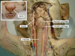
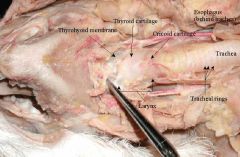
larynx |
pie |
|
|
epiglottis |
|
|
|
trachea |
windpipe, help open by incomplete rings of cartilage; continues caudally from the larynx |
|
|
esophagus |
collapsed muscular tube found dorsal to the trachea |
|
|
gullet |
the opening of the esophagus |
|
|
thyroid gland |
fatty area around the trachea |
|
|
intrinsic larynx muscles |
muscles running between various cartilages which compose the voice box |
|
|
thyroid cartilage |
|
|
|
arytenoid cartilages |

|
|
|
glottis |

|
|
|
*true and false vocal cords |
? |
|
|
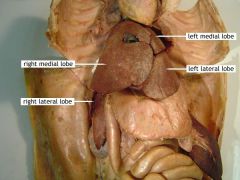
diaphragm, liver |

|
|
|
Right Lung |
on cats right, four lobes (cranial, middle, caudal, accessory) |
|
|
parietal pleura |
shiny, serous epithelium which lines all of the walls of the plueral cavity |
|
|
visceral pluera |
shiny, serous membrane on the outer surface of the lung |
|
|
root of the lung |
medial region of the lung where structures enter and leave the lung |
|
|
mediastinum |
the potential space between the right and left pleural cavities extending from the breastbone to the vertebral column. all of the chest structures except the lungs and pleural membranes are contained here. |
|
|
pleural folds (mediastinal septum)? |
delicate, transparent structure which runs from the ventral surface of the heart area to the breastbone. |
|
|
left lung |
divided into 3 lobes (cranial, middle, caudal) |
|
|
thymus gland |

|
|
|
phrenic nerves |
whitish strands which pass ventral to the root of the lung and extend to the diaphragm |
|
|
esophagus |
dorsal to the left lung. to see it, lift left lung entirely. it is also behind the trachea. |
|
|
aorta? ask to point out. |
a vessel seen adjacent and just dorsal to esophagus. |
|
|
peritoneal cavity |
the abdominal cavity |
|
|
parietal peritoneum |
shiny membrane which lines the abdominal wall |
|
|
visceral peritoneum |
serous membrane covering the viscera. where is the viscera? |
|
|
central tendon |
center portion of the diaphragm. can be seen by gently pulling the liver and diaphragm apart. |
|
|
falciform tendon |

|
|
|
Round ligament |
a slight thickening which is observable on the free edge of the falciform ligament. |
|
|
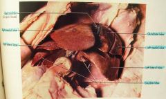
Lobes of the liver |
|
|
|
gall bladder |
gall bladder is a storage area for bile, is usually found in the lier |
|
|
lesser omentum |
|
|
|
stomach |
|
|
|
greater and lesser curvature, fundus, corpus, pyloric region and valve. |

|
|
|
spleen |

|
|
|
greater omentum |
yellowish gross stuff in cat |
|
|
gastrosplenic ligament |
the part of the greater omentum that is between stomach and spleen |
|
|
omental bursa |
the greater omentum that covers all of the intestines |
|
|
hepatic ducts |
carries bile from various parts of the liver |
|
|
cystic duct |
carries bile from the gallbladder |
|
|
common bile duct |
the two come together and form this |
|
|
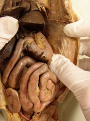
duodenum, jejunum, ileum |
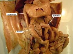
|
|
|
mesoduodenum |
the fatty substance of the mesentery |
|
|
mesentery |
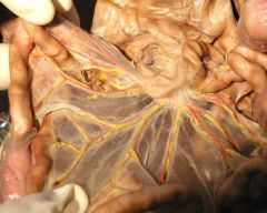
suspends both the jejunum and ileum |
|
|
pancreas |
sits on the small intestines |
|
|
cecum, ascending colon, transverse colon, descending colon, rectum |
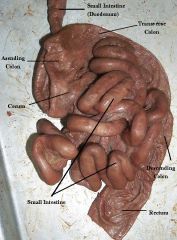
|